• 검색 결과가 없습니다.

A Case of Posterior Mediastinal Plasmacytoma Confounded by Community-Acquired Pneumonia

N/A
N/A
Protected

Academic year: 2021

Share "A Case of Posterior Mediastinal Plasmacytoma Confounded by Community-Acquired Pneumonia"

Copied!
5
0
0

로드 중.... (전체 텍스트 보기)

전체 글

(1)

A Case of Posterior Mediastinal

Plasmacytoma Confounded by Community- Acquired Pneumonia

Dahee Heo, M.D.

1

, Ki Yung Boo, M.D.

1

, Hyeyoung Jwa, M.D.

1

, Hwa Young Lee, M.D.

1

, Jihyun Kim, M.D.

1

, Seong Taeg Kim, M.D.

1

, Hye Mi Seo, M.D.

1

, Sang Hoon Han, M.D.

1

, Young-Hee Maeng, M.D.

2

and Jong Hoo Lee, M.D.

1

Departments of

1

Internal Medicine and

2

Pathology, Jeju National University Hospital, Jeju National University School of Medicine, Jeju, Korea

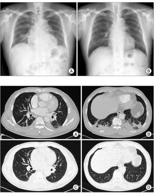
Plasmacytomas are extramedullary accumulations of plasma cells originating from soft tissue. Mediastinal plasmacytoma is a rare presentation. A 67-year-old man recovered after antibiotic treatment for community-acquired pneumonia. However, on convalescent chest radiography after 3 months, mass like lesion at the right lower lung field was newly detected. Follow-up chest computed tomography (CT) revealed an increase in the extent of the right posterior mediastinal mass that we had considered to be pneumonic consolidations on previous CT scans. Through percutaneous needle biopsy, we diagnosed IgG kappa type extramedullary plasmacytoma of the posterior mediastinum.

Keywords: Plasmacytoma; Hematopoiesis, Extramedullary; Pneumonia; Diagnosis

mopathy of unknown significance

1

. Plasmacytomas may be primary or secondary to disseminated multiple myeloma and may arise from medullary or extramedullary sites

1

.

The mediastinal involvement of EMP is rare, and diagnosis of plasmacytoma could be often delayed

2

. However, because EMP can be the sequence of proceeding for multiple myelo- ma, these tumors need to be diagnosed early and treated to prevent further morbidity. We describe a case of posterior me- diastinal plasmacytoma with delayed diagnosis due to clinical improvement after antimicrobial therapy for community- acquired pneumonia (CAP) with a review of the literature.

Case Report

A 67-year-old man was admitted to our hospital with febrile sensation and cough for 3 days. He had no other underlying chronic disease. He was an ex-smoker (20 pack-years) and did not have a history of alcohol abuse. His vital signs were as fol- lows: blood pressure, 140/82 mm Hg; heart rate, 68 beats per minute; respiratory rate, 20 per minute; and body tempera- ture, 38.5

o

C. Auscultation revealed coarse crackles in both lower lung fields.

Copyright © 2015

The Korean Academy of Tuberculosis and Respiratory Diseases.

All rights reserved.

Introduction

Extramedullary plasmacytomas (EMPs), a category of plasma cell malignancies, are characterized by localized monoclonal plasma cell proliferation forming a solitary lesion outside the bone marrow

1

. These are associated with the up- per aerodigestive tract in about 80% of cases, and manifest as multiple myeloma, primary amyloidosis, or monoclonal gam-

Address for correspondence: Jong Hoo Lee, M.D.

Department of Internal Medicine, Jeju National University Hospital, Jeju National University School of Medicine, 15 Aran 13-gil, Jeju 690-767, Korea

Phone: 82-64-717-1601, Fax: 82-64-717-1131 E-mail: [email protected]

Received: Aug. 12, 2014 Revised: Dec. 5, 2014 Accepted: Jan. 1, 2015

cc

It is identical to the Creative Commons Attribution Non-Commercial

License (http://creativecommons.org/licenses/by-nc/4.0/).

(2)

Chest radiography showed multiple pulmonary infiltrations in the right lung fields (Figure 1A). White blood cell count was 8,000/mm

3

with 76.0% segmented neutrophils, 13.7% lym- phocytes, and 1% eosinophils. C-reactive protein and procalci- tonin were 23.34 mg/dL and 0.579 mg/dL, respectively. Other routine blood chemistry tests were all normal. Arterial blood gas analysis revealed pH 7.439, PCO

2

36.3 mm Hg, PO

2

52.4 mm Hg, and HCO

3

24.9 mmol/L.

A computed tomography (CT) scan of the chest was per- formed, and revealed multiple pulmonary consolidations, ground-glass opacities, and interstitial septal thickenings in both lower lung fields (Figure 2A, B). Based on these observa- tions, we made a diagnosis of CAP and initiated antimicrobial therapy of intravenous ceftriaxone 2 g plus azithromycin 500 mg. After treatment of CAP, the clinical findings of the patient

stabilized, and the patient was discharged on hospital day 4.

After three months, we performed a routine check-up of the patient. The patient had no definitive symptoms. Follow-up chest radiography showed resolution of multiple pulmonary consolidations throughout most of the lung fields, but a mass- like lesion without the loss of right cardiac borders at the right lower lobe field was observed (Figure 1B). Follow-up CT scans of the chest revealed interval improvements in pneumonic consolidations in both lower lung fields, but an increased mass in the right posterior mediastinum at T6–7 level that we had considered to be pneumonic consolidations on the previous CT scans was visible (Figure 2C, D). This oval and elongated mass with mild enhancement of soft tissue density without any bone destruction was observed (Figure 3A, B).

Positron emission tomography using 2-[fluorine-18] fluoro-

Figure 1. Chest radiography images of the patient. (A) Initial chest radiography showing multiple pulmonary consolida- tions. (B) A mass-like lesion without the loss of right cardiac borders on a rou- tine follow-up chest radiography after 3 months.

Figure 2. Computed tomography (CT)

scan images of the chest. (A, B) Initial

chest CT scan showing multiple pulmo-

nary consolidations at both lung fields. (C,

D) Follow-up chest CT scan revealing an

increased mass in the right posterior me-

diastinum with improvements in other

pneumonic consolidations at other lung

fields.

(3)

2-deoxy-D-glucose (FDG) imaging and CT scans of the whole body except the brain revealed mild FDG uptake of maximum standardized uptake value 3.4 at the posterior mediastinal mass.

Percutaneous needle biopsy using ultrasound was per- formed. Lung ultrasound revealed an ill-defined, hypoechoic subpleural lesion measuring about 5.0 cm×2.7 cm at right paraspinal area of T6–7 level. And microscopic examination showed diffuse infiltrates of polymorphous and asynchronous plasma cells with large and eccentric nuclei, prominent nucle- oli, and abundant cytoplasm (Figure 4A). Immunohistochem- istry revealed the biopsy was positive for CD138, the cyto- plasm was diffusely reactive for monoclonal kappa-light chain (Figure 4B). Based on these findings, the posterior mediastinal mass was diagnosed as a IgG kappa type plasmacytoma.

We subsequently investigated the possibility of multiple myeloma. Laboratory tests showed an inverse albumin/

globulin ratio (total protein, 10.7 g/dL; albumin, 3.0 g/dL; A/G

ratio, 0.38). Serum hemoglobin, creatinine, and total calcium were 11.8 g/dL, 1.4 mg/dL, and 8.5 mg/dL, respectively. Im- munofixation of serum showed monoclonal gammopathy of IgG-kappa type. In serum protein electrophoresis, the M spike level was 4.58 g/dL. Serum Ig determination revealed a markedly increased IgG level (6,993.1 mg/dL), and the level of β2-microglobulin was 3,674.2 ng/mL. On bone marrow aspiration and biopsy, plasma cells were increased up to 7.8%

of all marrow nucleated cells. Those findings were consistent with monoclonal gammopathy of undetermined significance (MGUS) and insufficient for diagnostic criteria of symptom- atic multiple myeloma. The patient was finally diagnosed as a posterior mediastinal plasmacytoma (IgG-kappa type). The patient was referred to the hematology department for proper treatment. This report was approved by the Ethical Review Committee.

Figure 3. (A, B) Contrast-enhanced com- puted tomography (CT) scan images of the chest after treatment of pneumonia.

Chest CT scan revealing oval and elon- gated mass (arrow) with mild enhance- ment of soft tissue density without any bone destruction.

Figure 4. (A) Microscopic examination showing diffuse infiltrates of plasma cells with large and eccentric nuclei with prominent nucleoli and

abundant cytoplasm (H&E stain, ×200). (B) Immunohistochemistry showing tumor cells with diffuse reactivity to cytoplasmic monoclonal

kappa-light chain reactivity (×200).

(4)

Discussion

EMPs result from monoclonal proliferation of plasma cells in soft tissues or organs outside the bone marrow, and con- stitute about 4% of plasma cell malignancies. Although most tumors (approximately 82%) occur in regions of the upper respiratory tract such as the oronasopharynx and paranasal sinuses, these tumors can also occur in the gastrointestinal tract, spleen, liver, lymph nodes, kidneys, thyroid gland, adre- nal gland, ovary, testes, lung, pleura, pericardium, and skin

1

. The posterior mediastinal plasmacytoma as occurred in our case is rare, and most studies are case reports

3-5

.

While EMP of the upper respiratory tract develops fre- quently in men (male-to-female ratio ranges from 3:1 to 5:1), no predilection for thoracic involvement according to sex has been reported

6

. The most common age of onset is 50 to 60 years, and clinical signs depend on the location of the tumor

6

.

EMP can occur as a primary tumor without any bone mar- row plasmacytosis, or as a manifestation of multiple myeloma with extramedullary involvement

1

. Before multiple myeloma enters into an aggressive phase, the disease is mostly con- tained within the bone marrow

6

. However, during the trans- formation phase, extramedullary manifestations can develop

6

. The incidence of EMP associated with multiple myeloma ranges from 7% to 17% in patients at the time of diagnosis, and 6% to 20% during the clinical course of the disease

7

.

Otherwise, EMP can be the sequences of proceedings for multiple myeloma. Moran et al.

8

reported two cases of EMP presenting as a mediastinal mass, which preceded the onset of full-blown multiple myeloma. And the other report concerned that 10% to 30% of solitary EMP cases could be disseminated into multiple myeloma within the first 2 years

9

. Therefore, the presence of the EMP often provides a hint to the diagnosis of multiple myeloma.

In terms of posterior mediastinal manifestations of EMP, it should be differentiated from neurogenic tumor, lymphoma, and lymphangioma

10

. And EMP within thoracic cavity can present as lung nodule or mass, or as pulmonary consolida- tive patterns

11,12

. In practice, EMP is highly indistinguishable from other thoracic disease and it is a great challenge to diag- nose thoracic or mediastinal plasmacytoma.

In our case, we failed to detect underlying posterior medi- astinal plasmacytoma at the time of initial admission because the patient had pneumonia that subsequently responded to antimicrobial therapy. Follow-up chest CT scans revealed an irregular enhancing mass at the right posterior mediastinum.

Based on histology findings, we made a delayed diagnosis of posterior mediastinal plasmacytoma. In additioin, we sug- gested that EMP could be the sequences of proceedings for multiple myeloma. In our case, patient was diagnosed as a EMP coexisted with MGUS according to International myelo- ma working guidelines published in 2009

13

. This case is rare because there are only three cases in PubMed which reported

plasmacytoma of posterior mediastinum without multiple myeloma

3-5

. Furthermore, this is the second case report ever with rare IgG kappa type of plasmacytoma in posterior medi- astinum

3

.

The management of plasmacytoma is entirely different for both types of plasma cell dyscrasias. Most cases of primary EMP without multiple myeloma are treated with surgery and/or radiation therapy, and clinical responses are generally good

6

. However, if the diagnosis of EMP is concurrent with multiple myeloma, the prognosis is very poor and treatment includes chemotherapy or autologous hematopoietic cell transplantation which is directed towards the underlying dis- ease

14

.

In conclusion, this case suggests that plasmacytoma can manifest as a form of posterior mediastinal mass. And, be- cause this disease cannot be often differentiated from pneu- monic consolidations, careful monitoring may be required in some patients who recover after antimicrobial therapy of pneumonia.

Conflicts of Interest

No potential conflict of interest relevant to this article was reported.

Acknowledgements

This research was supported by the 2015 scientific promo- tion program funded by Jeju National University.

References

1. Alexiou C, Kau RJ, Dietzfelbinger H, Kremer M, Spiess JC, Sch- ratzenstaller B, et al. Extramedullary plasmacytoma: tumor occurrence and therapeutic concepts. Cancer 1999;85:2305- 14.

2. Bataille R, Sany J. Solitary myeloma: clinical and prognostic features of a review of 114 cases. Cancer 1981;48:845-51.

3. Masood A, Hudhud KH, Hegazi A, Syed G. Mediastinal plas- macytoma with multiple myeloma presenting as a diagnostic dilemma. Cases J 2008;1:116.

4. Lee SY, Kim JH, Shin JS, Shin C, In KH, Kang KH, et al. A case of extramedullary plasmacytoma arising from the posterior mediastinum. Korean J Intern Med 2005;20:173-6.

5. Ganesh M, Sankar NS, Jagannathan R. Extramedullary plas- macytoma presenting as upper back pain. J R Soc Promot Health 2000;120:262-5.

6. Montero C, Souto A, Vidal I, Fernandez Mdel M, Blanco M, Verea H. Three cases of primary pulmonary plasmacytoma.

Arch Bronconeumol 2009;45:564-6.

(5)

7. Oriol A. Multiple myeloma with extramedullary disease. Adv Ther 2011;28 Suppl 7:1-6.

8. Moran CA, Suster S, Fishback NF, Koss MN. Extramedullary plasmacytomas presenting as mediastinal masses: clinico- pathologic study of two cases preceding the onset of multiple myeloma. Mod Pathol 1995;8:257-9.

9. Husain M, Nguyen GK. Primary pulmonary plasmacytoma diagnosed by transthoracic needle aspiration cytology and immunocytochemistry. Acta Cytol 1996;40:622-4.

10. Agarwal PP, Seely JM, Matzinger FR. Case 130: mediastinal hemangioma. Radiology 2008;246:634-7.

11. Mohammad Taheri Z, Mohammadi F, Karbasi M, Seyfollahi L, Kahkoei S, Ghadiany M, et al. Primary pulmonary plas- macytoma with diffuse alveolar consolidation: a case report.

Patholog Res Int 2010;2010:463465.

12. Horiuchi T, Hirokawa M, Oyama Y, Kitabayashi A, Satoh K, Shindoh T, et al. Diffuse pulmonary infiltrates as a roentgeno- graphic manifestation of primary pulmonary plasmacytoma.

Am J Med 1998;105:72-4.

13. Palumbo A, Sezer O, Kyle R, Miguel JS, Orlowski RZ, Moreau P, et al. International Myeloma Working Group guidelines for the management of multiple myeloma patients ineligible for standard high-dose chemotherapy with autologous stem cell transplantation. Leukemia 2009;23:1716-30.

14. Chen HF, Wu TQ, Li ZY, Shen HS, Tang JQ, Fu WJ, et al. Ex-

tramedullary plasmacytoma in the presence of multiple

myeloma: clinical correlates and prognostic relevance. Onco

Targets Ther 2012;5:329-34.

수치

Figure 1. Chest radiography images of  the patient. (A) Initial chest radiography  showing multiple pulmonary  consolida-tions
Figure 3. (A, B) Contrast-enhanced com- com-puted tomography (CT) scan images of  the chest after treatment of pneumonia

참조

관련 문서

I t maintains that scenes from early infancy, such as are brought up by an exhaustive analysis of neuroses (as, for in- stance, in the present case), are not

한국형 Slow City 의 필요성과 시사점.. 10) Mass travel could only develop with improvements in technology allowed the transport of large numbers of people in a short space

2재화 2요소 헥셔-올린 모형에서는 어느 한 경제에서 어느 한 요소의 양이 증가하면, 그 요소를 집약적으로 사용하는 산업의 생산량은 증가하고 다른

다양한 번역 작품과 번역에 관한 책을 읽는 것은 단순히 다른 시대와 언어, 문화의 교류를 넘어 지구촌이 서로 이해하고 하나가

Zao Wou-Ki generated the best H1 result for the entire Asian continent in Hong Kong; 53% of Zhang Daqian’s turnover was hammered there, and lots of new

The index is calculated with the latest 5-year auction data of 400 selected Classic, Modern, and Contemporary Chinese painting artists from major auction houses..

management of community-acquired pneumonia: an evidence- based update by the Canadian Infectious Diseases Society and the Canadian Thoracic Society. The

1 John Owen, Justification by Faith Alone, in The Works of John Owen, ed. John Bolt, trans. Scott Clark, "Do This and Live: Christ's Active Obedience as the