DOI 10.17480/psk.2018.62.6.381
Notch1 발현이 NCI-H460 폐암원형체의 침윤성 및 항암제 내성 발현에 미치는 기전 분석
윤 지 은#
청주대학교 이공대학 제약공학과
(Received September 27, 2018; Revised November 9, 2018; Accepted November 23, 2018)
Notch1 Expression Regulates the Invasiveness and Drug Resistance in Sphere-forming NCI-H460 Cancer Cells
Jieun Yun#
Department of Pharmaceutical Engineering, Cheongju University
Abstract — Drug resistance is an obstacle to chemotherapy in non-small cell lung cancer caner (NSCLC). It has been reported cancer stem cells are regarded as the mediators of drug resistance, metastasis, and/or recurrence. Sphere-forming cells isolated from cancer cell lines are identified as cancer stem cells and broadly utilized for cancer stem cell studies. In this study, sphere-forming cells were isolated from NCI-H460 (H460) cells, the NSCLC cell line. The H460 spheres demon- strated increased invasion capacity as compared with H460 adherent cells and also appeared to be resistant to doxorubicin and cisplatin which are chemotherapy drugs. The mechanism involves in part through Notch signaling and Slug, known as a direct target of Notch1. The expression of Notch1 and Slug was remarkably higher in H460 spheres compared to H460 adherent cells. In addition, suppression of Notch1 expression, accompanied by decreased Slug expression, reversed the increased invasion capacity and the acquired drug resistance in H460 spheres. These findings suggest that Notch1 is a potential therapeutic target for cancer stem cells as a strategy for resolving metastasis and resistance to anticancer drugs.
Keywords Cancer stem cells, Notch signaling, Slug, Drug resistance, Invasion
서 론 (Introduction)
폐암치료를 위해 최근 효과적인 암치료제들이 개발되어 임상 에 사용되고 있으나 암전이, 재발, 항암치료 내성은 여전히 풀어 야 할 숙제로 남아 있다.1)암줄기세포(cancer stem cell)는 암세 포 중 특정한 성질을 지닌 세포들로 자기갱신(self-renewal), 분 화(differentiation), 종양원성(tumorigenesis)을 나타내며 암의 전 이유발, 암재발, 항암제 내성의 주요 원인으로 제시가 되어 왔다.2) 따라서 보다 효과적인 암치료를 위해서 암줄기세포의 특성을 파 악하고 생성, 유지 및 작용 기전에 대한 연구가 필요하다. 암원
형체(tumor spheres) 형성은 암줄기세포연구에 활용되는 방법으 로 기존 연구보고에 따르면 암원형체세포 표면의 CD133, CD44, Nanog, Sox2 발현을 마커로 하여 암줄기세포임을 확인한다.3,4) 비소세포폐암 세포주인 NCI-H460 (H460) 세포주에서 폐암원형 체를 분리하여 CD133과 Nanog의 과발현으로 암줄기세포를 확 인한 연구결과에서는 이들 폐암원형체가 플루오로우라실과 메토 트렉세이트 항암제에 대해 내성을 유발하였고 작용기전으로 IL- 6R의 역할을 제시하였다.5)또한 비소세포폐암 A549 세포주에서 분리한 폐암원형체의 특성을 분석한 연구에서는 A549폐암원형 체가 A549 세포주와 비교하였을 때 세포 침윤성 증가 및 독소 루비신 항암제 내성 발현을 보였고 기전으로 Notch 신호전달체 계가 관련되어 있을 것이란 가능성을 시사하였다.6)
Notch 신호전달은 유전적으로 잘 보존된 리간드-수용체 신호 체계로써 세포분열, 분화, 세포운명 결정, 세포생존을 조절하는 것으로 알려져 있다.7) Notch 단백질은 단일 막관통 단백질로 세 포표면에 수용체로 작용한다. Notch 리간드와 수용체가 결합하
#
Corresponding Author Jieun Yun
Department of Pharmaceutical Engineering, Cheongju University, 298 Daeseong ro, Cheongwon gu, Cheongju, 28503, Republic of Korea
Tel.: +82-43-229-7863 Fax.: +82-43-229-8577 E-mail: [email protected]
Short Report
종설면 Notch 수용체는 순차적인 두 번의 잘림을 거치게 된다. 첫 번 째 담당 효소는 TACE (TNF-α converting enzyme)로 Notch의 extracellular domain이 잘리고 두 번째는 gamma-secretase 복 합체에 의해 일어난다. gamma-secretase 효소의 작용으로 세포 막에서 분리된 Notch는 세포 핵 안으로 이동하여 타겟 유전자 (HES, HEY, CyclinD1, P21/WAF1, c-Myc 등)의 프로모터에 붙 어 전사조절의 역할을 한다.8-10 포유동물은 4개의 Notch 수용체 를(Notch1, 2, 3, 4)를 가지고 있으며 리간드로는 Jagged1, 2와 DLL1, 3, 4가 알려져 있다.9)암에서 Notch의 역할규명은 활성 화된 Notch 단백질을 쥐 조혈모세포에 발현시키면 T-cell leukemia와lymphoma를 일으킨다는 보고로 시작되었다.11)이후 Notch 신호전달체계가 다양한 고형암(자궁경부암, 폐암, 췌장암, 난소암, 유방암, 전립선암 등)과 혈액암(호지킨림프종, 급성 림프 구성 백혈병 등) 모델에서 암 유발을 촉진시킨다고 보고되었고 Notch 수용체와 리간드들은 암의 진단 마커로 제시되었다.11-13) 더 나아가 최근Notch 신호전달체계는 암줄기세포형성, 암전이, 항암제 내성을 유발하는 주요 기전으로도 제안이 되었다.14-17) Notch 신호전달체계가 암줄기세포형성을 유도하며 Snail, Slug, TGF-α 신호전달체계를 조절하여 EMT (Epithelial to Mesenchymal transition) 를 유발한다는 결과들이 보고되었고 항암제 내성과 관련해서 Notch와 CD133이 과발현되어 있는 폐암세포주가 파 클리탁셀과 독소루비신에 내성을 나타내며 이에 MDR1과 P- glycoprotein이 관여한 것으로 밝혀졌다.18,19)또한 비소세포폐암 세포주에서 분리한 암줄기세포들이 시스플라틴 내성을 보이며 그 작용기전에 Notch3이 연계된 autophagy의 활성화가 관여한 다고 알려졌다.20,21)이러한 연구결과를 근거로 Notch 신호전달 체계는 항암 및 암전이 표적치료제 개발에 주요 타겟으로 제안 이 되었고 Notch 신호전달체계를 활성화 시키는 주 효소인 gamma-secretase 억제제, 항 Notch 항체, 항 DLL4 항체 등이 개발되고 있다.22,23)하지만 이런 다양한 연구에도 불구하고 암줄 기세포에서 보이는 활성화된 암세포 전이능 및 항암제 내성에 대 한 정확한 작용기전 연구가 필요한 상황이며 또한 암줄기세포를 타겟으로 하는 치료법 개발에 주요 표적을 발굴해야 한다. 따라 서 이 연구에서는 비소세포폐암 세포주인 H460 세포주에서 H460 폐암원형체를 분리하여 세포 증식능과 세포 침윤성, 항암제 내 성 여부 파악하고 관련된 기전을 분석하는 연구를 수행하였다.
실험방법 (Experimental Methods)
세포배양
비소세포폐암 세포주인 NCI-H460 (H460)은 American Type Culture Collection (Manassas, VA, USA)에서 구입하였다. 세포 주는 10% FBS (fetal bovine serum; Thermo Fisher Scientific, Waltham, MA, USA), 100 U/mL의 penicillin (Invitrogen Life
Technologies, Carlsbad, CA, USA)과 100 μg/mL의 streptomycin (Invitrogen Life Technologies, Carlsbad, CA, USA)이 포함된 RPMI1640 (Invitrogen Life Technologies, Carlsbad, CA, USA) 을 배지로 하여 37oC, 95% humidified air/5% CO2 incubator (VISION Scientific Inc., 대전, 대한민국)에서 배양하였으며 2-3 일마다 새로운 배지로 계대 배양하여 세포를 유지하였다. H460 폐암원형체 분리는 무혈청배지인 BEGM (Lonza Group Ltd., Basel, Switzerland)에 hEGF (10 ng/mL; Peprotech Inc., Rocky Hill, NJ, USA)와 사람 bFGF (10 ng/mL; Peprotech Inc., Rocky Hill, NJ, USA))를 첨가하여 배양하였고 무코팅 배 양플레이트(Corning Inc., Corning, NY, USA)를 사용하였다.
H460 폐암원형체는 위와 같은 조건에서 배양하였을 때 5일 후 에 형성이 되었으며 hEGF와 bFGF는 주 2회씩 추가로 배양액 에 첨가해 주었다.
세포 증식 분석
세포 생존률 분석을 위해 8,000 개의 세포를 96-well 플레이 트(Corning Inc., Corning, NY, USA)에 분주하여 RPMI1640에 10% FBS를 첨가한 배지에서 배양하였고 Cell Proliferation Kit II (XTT assay; Roche, Basel, Switzerland)를 사용하여 정량화 하였다. 분석방법은 제조회사에서 제시한 실험방법을 준수하였 다. 각 실험 당 사용한 well의 개수는 4개 이상을 사용하였다.
세포 침윤성 분석
트랜스웰 필터(24-well 용; Corning Inc., Corning, NY, USA) 를 매트리젤(BD Biosciences, Franklin Lakes, NJ, USA)로 코 팅한 후 50,000개의 세포를 개수하여 RPMI1640 배지에 희석한 후 매트리젤이 코팅된 트랜스웰의 상부에 분주하였다. 필터를 24- well 플레이트(Corning Inc., Corning, NY, USA)에 삽입하고 필 터 하부가 10% FBS가 포함된 세포 배양액(RPM1640+10%
FBS)에 잠기도록 하였다. 배양 시작 24시간 후에 필터는 회수하 여 면봉으로 상부 매트리젤을 제거한 후 필터 아래 부분으로 이 동한 세포들을 현미경을 이용하여 개수하였다. 각 실험 당 트랜 스웰은 4개 이상을 사용하였다.
단백질발현 분석
세포 단백질은 PMSF (Sigma-Aldrich, Waltham, MA, USA) 와 proteinase inhibitor cocktail (Calbiochem, San Diego, CA, USA)이 포함된 RIPA buffer (50 mM Tris-Cl (pH 8.0), 5 mM EDTA, 150 mM NaCl, 1% NP-40, 0.1% SDS, 1 mM phenylmethylsulfonyl fluoride; Sigma-Aldrich, Waltham, MA, USA)를 사용하여 세포들을 모았고 얼음에서 30분 방치 후 15000 rpm, 4oC에서 15 분간 원심분리하여 상층액을 새로운 1.5 mL microcentrifuge tube에 나누어 담았다. 얻어진 단백질의 농도는
Bio-Rad protein assay kit(Bio-Rad, Hercules, CA, USA)를 이 용하여 분석하였고 제조회사에서 제시한 실험방법을 준수하였 다. 정량 후 20 μg의 전체 단백질을 Protein 5 × Sample Buffer (ELPIS BIOTECH, 대전, 대한민국)에 혼합하고 100oC에서 5 분 간 가열 한 후 10% SDS-PAGE gel을 이용해서 단백질을 크기 별로 분리하였다. 이후 PVDF막(Bio-Rad, Hercules, CA, USA)에 트랜스퍼한 후 발현된 단백질량은 enhanced chemiluminescent protein (ECL) detection system (Millipore, Burlington, MA, USA)을 사용하여 X-ray 필름에 현상하여 분석하였다. 사용한 항 체 중 Notch 1 (C-20), Notch 4 (H-225), Jagged 1 (C-20)는 Santa Cruz Biotechnology (Santa Cruz, CA, USA)에서 구입하였고, c- Myc (M5546)은 Sigma-Aldrich (Waltham, MA, USA)에서, GAPDH 는 Cell Signaling Technology (Beverly, MA, USA)에 서 구입하였다.
정량적 역전사 중합효소 연쇄반응(Quantitative real time PCR)
RNA 추출은 배양된 세포를 PBS로 세척한 후 세포 스크랩퍼 (Thermo Fisher Scientific, MA, USA)를 이용해서 세포를 모았 으며, 이를 12,000 rpm, 4oC에서 2 분간 원심 분리하여 상층액 을 버리고 세포 펠렛을 얻었다. 전체 RNA는 RNeasy Plus Mini Kit (Qiagen, Hilden, Germany)를 사용하여 제조사가 제공한 실 험법으로 추출하였다. DEPC-water로 녹인 RNA는 A260/280 ratio(Versa Max microplate reader, Molecular Devices, San Jose, CA, USA)로 흡광도를 측정해 순도와 농도를 확인하였다.
얻어진 RNA는 PrimeScript 1st strand cDNA synthesis kit (TaKaRa, Kusatsu, Japan)를 이용하여 역전사 반응시켰고 이때 1μg의 RNA를 사용하였다. Real-time PCR 분석은 7500 Fast Real-Time PCR System (Applied Biosystems, Foster City, CA, USA)을 활용하여 제조사에서 제공된 실험법에 따라 실험하 였다. GAPDH를 internal control로 사용하였고 유전자의 상대
적 발현을 ΔΔCt method로 계산하였다. 실험에 사용한 primer 서열은 Table I에 정리하였다.
Transfection
단백질 발현 억제를 위해 Control siRNA (scrambled siRNA;
sc-37007)와 Notch1 siRNA (sc-36095)를 Santa Cruz Biotechnology (Santa Cruz, CA, USA)에서 구입하여 세포에 transfection하였 다. Lipofectamin 2000 Transfection reagent (Invitrogen Life Technologies, Carlsbad, CA, USA)를 사용하였고 제조사에서 제 공된 실험법에 따라 실험하였고 최종 siRNA의 농도는 100 pmole 을 사용하였다.
통계분석
통계학적 유의성을 평가하기 위해 SigmaPlot 프로그램(Systat Software, Inc., San Jose, CA, USA)을 이용하여 Student’s t- test 또는 one-way ANOVA 방법으로 분석하였다. P 값이 0.05 이하 일 때 통계적으로 유의한 차이가 있다고 보았다.
결과 및 고찰 (Results and Discussion)
암줄기세포 이론은 암세포 중 특정 세포들이 자기갱신, 분화 등의 성질을 가지고 있으며 이러한 성질들이 암의 전이유발, 암
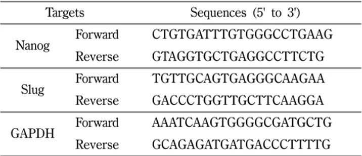
Table I – Sequences of primers used for quantitative real time PCR
Targets Sequences (5' to 3')
Nanog Forward CTGTGATTTGTGGGCCTGAAG Reverse GTAGGTGCTGAGGCCTTCTG
Slug Forward TGTTGCAGTGAGGGCAAGAA
Reverse GACCCTGGTTGCTTCAAGGA
GAPDH Forward AAATCAAGTGGGGCGATGCTG
Reverse GCAGAGATGATGACCCTTTTG
Fig. 1 – Isolation of lung cancer spheres from H460 cells. (A) H460 NSCLC cells were grown in RPMI1640 media with 10% FBS displaying
adherent phenotype. (B) H460 cells, cultured in serum-free BEGM media supplemented with EGF and bFGF, formed floating spheres
(X 100). (C) Nanog mRNA expression was analyzed in H460 adherent and spheres by quantitative real time PCR. GAPDH was used
as an internal control. ***p<0.001.
재발, 항암제 내성 등의 주요 원인이라는 가설을 제시한다.2,5)따 라서 효과적인 암치료를 위해서 우선적으로 암줄기세포의 특성 을 파악하고 이를 억제할 수 있는 타겟을 찾는 연구가 매우 중 요하다. 비소세포폐암 세포주인 NCI-H460세포주(H460)는 배양 플레이트에 붙어 자라는 세포로(Fig. 1A; H460 adherent cell) 기존의 연구에서 무코팅 배양플레이트에서 무혈청배지인 BEGM 에 hEGF와 bFGF를 첨가하여 배양하면 암줄기세포의 특성을 지 닌 폐암원형체가 형성된다는 것이 밝혀졌다.5)기존 연구에서 제 시한 배양조건인 무혈청배지인 BEGM에 hEGF와 bFGF첨가하 여 H460을 배양하였을 때 배양 시작 5일 후(Fig. 1B) H460 폐 암원형체가 형성되는 것을 확인하였고 이후 hEGF와 bFGF는 주 2회씩 추가로 배양액에 첨가해 주며 배양조건을 유지하였다. 형 성된 H460 폐암원형체의 암줄기세포적 특성을 확인하기 위해 배 아줄기세포 마커인 Nanog의 mRNA 발현 정도를 확인하였다. 기 존 연구에서 밝혀낸 바와 유사하게 H460 adherent cell (H460) 와 비교하였을 때 H460 폐암원형체(H460 sphere)에서 Nanog의 mRNA 발현이 2.7 배 과발현되어 있는 것을 확인하였다(Fig. 1A).
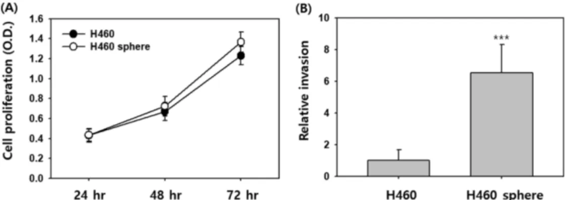
H460 세포주와 H460 폐암원형체의 세포 증식능에 차이가 있 는지를 평가하기 위해 같은 수(8,000 cells/well)의 세포를
RPMI1640에 10% FBS를 첨가 한 배지를 이용하여 희석한 후 96-well에 분주하였다. 세포 배양 시작 후 24, 48, 72시간 후에 각각 살아있는 세포를 XTT 분석법으로 평가하였다. Fig. 2A에 서 H460 세포주(H460)와 H460 폐암원형체(H460 sphere)의 세 포 증식 정도를 비교한 결과 두 세포 간 세포 증식 정도는 통계 적으로 유의한 차이를 나타내지 않았다. 다음으로 암줄기세포들 이 암의 전이유발 및 항암제 내성을 야기시키는 주된 원인이라 는 연구결과들을 바탕으로 암전이의 초기단계인 세포 침윤성 증 가 여부를 H460 세포주와 H460 폐암원형체에서 비교해 보았다 (Fig. 2B). 세포 침윤성을 비교하기 위해 같은 수(50,000 cells/
well)의 세포를 이용하여 매트리젤로 코팅한 트렌스웰에 세포들 을 분주하였고, 24시간 후에 매트리젤을 통과하여 트렌스웰 하 부로 침윤한 세포를 개수하여 평가하였다. Fig. 2B의 결과에서 H460 세포주(H460)와 비교하여 H460 폐암원형체(H460 sphere) 가 6.5배 높은 세포 침윤성을 보였다. 더 나아가 H460 폐암원형 체가 기존에 사용하는 항암제에 내성을 나타내는지 독소루비신 과 시스플라틴을 각각 처리하여 세포 증식능을 H460 세포주와 비교하여 평가하였다. H460 세포주에 독소루비신을 농도 별로 처리하였을 때 0.3 μM과 1 μM 농도 처리(48시간 처리) 각각 40,
Fig. 2 – Comparison of H460 adherent cells and spheres in proliferation and invasion. (A) Cells were seeded in 96-well plate and proliferation was assayed by XTT reagent at 24, 48, and 72 hours (hr). (B) Cells were seeded on the top side of Transwell filter coated with Matrigel in the top chamber of the 24-well plate and incubated for 24 hours. ***p<0.001.
Fig. 3 – Effect of doxorubicin and cisplatin on the proliferation of H460 adherent cells and spheres. Cells were treated with doxorubicin (A) or
cisplatin (B) for 48 hours and proliferation was assayed by XTT reagent. *p<0.05, ***p<0.001.
83%의 세포 증식 억제 효과를 나타냈다. 반면 같은 조건에서 H460 폐암원형체에 독소루비신을 처리하였을 때 0.3 μM과 1 μM 농도에서 각각 13, 17%의 세포 증식 억제 효과를 나타내어 통 계적으로 유의하게 H460 폐암원형체가 독소루비신의 세포 증식 억제 효과에 대한 내성을 나타냈다(Fig. 3A). 또한 시스플라틴을 H460 세포주에 처리하였을 때 10 μM과 30 μM 농도에서(48시 간 처리) 각각 56, 89%의 세포 증식 억제 효과를 나타낸 반면 같은 조건에서 H460 폐암원형체에 시스플라틴을 처리하였을 때 는 10 μM과 30 μM 농도에서 각각 30, 40%의 세포 증식 억제 효과를 나타내어 통계적으로 유의하게 H460 폐암원형체가 시스 플라틴의 세포 증식 억제 효과에 대한 내성을 나타냈다(Fig. 3B).
기존 연구에서 비소세포폐암 세포주인 A549와 H520에서 분 리한 암줄기세포들이 시스플라틴 내성을 보이며 그 작용기전에 Notch3의 역할을 제시한 결과가 있다.20)또한 시스플라틴 내성 이 야기되고 암줄기세포 마커인 CD133이 발현된 H460세포의 경우 파클리탁셀과 독소루비신에도 교차 내성이 생긴다는 결과 와 더불어 내성이 생기는 기전으로 Notch 신호체계의 활성을 제 안한 연구가 있다.19)따라서 H460 폐암원형체가 보이는 세포 침 윤성 증가 및 항암제 내성의 기전을 분석하기 위해 주요 Notch 수용체, 리간드 및 Notch 타겟들의 발현을 분석하였다. H460 세 포주와 비교 시 H460 폐암원형체에서 Notch 신호전달체계의 주 요 수용체인 Notch1의 단백질발현이 현저하게 높은 것을 확인 할 수 있었다(Fig. 4A). Notch4, 리간드인 Jagged1, 그리고 Notch 의 직접적 타겟으로 알려져 있는 c-Myc과 CylcinD1의 발현에는 큰 차이가 없었다. 반면 Notch의 직접적인 타겟으로 알려져 있고 암전이 초기단계에 작용하는 EMT (Epithelial to Mesenchymal transition) 마커로 알려진 Slug의 mRNA의 발현은 5.2 배 증가 된 것으로 확인이 되었다(Fig. 4B). Slug는 Snail과 함께 E-
cadherin 전사 억제인자로 알려져 있고 Slug와 Snail의 발현은 세 포의 EMT 현상을 유발시킨다고 알려져 있다.18)폐암 전이에 관 한 연구에서 보면 Slug는 SOX9를 안정화 시키는 인자로 작용하 고 폐암줄기세포 형성 및 폐암세포의 전이를 촉진하는 것으로 밝 혀졌다.24)또한 유방암 세포주에서 Slug의 발현 조절에 Notch1 단백질이 직접적으로 Slug 프로모터에 붙어 전사 수준에서 발현 을 조절하는 것으로 보고하였고 Slug를 Notch1으로 유도된 EMT 현상의 주요 매개체로 제안을 하였다.18)
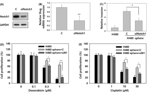
Notch1의 과발현이 H460 폐암원형체의 세포 침윤성 증가 및 항암제 내성야기에 주된 원인인지 확인하기 위하여 Notch1 siRNA 를 활용하여 H460 폐암원형체의 Notch1 발현을 억제하였다(Fig.
5A). 이때 대조군으로 scrambled siRNA (C)를 사용하였고 각각 100 pmole의 농도로 같은 조건에서 실험을 수행하였다. Fig. 5A 에서 보듯이 Notch1 siRNA 처리 H460 폐암원형체에 Notch1 발현이 억제됨을 확인할 수 있었고 이와 동시에 Slug의 mRNA 의 발현 또한 60% 억제됨을 보였다(Fig. 5B). H460 폐암원형체 에 Notch1 siRNA를 처리한 후 세포 침윤성을 분석한 결과, H460 세포주와 비교 시 scrambled siRNA(C)를 처리한 H460 폐 암원형체는 여전히 5.7 배의 세포 침윤성 증가를 나타냈으나 Notch1 siRNA를 처리한 H460 폐암원형체의 경우 증가된 세포 침윤성이 53% 정도 억제됨을 확인하였다(Fig. 5C). 따라서 Notch1 발현이 H460 폐암원형체의 세포 침윤성을 조절한다는 결론을 내릴 수 있었다. 이어서 Notch1이 H460 폐암원형체의 독소루비신과 시스플라틴 약 내성에 영향을 끼치는지 평가하기 위하여 H460 폐암원형체에 Notch1 siRNA와 항암제를 처리한 후 세포 증식 분석 실험을 실시하였다. H460 세포주와 비교 시 scrambled siRNA (C)를 처리한 H460 폐암원형체는 독소루비신 과 시스플라틴에 각각 Fig. 3A, 3B와 유사한 수준으로 내성을 나
Fig. 4 – Expression level of Notch signaling components in H460 adherent cells and spheres. (A) The level of proteins were determined by
Western blotting using the indicated antibodies. (B) Slug mRNA expression was analyzed in H460 adherent and spheres by quantitative
real time PCR. GAPDH was used as an internal control. ***p<0.001.
타내었다(Fig. 5D, 5E). 반면 Notch1 siRNA를 처리한 H460 폐 암원형체는 통계적으로 유의하게 각각의 항암제에 대한 내성이 감소되었고, 각 항암제 처리에 대한 세포 증식 억제 효과가 H460 세포주와 유사한 수준으로 나타났다(Fig. 5D, 5E). 따라서 Notch1 단백질의 과발현이 H460 폐암원형체의 독소루비신과 시스플라 틴 약 내성에 주된 요인으로 작용한다는 결론을 내릴 수 있었다.
결 론 (Conclusion)
비소세포폐암 세포주인 H460 세포주에서 H460 폐암원형체를 분리 한 후 그 특성을 분석 한 결과 H460 폐암원형체는 세포증 식에는 H460 세포주와 유의한 차이가 없는 반면 세포 침윤성 증 가와 독소루비신과 시스플라틴 약 내성이 유발되는 현상을 나타 냈다. H460 폐암원형체의 세포 침윤성 증가 및 항암제 내성유발 의 기전을 분석하기 위하여 H460 세포주와 H460 폐암원형체에 서 세포 침윤성, 항암제 내성에 관련된 주요 유전자 발현을 비교 한 결과 H460 폐암원형체에서 Notch1과 Slug의 발현이 증가됨 을 확인하였다. 더 나아가 Notch1 siRNA를 이용하여 H460 폐 암원형체에서의 Notch1 발현을 억제하였을 때 동시에 Slug의 mRNA 발현수준도 같이 저해됨을 보였고, 결과적으로 Notch1 발현 억제가 증가된 H460 폐암원형체에서의 세포 침윤성과 항 암제 내성을 H460 세포주 수준으로 되돌리는 결과를 나타냈다.
본 연구로 비소세포폐암 세포주인 H460 세포주에서 분리된 폐 암원형체의 세포 침윤성과 항암제 내성의 특성을 파악했으며 이 러한 현상에 Notch1-Slug의 신호전달체계가 주요한 조절 기전으 로 작용함을 밝혀냈다. 따라서 암전이와 항암제 내성 치료를 위 한 암줄기세포 연구에 있어 Notch1 신호전달체계를 치료 타겟 으로 제시한다.
감사의 말씀 (Acknowledgment)
이 논문은 2018학년도에 청주대학교 산업과학연구소가 지원 한 학술연구조성비(특별연구과제)에 의해 연구되었음.
References
1) Siegel, R., Naishadham, D. and Jemal, A. : Cancer statistics, 2012. CA-Cancer J. Clin. 62, 10 (2012).
2) Pardal, R., Clarke, M. F. and Morrison, S. J. : Applying the principles of stem-cell biology to cancer. Nat. Rev. Cancer 3, 895. (2003).
3) Eramo, A., Lotti, F., Sette, G., Pilozzi, E., Biffoni, M., Virgilio, A. D., Conticello, C., Ruco, L., Peschle, C. and Maria, R. D. : Identification and expansion of the tumorigenic lung cancer stem cell population. Cell Death Differ. 5, 504 (2008).
Fig. 5 – Effect of Notch1 siRNA on invasion capacity and the acquired drug resistance of H460 spheres. (A) H460 spheres were transfected
with either scrambled siRNA (as a control (C)) or Notch1 siRNA for 48 hours, and the expression level of Notch1 proteins was
determined by Western blotting. (B) Slug mRNA expression was analyzed in H460 spheres transfected with Notch1 siRNA by
quantitative real time PCR. GAPDH was used as an internal control. (C) Cells were seeded on the top side of Transwell filter coated
with Matrigel in the top chamber of the 24-well plate and incubated for 24 hours. (D, E) Cells were treated with doxorubicin or cisplatin
for 48 hours and proliferation was assayed by XTT reagent. *p<0.05, **p<0.01, ***p<0.001.
4) Ling, G. -Q., Chen, D. -B., Wang, B. -Q. and Zhang, L. -S. : Expression of the pluripotency markers Oct3/4, Nanog and Sox2 in human breast cancer cell lines. Oncol. Lett. 4, 1264 (2012).
5) Yi, H., Cho, H. -J., Cho, S. -M., Jo, K., Park, J. -A., Kim, N. - H., Amidon, G. L., Kim, J. -S. and Shin, H. -C. : Blockade of interleukin-6 receptor suppresses the proliferation of H460 lung cancer stem cells. Int. J. Oncol. 41, 310 (2012).
6) Yun, J. : A549 lung cancer spheres increase invasiveness and doxorubicin resistance. J. Ind. Sci., Cheongju Univ. 35, 173 (2018).
7) Miele, L. : Notch signaling. Clin. Cancer Res. 12, 1074 (2016).
8) Borggrefe, T. and Oswald, F. The Notch signaling pathway:
transcriptional regulation at Notch target genes. Cell. Mol. Life Sci. 66, 1631 (2009).
9) Espinoza, I., Pochampally, R., Xing, F., Watabe, K. and Miele, L. : Notch signaling: targeting cancer stem cells and epithelial- to-mesenchymal transition. OncoTargets Ther. 6, 1249 (2013).
10) Antonio, P., Kimberly, F., Rizzo, P., Osipo, C., Golde, T., Osborne, B. and Miele, L. : Targeting Cancer Stem Cells through Notch Signaling. Clin. Cancer Res. 16, 3141 (2010).
11) Weng, A. P., Ferrando, A. A., Lee, W., Morris, J. P., Silverman, L. B., Sanchez-Irizarry, C., Blacklow, S. C., Look, A. T. and Aster, J. C. : Activating mutations of NOTCH1 in human T cell acute lymphoblastic leukemia. Science 306, 269 (2004).
12) Armstrong, F., Brunet de la Grange, P., Gerby, B., Rouyez, M.
C., Calvo, J., Fontenay, M., Boissel, N., Dombret, H., Baruchel, A., Landman-Parker, J., Romeo, P. H., Ballerini, P. and Pflumio, F. : NOTCH is a key regulator of human T-cell acute leukemia initiating cell activity. Blood 113, 1730 (2009).
13) Yao, K., Rizzo, P., Rajan, P., Albain, K., Rychlik, K., Shah, S. and Miele, L. : Notch-1 and notch-4 receptors as prognostic markers in breast cancer. Int. J. Surg. Pathol. 19, 607 (2011).
14) Iglesias, V. D., Giuranno, L., Dubois, L. J., Theys, J. and Vooijs, M. : Drug Resistance in Non-Small Cell Lung Cancer: A Potential for NOTCH Targeting? Front. Oncol. 24, 267 (2018).
15) Espinoza, I. and Miele, L. : Deadly crosstalk: Notch signaling at the intersection of EMT and cancer stem cells. Cancer Lett.
341, 41 (2013).
16) Harrison, H., Farnie, G., Brennan, K. R. and Clarke, R. B. : Breast cancer stem cells: something out of notching? Cancer Res. 70, 8973 (2010).
17) Yun, J., Pannuti, A., Espinoza, I., Zhu, H., Hicks, C., Zhu, X., Caskey, M., Rizzo, P., D'Souza, G., Backus, K., Denning, M. F.,
Coon, J., Sun, M., Bresnick, E. H., Osipo, C., Wu, J., Strack, P.
R., Tonetti, D. A. and Miele, L. : Crosstalk between PKC? and Notch-4 in endocrine-resistant breast cancer cells. Oncogenesis 2, e60 (2013).
18) Shao, S., Zhao, X., Zhang, X., Luo, M., Zuo, X., Huang, S., Wang, Y., Gu, S. and Zhao, X. : Notch1 signaling regulates the epithelial–mesenchymal transition and invasion of breast cancer in a Slug-dependent manner. Mol. Cancer 14, 28 (2015).
19) Liu, Y. P., Yang, C. J., Huang, M. S., Yeh, C. T., Wu, A. T., Lee, Y. C., Lai, T. C., Lee, C. H., Hsiao, Y. W., Lu, J., Shen, C. N., Lu, P. J. and Hsiao, M. : Cisplatin selects for multidrug- resistant CD133+ cells in lung adenocarcinoma by activating Notch signaling. Cancer Res. 73, 406 (2013).
20) Ma, Y., Li, M., Si, J., Xiong, Y., Lu, F., Zhang, J., Zhang, L., Zhang, P. and Yang, Y. : Blockade of Notch3 inhibits the stem- like property and is associated with ALDH1A1 and CD44 via autophagy in non-small lung cancer. Int. J. Oncol. 48, 2349 (2016).
21) Barr, M. P., Gray, S. G., Hoffmann, A. C., Hilger, R. A., Thomale, J., O'Flaherty, J. D., Fennell, D. A., Richard, D., O'Leary, J. J. and O'Byrne, K. J. : Generation and characterization of cisplatin-resistant non-small cell lung cancer cell lines displaying a stem-like signature. PLOS One 8, 54193 (2013).
22) Lee, D., Kim, D., Choi, Y. B., Kang, K., Sung, E. S., Ahn, J. H., Goo, J., Yeom, D. H., Jang, H. S., Moon, K. D., Lee, S. H. and You, W. K. : Simultaneous blockade of VEGF and Dll4 by HD105, a bispecific antibody, inhibits tumor progression and angiogenesis. mAbs 8, 892 (2016)
23) Brana, I., Berger, R., Golan, T., Haluska, P., Edenfield, J., Fiorica, J., Stephenson, J., Martin, L. P., Westin, S., Hanjani, P., Jones, M. B., Almhanna, K., Wenham, R. M., Sullivan, D. M., Dalton, W. S., Gunchenko, A., Cheng, J. D., Siu, L. L. and Gray, J. E. : A parallel-arm phase I trial of the humanised anti-IGF- 1R antibody dalotuzumab in combination with the AKT inhibitor MK-2206, the mTOR inhibitor ridaforolimus, or the NOTCH inhibitor MK-0752, in patients with advanced solid tumours. Brit. J. Cancer 111, 1932 (2014).
24) Luanpitpong, S., Li, J., Manke, A., Brundage, K., Ellis, E., McLaughlin, S. L., Angsutararux, P., Chanthra, N, Voronkova, M., Chen, Y. C., Wang, L., Chanvorachote, P., Pei, M., Issaragrisil, S. and Rojanasakul, Y. : SLUG is required for SOX9 stabilization and functions to promote cancer stem cells and metastasis in human lung carcinoma. Oncogene 2, 2824 (2016).