The Utility of
18F-FDG PET/CT for Following Up Head and Neck Cancer
Eun-Jae Chung, PhD1, Seung-Kuk Baek, PhD2, Jeong-Soo Woo, PhD2, Soon-Young Kwon, PhD2, Kwang-Yoon Jung, PhD2
Department of Otolaryngology-Head and Neck Surgery,1 Ilsong Memorial Institute Head and Neck Cancer, Hallym University Medical Center, Seoul, Korea
Department of Otolaryngology-Head and Neck Surgery,2 Korea University College of Medicine, Seoul, Korea
두경부암 환자의 추적관찰에서
18F-FDG PET/CT의 유용성
한림대학교 의과대학 이비인후과학교실,1 고려대학교 의과대학 이비인후과학교실2
정은재1·백승국2·우정수2·권순영2·정광윤2
= 국 문 초 록 =
서 론
두경부암의 예후에 가장 중요한 인자는 진단시 병기와 치료 후 재발의 유무이다. 따라서, 재발 병변을 조기에 발견 하는 것이 매우 중요하다. 본 연구에서는 두경부암 환자의 추적 관찰에서 18F-FDG PET/CT의 유용성에 대해 알아 보고자 하였다.
대상 및 방법
2004년 4월부터 2008년 8월까지 고려대 안암병원에서 두경부암으로 치료를 완료한 후 추적관찰이 가능하였던 88 예의 환자를 대상으로 하였다. 환자는 본원의 두경부암 추적관찰 양식에 따라 술 후 6, 12개월째 18F-FDG PET/
CT를 시행하였으며, 원발부위 및 경부 재발, 원격전이와 중복암의 진단에 있어 18F-FDG PET/CT의 민감도, 특이도, 양성예측율, 음성예측율을 평가하였고, 기존의 추적관찰 방법과 이를 비교하였다.
결 과
원발부위 재발의 진단에 있어 민감도, 특이도, 양성예측율, 음성예측율은 각각 95%, 97%, 90%, 98% 였다. 경부전 이 재발의 진단에 있어서는 각각 94%, 98%, 83%, 99%였으며, 원격전이 및 이차암 진단에 있어서는 94%, 100%, 100%, 99%였다. 원발부위 재발은 고식적인 진단 방법, 원격전이와 이차암은 18F-FDG PET/CT가 진단에 의미있게 유리하였다.
결 론
18F-FDG PET/CT는 두경부암의 추적관찰 과정에서 재발 여부를 판정하는데 있어 매우 유용하며, 특히 원격전이 및 이차암의 발견에 있어 유용하다. 그러나, 위양성 및 위음성의 가능성으로 고식적인 진단 방법과 병행해야 진단의 효과를 높힐 것으로 생각된다.
중심 단어:양전자방출 단층촬영·재발·두경부암.
Introduction
The early detection of recurrent head and neck cancer
may improve patient survival, so careful monitoring when follow-up these patients is essential. However, making the early diagnosis of recurrent head and neck cancer is difficult.
Asymptomatic recurrence, and especially that for distant me- tastasis, is more difficult to identify with using the conven- tional methods.
교신저자:정광윤, 136-705 서울 성북구 안암동 5가 126-1 고려대학교 의과대학 이비인후과학교실
전화:(02) 920-5486·전송:(02) 925-5233 E-mail:[email protected]
Many physicians have used physical examinations and structural imaging modalities such as computed tomogra- phy(CT) and magnetic resonance imaging(MRI) as the con- ventional methods. However, these methods had been found to be too equivocal in detecting recurrence because of the anatomical changes induced by therapy, and these changes may be further obscured by flap reconstruction.1)
PET-CT imaging, for which CT is performed in conjunc- tion with PET, provides matching metabolic and anatomical images, and this allows precise localization of the foci of high FDG uptake. Improving the localization of high uptake foci results in better differentiation of the physiological and pathological FDC uptake.2) Several retrospective and pro- spective studies3-8) have shown that 18F-FDG PET/CT is more effective in detecting recurrence than conventional methods.
However, in these previous studies, patients were enrolled if recurrence was suspected.
The objective of the current study was to perform a retro- spective evaluation of the accuracy of performing routine
18F-FDG PET/CT surveillance for follow-up head and neck cancer patients.
Materials and Methods
1. Patients
From April 2004 to April 2008, whole-body 18F-FDG PET/
CT were performed on 84 patients with head and neck cancer after completion of treatment to detect residual cancer or tumor recurrence. There were 61 men and 23 women, aged from 30 to 84years(mean, 59years). The patients’ character- istics are given in Table 1.
A physical examination was done every month for 2 years, and then every 6 months for year and then annually if the patient was disease free during this time. Periodic neck CT scan and chest X-ray(at the 6th and 12th post-operative month, and then annually) and 18F-FDG PET/CT(at the 6th and 12th month after the initial treatment and then annually) were done according to our institutional follow-up protocol. The recurrences were divided according to the location of le- sion:local recurrence, recurrence in the regional area and distant metastasis/ secondary malignancy.
The conventional detection methods along with histolo- gical verification, were performed for the patients with sus- pected recurrence observed on their 18F-FDG PET/CT. The
18F-FDG PET/CT was carried out within an interval of no longer than 2 weeks from the time of finding a suspected re- currence on the conventional detection methods.
A total of 111 whole-body 18F-FDG PET/CT were perform- ed. Two scans were performed on 19 patients, three scans were performed on 3 patients and four scans were perform- ed on 1 patient. Recurrence was confirmed histopathologi- cally or by at least 3 months of clinical follow-up.
2. Whole-Body 18F-FDG PET/CT
Scans were performed on a Gemini TF PET/CT scanner (Philips). Patients fasted 6 h before PET acquisitions, and the blood glucose level had to be less than 7mmol/L before injection of 370 MBq(5 MBq/kg) of 18F-FDG. Intravenous injection was followed by a period of approximately 60 min when the patients remained in a quiet room. No muscle relax- ants were administered. The 18F-FDG PET/CT images were obtained over the entire body from the base of the skull to the mid-thigh. PET data were acquired in the 3-dimensional mode. Transmission CT imaging was performed for attenua- tion correction and anatomical localization with using the integrated PET-CT system, and the accompanying software provides the transmission CT, FDG-PET and fusion images.
The Gemini scanner consists of a 16-slice multi-detector-row spiral CT scanner with a transverse field of view of 600mm.
Table 1. The patients’ demographics and the tumor character- istics
Factors No.
Larynx 19 Oropharynx 18
Oral cavity 17
Nasopharynx 10 Hypopharynx 10
Sinonasal 06
Salivary gland 02
Primary tumor site
Others 02
Squamous cell carcinoma 73 Low grade mucoepidermoid ca 01 Salivary duct ca 01 Nonkeratinizing carcinoma 05 Undifferentiated carcinoma 02 Cell type
Melanoma 02
Stage I 13
Stage II 18
Stage III 18
Stage
Stage IV* 35
Surgery+postoperative radiotherapy 51
Surgery alone 10
Chemoradiotherapy 14 Neoadjuvant chemotherapy+salvage
surgery+radiotherapy
04 Treatment
modality
Radiotherapy alone 05 Range 3-105 Follow-up
months Median 29.3
*:No distant metastasis patient among the stage IV group
The CT parameters-a collimation of 6×5mm, tube voltage of 120 kV, and effective tube current of 100 mAs-are standard for PET/CT studies and permit differentiation between tissues with good spatial resolution while ensuring that the patient does not receive a high radiation dose.
3. Analysis
A study was considered false-positive if no recurrence was demonstrated by biopsy or there was no disease progression on multiple following studies, or if the subsequent studies became negative without administering any therapy for re- current disease. A study was considered false-negative if there was clinical progression of disease, or if disease was detected on another imaging modality and this was subsequently pro- ven by biopsy or the clinical progression after the first false- negative study. Residual disease after chemoradiation(n=
5) was regarded as a recurrence for statistical purpose.
The sensitivity, specificity, positive predictive value(PPV) and negative predictive value(NPV) of the 18F-FDG PET/CT were calculated. Statistical analysis was performed using the McNemar test and chi-square test. Statistical analysis was per- formed by using SPSS for Windows(version 12.0;SPSS Inc., Chicago, IL, USA). Statistical significance was set at p<0.05.
Results
1. Confirmation of the diagnosis
Recurrent disease was diagnosed in 48 cases, while there was no evidence of recurrence in 36 cases. The definitive diagnosis was obtained by means of the histology or cyto- logy for 47 cases and the definitive diagnosis was based on the clinical follow-up for 1 case(multiple bone metastasis).
1. The diagnostic accuracy of 18F-FDG PET/CT The diagnostic accuracy of 18F-FDG PET/CT is summa- rized in Table 2. The overall diagnostic sensitivity, specifi- city, PPV, NPV and accuracy for local recurrence were 95%,
97%, 90%, 98% and 96% respectively, and those for regional recurrence were 94%, 98%, 83%, 99% and 95% respecti- vely, and those for distant metastasis/second primary cancer were 94%, 100%, 100%, 99% and 99% respectively(Table 2). There were 5 false positive cases of 18F-FDG PET/CT for locoregional recurrence- Two cases of primary lesion:1) T4a floor of mouth, surgery+postoperative radiotherapy;
2) T4a hypopharynx, concurrent chemoradiotherapy. Three cases of regional lesion:1) N0 tongue, elective neck dissec- tion, SUV 3.85;2) N0 tonsil, surgery+postoperative radio- therapy, SUV 3.3;3) N2 Nasopharynx, concurrent chemo- radiotherapy, SUV:3.5. There were 2 cases of false nega- tive scans for locoreginal recurrence- Primary lesion:T1b glottis cancer, surgery+postoperative radiotherapy;Region- al lesion:N1 Nasopharynx, concurrent chemoradiotherapy and 1 case of a false negative scan for distant metastasis/se- cond primary cancer- N2a unknown primary, surgery+post- operative radiotherapy(Fig. 1). The overall sensitivity was significantly higher with 18F-FDG PET/CT than conven- tional methods(p=0.035). However, there was no significant difference for overall specificity(p=1.0). Most of subclinical recurrent patients with routine surveillance were shown to have distant metastasis/second primary tumor with the aid of 18F-FDG PET/CT(Fig. 2).
We detected primary recurrence with using the conven- tional methods significantly more often than we did with using 18F-FDG PET/CT(p=.007). Distant metastasis/second primary tumor was detected significantly more often with
18F-FDG PET/CT(p=.001). No significant difference was found for the detection of regional recurrence between 18F- FDG PET/CT and the conventional methods(p=.725).
Discussion
The early, accurate detection of recurrent head and neck cancer is critically important for achieving successful sal-
Table 2. The results of routine 18F-FDG PET/CT surveillance for follow-up head and neck cancer patients
Local recurrence Regional recurrence Distant metastasis/ Secondary malignancy
True-positives 19 15 017
False-positives 02 03 000
False-negatives 01 01 001
True-negatives 62 65 066
Sensitivity(%) 95 94 094
Specificity(%) 97 98 100
PPV(%) 90 83 100
NPV(%) 98 99 099
Accuracy(%) 96 95 099
*:Sensitivity= TP/TP+FN, Specificity=TN/FP+TN, PPV=TP/TP+FP, NPV=TN/FN+TN. PPV:positive predictive value, NPV:negative predictive value, TP:true positive, TN:true negative, FP:false positive, FN:false negative
vage treatment. The patients with recurrent head and neck cancer and who undergo salvage surgery at early disease stages have a 70% 2-year relapse-free survival rate, whereas those with recurrent, advanced-stage head and neck cancer have just a 22% 2-year relapse-free survival rate after salvage surgery.9)
Conventional imaging such as CT and MRI has been of limited value for the early detection of recurrence due to the modalities’ relatively low sensitivity and specificity. The anat-
omical changes due to therapy and flap reconstruction are some of the main reasons for the difficulty to detect recur- rent tumor. 18F-FDG PET/CT, however, is able to detect areas of recurrent disease very early by demonstrating the increas- ed FDG metabolism in tumor, and 18F-FDG PET/CT is able to accurately differentiate recurrent tumor from the postop- erative changes. Therefore, 18F-FDG PET/CT can increase the diagnostic accuracy of the conventional methods and it
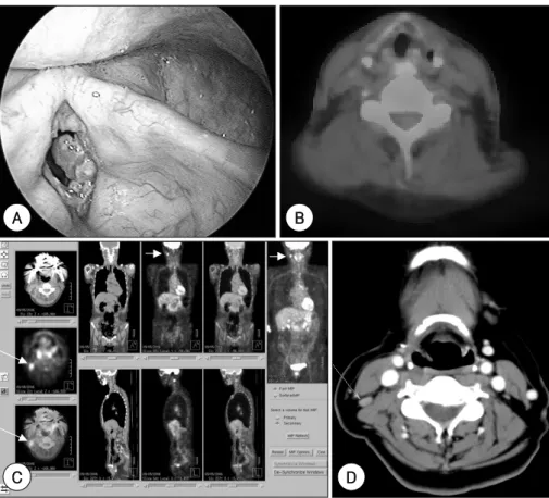
Fig. 1. False negative(A, B) and false positive(C, D) 18F-FDG PET/CT. A:A 75 year-old male with glottic cancer(initial stage T1bN0M0) at 4 years after undergoing anterior commisure laryngectomy. Biopsy confirmed the recurrence. B:There is no hypermetabolic lesion in the recurrent site. C:A 58 year-old female with tongue cancer(T2N0M0) at 2 years after undergoing partial glossectomy, SND(I-III). A hypermetabolic lesion that was suspicious for recurrence was detected on the 18F-FDG PET/CT scan (arrow:SUV 3.85).
D:The CT scan image. The ultrasound-guided fine needle aspiration cytology and clinical follow-up(>2years) showed no evidence of recurrence.
A
D B
C
Fig. 2. A 74-year-old female with supraglottic cancer(T3N2cM0) after extended supraglottic laryngectomy, (B) MRND, and post- operative radiotherapy. Routine follow-up(A) 18F-FDG PET/CT and (B) Chest X-ray at 6 months after treatment. 18F-FDG PET/CT demonstrates focal hypermetabolic lesion in left upper lung field interpreted as lung metastasis(arrow). However, no evidence of lung metastasis was suspected on the chest X-ray at the same time.
A B
may be helpful for detecting tumor recurrence at an earlier stage. Moreover, 18F-FDG PET/CT is able to detect recurrence by viewing the whole body, which can not done with the conventional methods, and this whole body 18F-FDG PET/
CT is valuable for detecting distant metastasis or a second primary lesion.1,10-13)
We evaluated the ability of 18F-FDG PET/CT to identify tumor recurrence in patients seen routinely and sequentially follow-up after completion of therapy. This was done in an at- tempt to demonstrate any subclinical recurrent disease. Such detection may precede detection of other clinical signs or symptoms, and therefore there may be reasonable cause for using PET routinely in such patients. In the earlier studies, the sensitivity of 18F-FDG PET ranged from 71% to 100%, its specificity ranged from 43% to 100%, its positive predic- tive value ranged from 64% to 100% and its negative pre- dictive value ranged from 66% to 100%.1) In our study, 18F- FDG PET/CT showed the high sensitivity, specificity, PPV and NPV, and especially for detecting distant metastasis/se- cond primary lesion. However, a negative PET/CT result does not provide absolute assurance of a disease-free status. In our series, 3 cases with negative PET/CT scans eventually presented with recurrence. We can guess the false negative results were well-differentiated cancers. Other potential causes for these false negatives were related to the time interval after treatment, the necrotic tissue around tumor and small size that were under the detection threshold.1,13) We found that
18F-FDG PET/CT was very sensitive, but it also displayed false-positivity. Five patients in our study had false-positive PET/CT scans. False-positive scans are usually associated with infection or inflammation.2) These results suggest that
18F-FDG PET/CT should not completely replace the conven- tional evaluation methods.
In summary, a 18F-FDG PET/CT can detect head and neck tumor recurrence when this tumor recurrence may be un- detectable by other clinical methods. It permits highly ac- curate detection of head and neck cancer recurrence in the post-therapy period. With its high sensitivity, specificity, PPV and NPV, 18F-FDG PET/CT may be a useful tool for per- forming routine surveillance for detecting the recurrence of head and neck cancer, and especially for detecting distant metastasis/second primary lesion.
References
1) Lee JC, Kim JS, Lee JH, Nam SY, Choi SH, Lee SW, et al. F-18 FDG-PET as a routine surveillance tool for the detection of re- current head and neck squamous cell carcinoma. Oral Oncol.
2007 Aug;43(7):686-692.
2) Rhodes MM, Delbeke D, Whitlock JA, William Mohn, Kuttesch JF, Frangoul HA, et al. Utility of FDG-PET/CT in Follow-Up of Children Treated for Hodgkin and Non-Hodgkin Lymphoma. J Pediatr Hematol Oncol. 2006 May;28(5):300-306.
3) Fischbein NJ, AAssar OS, Caputo GR, Kaplan MJ, Singer MI, Price DC, et al. Clinical utility of positron emission tomography with 18F-fluorodeoxyglucose in detecting residual/recurrent squa- mous cell carcinoma of the head and neck. Am J Neuroradiol.
1998 Aug;19(7):1189-1196.
4) Wong RJ, Lin DT, Schoder H, SG Patel, M Gonen, S Woldenet, et al. Diagnostic and prognostic value of [18F]fluorodeoxyglu- cose positron emission tomography for recurrent head and neck squamous cell carcinoma. J Clin Oncol. 2002 Oct;20(20):4199- 4208.
5) Kunkel M, Forster GJ, Reichert TE, JH Jeong, Peter Benz, Peter Bartenstein, et al. Detection of recurrent oral squamous cell car- cinoma by [18F]-2-fluorodeoxyglucose-positron emission tomo- graphy: implications for prognosis and patient management.
Cancer. 2003 Nov;98(15):2257-2265.
6) Kitagawa Y, Nishizawa S, Sano K, Ogasawara T, Nakamura M, Sadato N, et al. Prospective comparison of FDG PET with con- ventional imaging modalities(MRI, CT, and 67Ga scintigraphy) in assessment of combined intraarterial chemotherapy and ra- diotherapy for head and neck carcinoma. J Nucl Med. 2003 Feb; 44(2):198-206.
7) Stokkel MP, Terhaard CH, Hordijk GJ, van Rijk PP. The detec- tion of local recurrent head and neck cancer with fluorine-18 fluorodeoxyglucose dual-head positron emission tomography. Eur J Nucl Med. 1999 Jul;26(7):767-773.
8) Terhaard CH, Bongers V, van Rijk PP, Hordijk GJ. F-18-fluoro- deoxy-glucose positron-emission tomography scanning in detec- tion of local recurrence after radiotherapy for laryngeal/pharyn- geal cancer. Head Neck. 2001 Nov;23(11):933-941.
9) Wong RJ, Lin DT, Schoder H, Patel SG, Gonen M, Wolden S, et al. Diagnostic and prognostic value of 18Ffluorodeoxyglucose positron emission tomography for recurrent head and neck squa- mous cell carcinoma. J Clin Oncol. 2002 Oct;20(20):4199-4208.
10) Lowe VJ, Boyd JH, Dunphy FR, Han Kim, Dunleavy T, Collins BT, et al. Surveillance for Recurrent Head and Neck Cancer Using Positron Emission Tomography. J Clin Oncol. 2000 Feb;18(3): 615-618.
11) Zimny M, Siggelkow W, Schroder W, Nowak B, Biemann S, Rath W, et al. 2-[Fluorine-18]-fluoro-2-deoxy-D-glucose positron emission tomography in the diagnosis of recurrent ovarian cancer.
Gynecol Oncol. 2001 Nov;83(2):310-315.
12) Ruiz-Hernandez G, Delgado-Bolton RC, Fernandez-Perez C, La- pena-Gutierrez L, Carreras-Delgado JL. Meta-analysis of the diag- nostic efficacy of FDG-PET in patients with suspected ovarian cancer recurrence. Rev Esp Med Nucl. 2005 May-Jun;24(3): 161- 173.
13) Garcia-Velloso MJ, Jurado M, Carolina Ceamanos, Aramendia JM, Garrastachu MP, Guillermo Lopez-Garcia, et al. Diagnostic accuracy of FDG PET in the follow-up of platinum-sensitive epi- thelial ovarian carcinoma. Euro J Nucl Med Mol Imaging. 2007 Sep;34(9):1396-1405.