ORIGINAL ARTICLE
Significance of SUV on Follow-up F-18 FDG PET
at the Anastomotic Site of Gastroduodenostomy after Distal Subtotal Gastrectomy in Patients with Gastric Cancer
Byung Wook Choi&Seok Kil Zeon&Sung Hun Kim&
Il Jo&Hae Won Kim&Kyoung Sook Won
Received: 24 March 2011 / Revised: 16 August 2011 / Accepted: 22 August 2011 / Published online: 9 September 2011
# Korean Society of Nuclear Medicine 2011
Abstract
Purpose The aim of this study was to characterize the patterns of fluorodeoxyglucose (FDG) uptake on F-18 FDG positron emission tomography/computed tomography (FDG PET/CT) at the anastomotic site of gastroduodenos- tomy after distal subtotal gastrectomy in patients with gastric cancer.
Methods From May 2007 to May 2010, two or more follow-up measurements using FDG PET/CT scans were done for 19 patients (11 men, 8 women; mean age, 62.0±
10.3 years) who underwent distal subtotal gastrectomy with gastroduodenostomy between February 2006 and March 2008 for detecting gastric cancer recurrence at our medical center. The FDG PET/CT images were retrospectively reviewed. Patients with local recurrence, regional nodal metastasis or distant metastasis on follow-up studies were excluded. CT and endoscopy were done within 1 month before or after the FDG PET/CT scan. Eight patients had two follow-ups of FDG PET/CT, and 11 patients had three follow-ups. The mean interval between surgery and the first follow-up FDG PET/CT was 12.9±0.8 months (n=19);
between the first and second it was 12.3±1.0 months (n=
19); between the second and third it was 11.6±0.7 months (n=11). The F-18 FDG uptakes at the anastomotic site and fundus in the remnant stomach were measured by maximum standardized uptake value (SUVmax) using a region of interest technique.
Results The SUVmax at the anastomotic site was signifi- cantly higher than that of the fundus on all series of first, second and third follow-up studies (3.3 ± 1.1 vs. 2.1 ± 0.7, p < 0.001: 3.1 ± 0.9 vs. 2.2 ± 0.7, p = 0.001: 3.0 ±0.6 vs.
2.1 ± 0.7, p = 0.006, respectively). The SUVmax for the anastomotic site and fundus, and SUVmax ratio for the anastomotic site over the fundus were not significantly different throughout the series.
Conclusion The SUVmax at the anastomotic site is significantly higher than that of the fundus and does not decrease significantly over time. Therefore, the local recurrence of gastric cancer after surgery could not be definitely differentiated from physiologic uptake or postop- erative inflammatory change.
Keywords F-18 fluorodeoxyglucose . PET . Stomach cancer . Billroth I . Gastroduodenostomy . Anastomotic site
Introduction
Gastric cancer is the fourth most common cancer worldwide and a very aggressive neoplasm with a poor prognosis [1, 2]. In Korea, gastric cancer is the most common malignancy and a major cause of cancer death despite improved prognosis as a result of early diagnosis, radical operations and the advancement of adjuvant therapy [1,3–5].
The primary curative therapy for early and advanced gastric cancer is radical surgery [1,6–8]. Total gastrectomy is commonly performed for advanced gastric cancer of the proximal or middle third of the stomach, while subtotal gastrectomy is preferred for patients with distal gastric cancer because no survival benefit has been proven for total gastrectomy [9].
B. W. Choi:S. K. Zeon (*):S. H. Kim:I. Jo:H. W. Kim:
K. S. Won
Department of Nuclear Medicine, Keimyung University, School of Medicine,
#194, Dongsan-Dong, Jung-Gu, Daegu, Korea e-mail: [email protected] DOI 10.1007/s13139-011-0105-9
However, many patients with gastric cancer present recurrent disease in the follow-up period even after complete resection [5]. When a recurrent tumor develops in the remnant stomach after subtotal gastrectomy, treat- ments such as chemotherapy and an additional operation with curative intent can be offered. However, despite advances in the management of recurrent gastric cancer, several studies have reported a poor prognosis for patients with recurrent gastric cancer [2, 5, 7, 9, 10]. Imaging modalities, including contrast-enhanced computed tomog- raphy (CECT) and endoscopy, have been used for the detection of gastric cancer recurrence [4,11,12]. However, a gold standard modality has not been established.
Endoscopy is one of the most commonly used modalities for detecting recurrent gastric cancer after subtotal gastrectomy. Its utility has been well demon- strated in many studies, especially in cases of intra- luminal recurrence [13–17]. However, there are some limitations to endoscopy. Anatomical deterioration of the remnant stomach after the operation and mucosal changes at the gastric stump due to bile reflux are commonly observed [13, 14, 17]. Also, endoscopy cannot detect extraluminal recurrence.
On the other hand, CECT has been the modality of choice for detection of extraluminal recurrence in patients with gastric cancer, recurrences such as regional lymph node metastasis, hepatic metastasis and peritoneal seeding [4,18]. However, CECT also has limitations to its ability to detect recurrence because it uses size criteria [1, 18].
Furthermore CECT, similar to endoscopy, could not differentiate locoregional tumor recurrence from morphologic changes after surgery [18].
Recently, F-18 FDG PET, which is a non-invasive imaging modality that reflects glucose metabolism in the region of interest, especially in malignant cells, has been widely used for staging and restaging of a malignancy, determination of treatment response and detection of tumor recurrence in various cancers [2,12,19,20]. It is generally accepted that integrated PET/CT provides superior perfor- mance in the evaluation of a malignancy compared to PET or CT alone [12,19]. Several FDG PET and PET/CT studies have reported variable FDG uptake of primary gastric malignancies according to their histological subtypes [6].
However, only a few studies have been reported regarding the use of PET in recurrent gastric cancer [2,6,12,19]. In addition, no reports have focused on the anastomotic site, the most common site of local recurrence [5], with F-18 FDG PET/CT.
The aim of this study, therefore, was to evaluate, on follow-up FDG PET/CT, the pattern of FDG uptake at the anastomotic site of the gastroduodenostomy after distal subtotal gastrectomy in patients with gastric cancer.
Materials and Methods
Patients
We retrospectively analyzed date for 306 patients who underwent curative gastrectomy and two or more annual FDG PET/CT follow-up scans for detecting gastric cancer recurrence at our medical center between May 2007 and May 2010. Patients were included if they had two or more FDG PET/CT follow-ups by 1 year after distal subtotal gastrectomy with gastroduodenostomy and referral CECT and endoscopy within 1 month before or after a FDG PET/
CT scan. We excluded patients with regional nodal metastasis, distant metastasis and local recurrence on follow-up studies.
Ultimately, 19 patients (11 men, 8 women) were enrolled in our study. Mean age was 62 years old (range: 39–
79 years) (Table 1). These patients had surgery between February 2006 and March 2008. Histological type, clinico- pathologic tumor stage (following to AJCC 7th edition) and follow-up periods are shown in Tables1and 2.
F-18 FDG PET/CT Technique
All patients were asked to fast for at least 6 h. Blood glucose levels of patients were checked before F-18 FDG injection. The mean blood glucose level at the time of FDG PET/CT scanning was 97.6±12.1 mg/dl (range 72–137).
Three patients had a history of non-insulin-dependent diabetes mellitus, but the blood glucose level did not exceed 140 mg/dl. Patients received an intravenous injec-
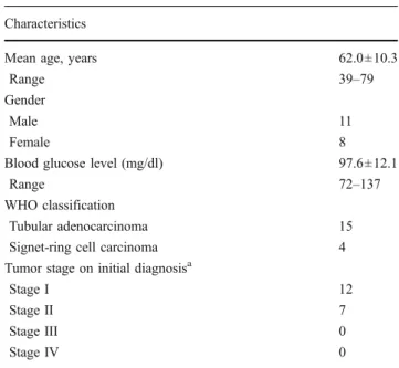
Table 1 Patient characteristics (N=19) Characteristics
Mean age, years 62.0±10.3
Range 39–79
Gender
Male 11
Female 8
Blood glucose level (mg/dl) 97.6±12.1
Range 72–137
WHO classification
Tubular adenocarcinoma 15
Signet-ring cell carcinoma 4
Tumor stage on initial diagnosisa
Stage I 12
Stage II 7
Stage III 0
Stage IV 0
aFollowing the AJCC 7th edition
tion of 7.0 MBq/kg of F-18 FDG and rested in bed for 60 min.
PET image data acquisition was done using an integrated PET/CT system (Discovery STE, GE Healthcare, Milwaukee, WI) 60 min after F-18 FDG injection. The data acquisition protocol was as follows: (1) we did a low dose CT scan; we then did a PET scan, from the skull base or top of the brain to mid-thigh, with 140 kV and 3.75 mm thickness at 30 mA (adjusted for PET section thickness). All patients were allowed normal breathing during CT scanning, and no oral or intravenous contrast was administered. After CT scanning, a PET scan of the identical transverse field of view was obtained without a change in the patient's position. Acquisition time was 3 min per table bed. PET images were reconstructed iteratively by an ordered subset expectation maximization (OS-EM). CT data were used for attenuation correction.
All fused images were viewed using dedicated software (eN-TEGRA; GE Healthcare) on a work station.
PET/CT Image Interpretation
For visual analysis, all PET and fused images were retrospectively reviewed by two experienced nuclear medicine physicians. These observers were fully aware of the patient’s medical records, including age of patients, histological type of tumor, type of operation, levels of tumor marker at the time of FDG PET/CT scanning and results of available imaging studies. In patient with mis-registration of PET and CT images, interpretation was made using PET images that corre- lated with anatomic positions on CT. For semi- quantitative analysis, we calculated the maximum standardized uptake value (SUVmax) using a region of interest (ROI) technique. The ROI was drawn as a circle over the anastomotic site. A background ROI was drawn as a circle of the same diameter as that of the lesion-based ROI at the fundus in the remnant stomach (Fig. 1).
Data Analysis
The SUVmax of the anastomotic site and fundus in the remnant stomach of each patient was measured, and the
SUVmax was calculated as the mean ± standard deviation.
The difference between the mean SUVmax values for the anastomotic site and that for the fundus was evaluated using the Mann-Whitney U test.
The repeated measures analysis of variance (RM ANOVA) was used to compare SUVmax values of the anastomotic site and fundus at different follow-up times.
Also, the ratio of SUVmax values for the anastomotic site over the fundus was obtained, and the difference for each of the follow-ups was evaluated using the RM ANOVA.
Statistical significance was set at p<0.05. Statistical calculations were done using Predictive Analytics software (PAWS)(version 18.0, IBM Corp., Somers, NY).
Results
A total of 49 FDG PET/CT studies for 19 patients were reviewed, and SUVmax values for 98 ROIs at the anastomotic site or fundus in the remnant stomach were measured.
The SUVmax at the anastomotic site was significantly higher than at the fundus on all series of first, second and third follow-up studies (3.3 ± 1.1 vs. 2.1 ± 0.7, p<0.001:
3.1 ± 0.9 vs. 2.2 ± 0.7, p=0.001: 3.0±0.6 vs. 2.1±0.7, p=
0.006, respectively). In addition, RM ANOVA with a sphericity assumed correction determined that the SUV- max at the anastomotic site of each patient was not significantly different throughout the series [F(2, 20)=
1.234, p=0.312]; also the SUVmax at the fundus of each patient was not differed statiscally significantly between time points [F(2, 20)= 0.864, p=0.079](Table3).
The ratio of the SUVmax at the anastomotic site over that of the fundus was calculated, and a RM ANOVA with a Greenhouse-Geisser correction determined that there was no significant difference between all follow-up series [1.5±
0.7, 1.3±0.6 and 1.5±0.4, respectively, F(2, 20)= 0.728, p=
0.441] (Table3).
Discussion
The high diagnostic performance of FDG PET/CT has been demonstrated in the diagnosis of recurrent gastric cancer.
Nakamoto et al. [21] reported that the sensitivity, specificity, positive predictive value, negative predictive value and accuracy of FDG PET/CT for recurrent gastric cancer were 78%, 91%, 90%, 78 and 84%, respectively. Park et al. [19]
reported that the sensitivity, specificity, positive predictive value, negative predictive value and accuracy of PET/CT for diagnosing true recurrence of gastric cancer were 75%, 77%, 89%, 55% and 75%, respectively. In a study of contrast-enhanced FDG PET/CT, Bilici et al. [12] reported Table 2 Mean interval of follow-up F-18 FDG PET/CT after surgery
Follow-ups Interval (months)
Operation and first (n=19) 12.9±0.8
First and second (n=19) 12.3±1.0
Second and third (n=11) 11.6±0.7
superior diagnostic performance of FDG PET/CT over CECT.
Although FDG PET/CT has been used for detecting recurrent gastric cancer, several limitations have been suggested for evaluating the remnant stomach. Mild F-18 FDG uptake is commonly seen in the normal stomach [8, 22]. In addition, a variable degree of contracted or non- uniform F-18 FDG uptake is occasionally observed in the stomach, and it could be misinterpreted as a pathological finding. The same is true for the remnant stomach in subtotal gastrectomy patients [8]. Accumulation of F-18 FDG in the gastrointestinal smooth muscle due to acceler- ated gastric peristalsis or inflammatory changes have been suggested as possible causes for physiological and diffuse
F-18 FDG uptake in the remnant stomach [23]. Also, the mechanism is not clear. Several studies suggested that specific or non-specific gastritis is one of the causes of diffuse F-18 FDG uptake in the stomach, and these findings have a positive relationship with Helicobacter pylori infection [23, 24]. Yun et al. [8] reported that gastric distension due to ingestion of a glass of water is an effective way to diagnose in a patient with suspected gastric tumor recurrence. In their study, physiological F-18 FDG uptake in a linear or curvilinear configuration disappears after water ingestion.
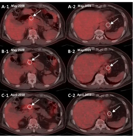
Yoo et al. [5] reported that the most common site of locoregional recurrence was the anastomotic site. In their study of 501 patients with recurrent gastric cancer, 80 Fig. 1 A 63-year-old male who
underwent gastroduodenostomy after distal subtotal gastrectomy in April 2007 for gastric cancer had three annual follow-up FDG PET/CT scans in May 2008, May 2009 and April 2010 for detecting tumor recurrence. In fusion images of the first (A-1 and A-2), second (B-1 and B-2) and third (C-1 and C-2) follow- up FDG PET/CT scans, the SUVmax values at the anasto- motic site were 4.2, 4.0 and 4.1, and the SUVmax values at the fundus were 3.0, 3.7 and 2.5, respectively. The ratios of SUVmax values at the anastomotic site over the fundus of this patient were 1.4, 1.1 and 1.5, respectively.
There was no evidence of tumor recurrence during follow-up (37 months)
Table 3 The mean SUVmax at the anastomotic site and the fundus on follow-up series
aRatio of the SUVmax of the anastomotic site over fundus
First (n=19) Second (n=19) Third (n=11) p-value
Anastomotic site 3.3±1.1 3.1±0.9 3.0±0.6 0.312
Fundus 2.1±0.7 2.2±0.7 2.1±0.7 0.079
RAFa 1.5±0.7 1.3±0.6 1.5±0.4 0.441
showed local recurrence at the anastomotic site. Therefore, evaluation of the anastomotic site of gastroduodenostomy in the remnant stomach is important for detection of locoregional recurrence of gastric cancer. No available data focus on the anastomotic site, and the reported studies of FDG PET/CT for detecting gastric cancer recurrence focused only on the focal increased F-18 FDG uptake lesion relative to the normal biodistribution of background F-18 FDG uptake in the remnant stomach [3,6,8,12,19, 25].
In this study, we focused on the anastomotic site in the remnant stomach, and to our knowledge, this study is the first to evaluate SUVmax of the anastomotic site using FDG PET/CT. At the beginning of this study, we expected that F-18 FDG uptake into the anastomotic site would be decreased with time, but SUVmax of the anastomotic site showed no significant difference on follow-up. In addition, the SUVmax of the anastomotic site was significantly higher than that of the fundus of the remnant stomach at all follow-up times. Such patterns of F-18 FDG uptake at the anastomotic site could help in the differential diagnosis of locoregional recurrence.
The normal physiologic biodistribution of F-18 FDG was reported for brain, reactive lymphoid tissues, liver, spleen, stomach, bowel, kidney, bladder, ureter, uterus, ovary, blood vessels, muscles, etc. [22,26]. Increased F-18 FDG uptake in inflammatory and infectious lesions is well established [23,27,28]. It is well documented that patients with a gastroduodenostomy have reflux of duodenal contents into the remnant stomach due to the absence of the pyloric sphincter, leading to gastritis [14, 17]. There- fore, several factors such as normal physiologic uptake, inflammation and fibrosis after surgery could contribute to the persistent increase in F-18 FDG uptake at the anastomotic site.
In this study, F-18 FDG uptake at the anastomotic site in the remnant stomach of patients without recurrent gastric cancer showed a diffusely increased uptake pattern. A few studies reported that most cases of locally recurrent gastric cancer showed a focal increased F-18 FDG uptake pattern [8,12,19]. Bilici et al. [12] reported SUVmax values for 23 patients with true-positive FDG PET/CT findings and 1 patient with a false-negative FDG PET/CT: the mean was 11.1±4.8 (range, 4.3–20.3). In a follow-up series to this study, the mean SUVmax value at the anastomotic site was 3.1±0.9 (range 1.3–5.1), and it was lower than that of the aforementioned study [12]. These imaging findings and analyses are compatible with those in the literature.
The present study has certain limitations such as the small sample size and being a retrospective study. A prospective study with a large number of patients and histopathological confirmation of the anastomotic site is recommended for further evaluation of the F-18 FDG
uptake pattern on follow-up FDG PET/CT after gastric cancer surgery.
In conclusion, the SUVmax of the anastomotic site in follow-up FDG PET/CT after gastric cancer surgery is significantly higher than that of the fundus in the remnant stomach and does not decrease significantly over time. Therefore, early detection of the local recurrence at the anastomotic site of the gastric cancer patient after surgery could not be definitely differentiat- ed from physiologic uptake or postoperative inflammatory change.
References
1. Sim SH, Kim YJ, Oh DY, Lee SH, Kim DW, Kang WJ, et al. The role of PET/CT in detection of gastric cancer recurrence. BMC Cancer. 2009;9:73.
2. De Potter T, Flamen P, Van Cutsem E, Penninckx F, Filez L, Bormans G, et al. Whole-body PET with FDG for the diagnosis of recurrent gastric cancer. Eur J Nucl Med Mol Imaging. 2002;29 (4):525–9.
3. Yoshioka T, Yamaguchi K, Kubota K, Saginoya T, Yamazaki T, Ido T, et al. Evaluation of18F-FDG PET in patients with advanced, metastatic, or recurrent gastric cancer. J Nucl Med. 2003;44 (5):690–9.
4. Eom BW, Ryu KW, Lee JH, Choi IJ, Kook MC, Cho SJ, et al.
Oncologic effectiveness of regular follow-up to detect recurrence after curative resection of gastric cancer. Ann Surg Oncol. 2011;18 (2):358–64.
5. Yoo CH, Noh SH, Shin DW, Choi SH, Min JS. Recurrence following curative resection for gastric carcinoma. Br J Surg.
2000;87(2):236–42.
6. Sun L, Su XH, Guan YS, Pan WM, Luo ZM, Wei JH, et al.
Clinical role of18F-fluorodeoxyglucose positron emission tomog- raphy/ computed tomography in post-operative follow up of gastric cancer: initial results. World J Gastroenterol. 2008;14 (29):4627–32.
7. Mochiki E, Kuwano H, Katoh H, Asao T, Oriuchi N, Endo K.
Evaluation of 18F-2-deoxy-2-fluoro-D-glucose positron emission tomography for gastric cancer. World J Surg. 2004;28(3):247–53.
8. Yun MJ, Choi HS, Yoo E, Bong JK, Ryu YH, Lee JD. The role of gastric distention in differentiating recurrent tumor from physio- logic uptake in the remnant stomach on18F-FDG PET. J Nucl Med. 2005;46(6):953–7.
9. Fuchs CS, Mayer R. Gastric carcinoma. N Engl J Med. 1995;333 (1):32–41.
10. Akoh JA, Macintyre IM. Improving survival in gastric cancer:
review of 5-year survival rates in English language publications from 1970. Br J Surg. 1992;79(4):293–9.
11. Whiting J, Sano T, Saka M, Fukagawa T, Katai H, Sasako M.
Follow-up of gastric cancer: a review. Gastric Cancer. 2006;9 (2):74–81.
12. Bilici A, Ustaalioglu BB, Seker M, Kefeli U, Canpolat N, Tekinsoy B, et al. The role of (18)F-FDG PET/CT in the assessment of suspected recurrent gastric cancer after initial surgical resection: can the results of FDG PET/CT influence patients' treatment decision making? Eur J Nucl Med Mol Imaging. 2011;38(1):64–73.
13. Park JH, Lee JH, Rhee PL, Kim JJ, Rhee JC, Kim S, et al.
Endoscopic screening for remnant gastric cancer: points to be considered. Gut Liver. 2007;1(1):22–6.
14. Kubo M, Sasako M, Gotoda T, Ono H, Fujishiro M, Saito D, et al.
Endoscopic evaluation of the remnant stomach after gastrectomy:
proposal for a new classification. Gastric Cancer. 2002;5(2):83–9.
15. Lee SY, Lee JH, Hwang NC, Kim YH, Rhee PL, Kim JJ, et al. The role of follow-up endoscopy after total gastrectomy for gastric cancer. Eur J Surg Oncol. 2005;31(3):265–9.
16. Johannesson KA, Hammar E, Staël von Holstein C. Mucosal changes in the gastric remnant: long-term effects of bile reflux diversion and Helicobacter pylori infection. Eur J Gastroenterol Hepatol. 2003;15(1):35–40.
17. Nunobe S, Okaro A, Sasako M, Saka M, Fukagawa T, Katai H, et al. Billroth 1 versus Roux-en-Y reconstructions: a quality-of-life survey at 5 years. Int J Clin Oncol. 2007;12(6):433–9.
18. Lim JS, Yun MJ, Kim MJ, Hyung WJ, Park MS, Choi JY, et al. CT and PET in stomach cancer: preoperative staging and monitoring of response to therapy. Radiographics. 2006;26(1):143–56.
19. Park MJ, Lee WJ, Lim HK, Park KW, Choi JY, Kim BT. Detecting recurrence of gastric cancer: the value of FDG PET/CT. Abdom Imaging. 2009;34(4):441–7.
20. Antoch G, Kanja J, Bauer S, Kuehl H, Renzing-Koehler K, Schuette J, et al. Comparison of PET, CT, and dual-modality PET/
CT imaging for monitoring of imatinib (STI571) therapy in patients with gastrointestinal stromal tumors. J Nucl Med.
2004;45(3):357–65.
21. Nakamoto Y, Togashi K, Kaneta T, Fukuda H, Nakajima K, Kitajima K, et al. Clinical value of whole-body FDG-PET for
recurrent gastric cancer: a multicenter study. Jpn J Clin Oncol.
2009;39(5):297–302.
22. Shreve PD, Anzai Y, Wahl RL. Pitfalls in oncologic diagnosis with FDG PET imaging: physiologic and benign variants. Radio- graphics. 1999;19(1):61–77. quiz150-1.
23. Takahashi H, Ukawa K, Ohkawa N, Kato K, Hayashi Y, Yoshimoto K, et al. Significance of18F-2-deoxy-2-fluoro-glucose accumulation in the stomach on positron emission tomography.
Ann Nucl Med. 2009;23(4):391–7.
24. Lin CY, Liu CS, Ding HJ, Sun SS, Yen KY, Hsieh TC, et al.
Positive correlation between standardized uptake values of FDG uptake in the stomach and the value of the C-13 urea breath test.
Clin Nucl Med. 2006;31(12):792–4.
25. Jadvar H, Tatlidil R, Garcia AA, Conti PS. Evaluation of recurrent gastric malignancy with [F-18]-FDG positron emission tomogra- phy. Clin Radiol. 2003;58(3):215–21.
26. Wang X, Koch S. Positron emission tomography/computed tomography potential pitfalls and artifacts. Curr Probl Diagn Radiol. 2009;38(4):156–69.
27. Balink H, Collins J, Bruyn GA, Gemmel F. F-18 FDG PET/CT in the diagnosis of fever of unknown origin. Clin Nucl Med. 2009;34 (11):862–8.
28. Imperiale A, Federici L, Lefebvre N, Braun JJ, Pfumio F, Kessler R. F-18 FDG PET/CT as a valuable imaging tool for assessing treatment efficacy in inflammatory and infectious diseases. Clin Nucl Med. 2010;35(2):86–90.