Antimicrobial Effect on Streptococcus mutans in Photodynamic Therapy using Different Light Source
Jaeyong Kim
1, Howon Park
1, Juhyun Lee
1, Hyunwoo Seo
1, Siyoung Lee
21
Department of Pediatric Dentistry, Oral Science Research Center, College of Dentistry, Gangneung-Wonju National University
2
Department of Oral Microbiology, Oral Science Research Center, College of Dentistry, Gangneung-Wonju National University
In a photodynamic therapy, the difference of antibacterial capacity was compared according to the type of source of light when the same quantity of energy is irradiated.
After S. mutans is formed in planktonic state and biofilm state, erythrosine diluted to 40 μM was treated for 3 minutes, and as the type of light source, Halogen, LED, and Plasma arc were used, which were irradiated for 30 seconds, 15 sec- onds and 9.5 seconds, respectively.
After the completion of the experiment, CFU of each experiment arm was measured to compare the photodynamic therapeutic effects according to each condition.
The CFU of each experiment arm had no statistically significant difference.
Under the same quantity of energy, the photodynamic therapeutic effect can be said to be the same regardless of types of light source, which is a useful result in the clinical field with various light irradiators.
Key words : Photodynamic therapy, Erythrosine, Streptococcus mutans , Biofilm, Light source, Energy, Halogen, Light-Emitting Diode, Plasma arc
Abstract
Corresponding author : Hyunwoo Seo
Department of Pediatric Dentistry, College of Dentistry, Gangneung-Wonju National University, 7 Jukheon-gil, Gangneung, 25457, Korea Tel: +82-33-640-2452 / Fax: +82-33-640-3113 / E-mail: [email protected]
Received August 14, 2017 / Revised September 16, 2017 / Accepted September 12, 2017
※이 논문은 2016년도 강릉원주대학교 학술연구조성비 지원에 의하여 수행되었음.
Ⅰ. Introduction
Oral diseases mainly occurring in childhood and adoles- cence include malocclusion, periodontal disease, dental caries, dental erosion, and temporomandibular joint disease. Tradi- tionally, dental caries is one of the biggest contributors to these diseases. Dental caries is a hard tissue disease involving demineralization of the enamel and dentin, which is caused by bacterial metabolism of sugar. Dental caries is one of the two major oral diseases that occur within the oral cavity with
periodontal disease. Streptococcus mutans , a gram-positive bacterium, is directly related to dental caries among the many related bacteria[1-3].
Bacteria involved in dental caries grow as surface adher-
ent biofilms. The biofilms are most effectively removed by
mechanical methods, such as tooth brushing. The success of
tooth brushing depends on compliance by the person. There
have also been attempts to use vaccines to inhibit dental
caries-inducing bacteria. This strategy has been hampered by
technical difficulties in developing vaccines[1]. The use antibi-
otics has the risks of encouraging antibiotic-resistant bacteria and disruption of the normal microflora[4].
Recently, photodynamic therapy (PDT) has been studied to prevent dental caries[5]. In this approach, active oxygen or free radicals with an affinity for bacterial cell walls and which that can damage bacteria by absorbing light of a specific wave- length are used[6].
PDT is an effective antibacterial therapy that acts only on the dental plaque attached to the photosensitizer and has little ef- fect on the normal bacterial flora in the oral cavity[7]. Previous studies have shown that PDT in the oral cavity is effective for many oral bacteria as well as S. mutans . However, PDT requires special light source, such as lasers or light emitting diodes (LED) [8-11]. Recently, however, antimicrobial activity against oral bacteria has been reported to be effective in PDT using eryth- rosine as a photosensitizer and a halogen or LED light curing unit, which is commonly used in clinical practice[12-14].
During PDT, the light source must match the activation spec- trum (the longest wavelength peak) of the photosensitizer to produce the appropriate light potency in this wavelength[15].
Most studies have focused on the absorption spectra of wave- lengths and photosensitizer. Little is known of the amount of energy in the light source itself and on the outcome of PDT.
Therefore, the purpose of this study was to investigate the results of PDT using various light sources and to apply the same amount of energy to the planktonic and biofilm popula- tions of S. mutans .
Ⅱ. Materials and methods
1. Bacterial culture
S. mutans ATCC 25175 was incubated under aerobic condi- tions in brain heart infusion broth (BHI; Becton, Dickinson and Company, Sparks, MD, USA) supplemented with 5% CO
2at 37°C for 18 hours. A Smart Plus 2700 spectrophotometer (Young - Woo Inst. Seoul, Korea) was used to measure the turbidity of the bacterial suspension. A standard curve related to turbidity and bacterial counts was used for estimating the colony form- ing units (CFU) of bacterial suspensions. The bacteria were diluted with phosphate buffered saline (PBS) to 10
5CFU/mL.
2. Biofilm formation
The CDC Biofilm Reactor was used to prepare S. mutans
biofilms. Hydroxyapatite coupons used as the surface for de- veloping biofilms were mounted into eight rods (each rod can hold three coupons) that could be aseptically removed and replaced through the lid. The rod equipped with the specimen was sterilized using ethylene oxide gas to avoid temperature changes that could affect the micro-hardness of the specimen.
The CDC Biofilm Reactor was filled with 100 mL S. mutans sus- pension (1 × 10
5CFU/mL) and 300 mL BHI broth, and placed on a stir plate at 50 rpm. During the initial 24 hours, only the vortex was formed and the shear stress was maintained without media flow. After 24 hours, the inflow and outflow of BHI medium were induced using a peristaltic pump (Jenie Well, Seoul, South Korea) at a rate of 18.6 mL/min for 72 h.
The block was washed twice with 2 ml phosphate buffered sa- line (PBS) to remove unattached bacteria and the back of the specimen was wiped with sterile gauze.
3. Photosensitizer
Erythrosine was used as the photosensitizer for PDT. A stock solution of 1 mM/L erythrosine (Sigma - Aldrich, St Louis, MO, USA) was prepared with PBS. The erythrosine solution was filtered-sterilized and stored at -20°C. Working solutions were obtained by diluting the stock solutions with PBS to 20 mM/L.
The application time of erythrosine solution was set to 3 min.
4. Light source
Halogen (XL 3000; 3M ESPE, St. Paul, MN, USA), LED (Blue- phase; Ivoclar Vivadent, Liechtenstein, Austria), and plasma arc (Flipo, Lokki, Les Roches de Condrieu, France) curing unit, which are used in dentistry, were used as light sources. The light irradiation diameter of all the light curing units was set equal to 8 mm. The outputs of halogen, LED, and plasma arc were 600, 1200, and 1800 mW/cm
2, respectively. To verify the output of each light source, we checked the output by send- ing a curing unit to the manufacturer before the experiment.
The power output of each curing units were checked for every
experiment using a radiometer (Light Intensity Meter; Denta-
merica, San Jose, CA, USA) and applying the formula: 1 W=1
J/s, 1 J = 1 W x 1 s. Using this formula, the irradiation time of
halogen, LED, and plasma arc was set to 30, 15, and 10 s, re-
spectively, to irradiate with the same amount of energy.
5. PDT of planktonic and biofilm populations of S. mutans
PDT was carried out using planktonic and adherent (biofilm) populations of S. mutans . All processes proceeded under natu- ral light. For planktonic samples, 50 μL S. mutans culture was added to each well of a sterile flat-bottomed 24 - well plate.
Erythrosine solution (40 μL) was added for groups Ⅱ, Ⅵ, Ⅶ, and Ⅷ. PBS was added to a final volume of 1000 μL. Samples were divided into eight test groups. Six experiments per group were repeated. In group Ⅰ, irradiation and PDT were not per- formed (P-L-). In group Ⅱ, photosensitizer treatment was done but irradiation was not (P+L-). In group Ⅲ (P-LH+) halogen irradiation was done 30 s (P-H+). In group Ⅳ, LED irradiation was done for 15 s (P-LL+). In group Ⅴ, plasma arc irradiation was done for 10 s (P-LP+). In group Ⅵ, halogen irradiation was done for 90 s and 30 s photosensitizer treatment was done (P+LH+). In group Ⅶ, LED irradiation was done for 15 s and photosensitizer treatment was done (P+LL+). Finally, in group Ⅷ, plasma arc irradiation was done for 10 s and photo- sensitizer treatment was done (P+LP+). The distance between the light source and the sample was 1 cm. After PDT, diluted sample solution was spread on blood agar (Hanil - KOMED, Seongnam, Gyeonggi - do, Korea) using an Eddy Jet spiral plater (IUL Instruments, Barcelona, Spain). CFU was determined using a Flash & Go colony counter (IUL Instruments) after in- cubation for 72 h at 37°C in an aerobic condition in an atmo- sphere of 5% CO
2. The viable count was expressed per mL.
PDT of biofilms was done using the same as for the plank- tonic samples. Three hydroxyapatite coupons per group were used. In groups Ⅰ, Ⅲ, Ⅳ, and Ⅴ, 1000 μL of PBS was added to each well. In groups Ⅱ, Ⅵ, Ⅶ, and Ⅷ, 40μL of 1 mM/L erythrosine and 960 μL of PBS were applied to each well. After the experiment, the blocks were transferred to 2 mL PBS and sonicated with a VC 100 ultrasonic device (Sonics & Materials Inc., Danbury, CT, USA) twice for 10 s to dissipate the biofilm.
Each sample was diluted with PBS, spread on duplicate blood agar plates, and incubated for 24 h at 37℃ in a CO
2incubator.
Viable cells were determined as CFU/mL.
6. Statistical analysis
Analysis of the PDT effect was performed in duplicate, and all procedures were independently repeated on different days.
Statistical analysis was performed using one - way ANOVA (SPSS version 21.0; SPSS Inc., Armonk, New York, USA) with
95% reliability. The differences between the study groups were compared and Scheffe’s method was performed for multiple comparison procedures.
Ⅲ. Results
The antimicrobial effects of 3 light sources with the same energy in the planktonic state and the biofilm state of S. mu- tans were compared with the photosensitizer.
1. Planktonic PDT
The mean and standard deviation of the experimental results for planktonic samples are presented in Table 1 and Fig. 1.
No statistical significance was observed in the group treated without both the photosensitizer and the light source, the group treated with the photosensitizer only, and the group treated with only the three kinds of light sources. No statisti- cal significance was observed in the three groups treated with photosensitizer and light source. However, statistical signifi- cance was found between the 3 groups treated with photo- sensitizer and light source and the rest of the groups.
2. Biofilm PDT
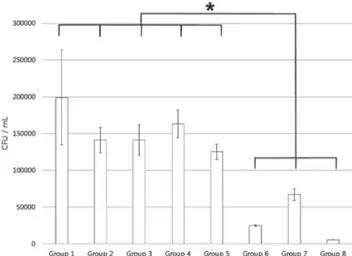
The mean and standard deviation of the experimental results in the biofilm state are specified in Table 2 and Fig. 2.
Similar to the planktonic state, the CFU of the S. mutans was significantly decreased when the photosensitizer and 3 kinds of light source were used at the same time as the other groups. In addition, the same amount of energy was irradiated in these 3 groups, but no statistical significance was observed in these 3 groups. When compared to the planktonic state, S.
mutans in the biofilm state showed a difference of about 100 to 1000 times CFU. It was confirmed that the same degree of antimicrobial effect could be obtained when using the same amount of light source in the planktonic state and the biofilm state of S. mutans .
Ⅳ. Discussion
Many methods for preventing dental caries have been de- veloped. PDT has been in operation since the 1990s[7,16,17].
PDT is a less invasive and less toxic method of reducing bio-
films that cause intraoral diseases. In PDT, the light source,
photosensitizer, and singlet oxygen interact to reducing the number of biofilms in the oral cavity[18].
The photosensitizer used in PDT has an affinity for bacterial cell walls and is activated by light irradiation, which damages the cell wall. Activated photosensitizer molecules can transfer energy to neighboring cell wall molecules, producing active oxygen or free radicals that can damage or kill bacteria[19]. In this study, the photosensitizer was erythrosine. The compound is commonly used as a disclosing agent in dental practice.
Erythrosine is effective in the treatment of intraoral bio-
films[12]. PDT using a halogen curing unit and erythrosine for S.
mutans biofilms optimized the concentration of erythrosine (>
20 - 40 μM) and application time (>2.5 min) for the prevention of dental caries[20].
Previous studies on PDT have used lasers as a light source for activating photosensitizers[21-23]. Use of a laser has ad- vantages that include monochromaticity and high potency.
However, the procedure is expensive, involves only a single wavelength, and requires a separate unit for each photosensi- tizer. To circumvent these disadvantages, PDT uses an easy-to- Fig. 1. Mean and standard deviation of Streptococcus mu-
tans planktonic cell count. One way ANOVA test (*: p < 0.05)
Fig. 2. Mean and standard deviation of Streptococcus mu- tans biofilm cell count. One way ANOVA test (*: p < 0.05)
Table 1. Streptococcus mutans planktonic viable counts
Group (n = 4) Bacterial count
(mean ± standard deviation CFU/ml) Group Ⅰ (P- L-) 6.01 × 10
9± 4.68 × 10
8 aGroup Ⅱ (P+L-) 6.28 × 10
9± 1.05 × 10
8 aGroup Ⅲ (P-LH+) 4.65 × 10
9± 1.20 × 10
8 aGroup Ⅳ (P-LL+) 4.26 × 10
9± 1.27 × 10
8 aGroup Ⅴ (P-LP+) 4.25 × 10
9± 1.25 × 10
8 aGroup Ⅵ (P+LH+) 3.36 × 10
6± 9.85 × 10
4 bGroup Ⅶ (P+LL+) 4.44 × 10
6± 4.45 × 10
4 bGroup Ⅷ (P+LP+) 1.90 × 10
6± 7.39 × 10
4 ba ,b: statistically significant at p < 0.05
a: No Significance difference between groups Ⅰ to Ⅴ b: No Significance difference between groups Ⅵ to Ⅷ
Table 2. Streptococcus mutans biofilm count
Group (n = 4) Bacterial count
(mean ± standard deviation CFU/ml) Group Ⅰ (P- L-) 1.99 × 10
5± 6.47 × 10
4 aGroup Ⅱ (P+L-) 1.41 × 10
5± 1.73 × 10
4 aGroup Ⅲ (P-LH+) 1.41 × 10
5± 2.10 × 10
4 aGroup Ⅳ (P-LL+) 1.63 × 10
5± 1.91 × 10
4 aGroup Ⅴ (P-LP+) 1.25 × 10
5± 1.04 × 10
4 aGroup Ⅵ (P+LH+) 2.47 × 10
4± 1.29 × 10
3 bGroup Ⅶ (P+LL+) 6.67 × 10
4± 8.12 × 10
3 bGroup Ⅷ (P+LP+) 5.36 × 10
3± 2.30 × 10
2 ba ,b: statistically significant at p < 0.05
a: No Significance difference between groups Ⅰ to Ⅴ
b: No Significance difference between groups Ⅵ to Ⅷ
use and low-cost light source. The three light sources (halogen, LED, and plasma arc) used in this study are also commonly used in dental practice. The photodynamic response of eryth- rosine induced by a halogen light source produces a sufficient amount of reactive oxygen that is lethal to S. mutans [24].
However, disadvantages of a halogen curing unit is the de- creased light output over time, heat generation, and reduction of curing efficiency over time.
To overcome these drawbacks, plasma arc curing units and blue light LED devices have begun to be used. Plasma arc lighting devices have been introduced that provide high light output for fast curing. The development of LEDs operating at 470 nm wavelength has provided another alternative to stan- dard halogen curing units. These light-curing units have re- cently become commercially available. In addition, LED curing devices have been developed as an alternative to halogen cur- ing devices, and these two types of curing units have recently been widely used[25]. A study on PDT using halogen as a light source and erythrosine as a photosensitizer for planktonic S.
mutans reported an effective reduction in viability[26].
Presently, compared with no treatment (Group Ⅰ), photo- sensitizer alone (Group Ⅱ), and light source alone (Groups
Ⅲ - Ⅴ), statistically significant decrease in the number of microorganisms was observed in the group using photosen- sitizer and light source at the same time (Group Ⅵ - Ⅷ) in both planktonic and biofilm state. This confirms the useful- ness of the antibacterial effect of PDT in both conditions[17].
There was no statistical significance in the three groups using both light source and photosensitizer. The PDT light source should match the activation spectrum of the photosensitizer and produce the appropriate light effects at this wavelength.
At the end of the 19th century, Ewald developed the oppo- nency theory[27]. The theory is consistent with PDT, in which the photosensitizer should have the maximum absorbance achieved by a complementary and appropriate light source;
for example, a blue photosensitizer must be irradiated by a red light, which is more absorbed, and both of which are com- plementary colors[27]. In this study, red erythrosine agent and a blue light source were used. The effect of the light source on PDT is influenced by affinity with the photosensitizer, wave- length, and power density[17]. Although the wavelengths of the three kinds of light sources differed from each other, the amount of energy was set to be the same. The same amount of PDT treatment results when the amount of energy applied is the same. Additional studies with the same wavelength and
energy will be needed.
Although there have been a few studies on PDT with differ- ent types of light sources, most did not quantify the amount of energy applied. One study used a 650 mW/cm
2halogen curing unit as an experimental group and 67 mW/cm
2LED as a control group. The irradiated energy was 36 J/cm
2and 4 J/
cm
2, respectively. Due to the differences in the amount of en- ergy irradiated, the LEDs failed to show the results of effective photodynamic therapy[24]. Others conducted a study on PDT using 600 mW/cm
2of halogen and 900 mW/cm
2of LED, and irradiated 18 J/cm
2and 27 J/cm
2respectively[26]. Both light sources were effective.
In this study, 18 J/cm
2of energy was irradiated in all light sources. The optimal dental caries preventive effect in PDT us- ing erythrosine and halogen has been determined to be an irradiation time exceeding 30 s, with an energy output of the halogen of 600 mW/cm
2[20]. Presently, the result of PDT was the same for the three groups with the same amount of en- ergy was used along with photosensitizer. Further studies on the results of photodynamic therapy with different energy ir- radiation will be needed.
The energy of the light source is affected not only by the intensity of the light source but also by the irradiation time.
Choi et al .[20] reported that the PDT was performed while changing the light irradiation time to the S. mutans biofilm us- ing a 600 mW/cm
2halogen light source and the antibacterial effect was significantly increased when the irradiation time was 30 seconds or more. On the basis of this result, 30 seconds of light irradiation was performed using the same Halogen cur- ing unit as used in Choi et al. The output of the Halogen cur- ing unit used was 600 mW/cm
2and the total energy applied was 18000 mW/cm
2. In order to irradiate the same amount of energy, an LED and a plasma arc curing unit with an energy amount of 1200 and 1800 mW/cm
2were used, respectively, and the irradiation time was set to 15 seconds and 10 sec- onds, respectively.
PDT outcome differed between the planktonic and biofilm states. In the planktonic state, compared with the group that had no treatment, the use of both light source and photosen- sitizer reduced bacterial viability by about 1000 times more.
However, in the biofilm state, the reduction was only 10 to 100 times. Other studies have shown similar experimental results in Aggregatibacter actinomycetemcomitans ,[28] Porphyromonas gingivalis and Fusarium nucleatum [29].
Planktonic oral bacteria are sensitized by PDT. However,
the microorganisms responsible for oral diseases are formed in the biofilm state, which is different from that of planktonic bacteria, such as the presence of extracellular polymeric mate- rial, differences in cell wall composition, metabolism activity, growth, and gene expression[30]. The bacteria grown in the biofilm are more resistant to antimicrobial agents like antibiot- ics and biocides. The use of PDT could be prudent in reducing viability of biofilms of bacteria involved in dental caries.
Ⅴ. Conclusion
In the planktonic and biofilm states of S. mutans , erythrosine was used as a photosensitizer and the antimicrobial effects of photodynamic therapy were compared using Halogen, LED, and plasma arc with the same energy as light source.
In the planktonic and biofilm states of S. mutans , the CFU of S. mutans was significantly reduced only when the light source and photosensitizer were simultaneously applied. The same energy was applied considering the output of the light source and the irradiation time, and the total amount of en- ergy applied was 18000 mW/cm
2. There was no statistically significant difference in the CFU of S. mutans among the three experimental groups using the photosensitizer and halogen, LED, and plasma arc sources, respectively.
Therefore, the effect of PDT on the same amount of energy in both planktonic and biofilm states of S. mutans was the same regardless of the type of light source.
This is a useful result in a clinical practice with various dental curing units.
References
1. Smith DJ : Dental caries vaccines: prospects and concerns.
Crit Rev Oral Biol Med , 13:335-349, 2002.
2. Marsh PD : Microbiologic aspects of dental plaque and dental caries. Dent Clin North Am , 43:599-614, 1999.
3. Loe H : Oral hygiene in the prevention of caries and peri- odontal disease. Int Dent J , 50:129-139, 2000.
4. Hoyle BD, Costerton JW : Bacterial resistance to antibiotics:
the role of biofilms. Prog Drug Res , 37:91-105, 1991.
5. Paschoal MA, Lin M, Santos-Pinto L, Duarte S : Photo- dynamic antimicrobial chemotherapy on Streptococcus mutans using curcumin and toluidine blue activated by a
novel LED device. Lasers Med Sci , 30:885-890, 2015.
6. Dougherty TJ : An update on photodynamic therapy appli-
cations. J Clin Laser Med Surg , 20:3-7, 2002.
7. Konopka K, Goslinski T : Photodynamic therapy in dentistry.
J Dent Res , 86:694-707, 2007.
8. Yildirim C, Karaarslan ES, Ozsevik S, et al . : Antimicrobial ef- ficiency of photodynamic therapy with different irradiation durations. Eur J Dent , 7:469-473, 2013.
9. Diniz IM, Horta ID, Azevedo CS, et al . : Antimicrobial pho- todynamic therapy: a promise candidate for caries lesions treatment. Photodiagnosis Photodyn Ther , 12:511-518, 2015.
10. Quishida CC, Mima EG, Dovigo LN, et al . : Photodynamic inac- tivation of a multispecies biofilm using Photodithazine((R)) and LED light after one and three successive applications.
Lasers Med Sci , 30:2303-2312, 2015.
11. Ricatto LG, Conrado LA, Turssi CP, et al . : Comparative evaluation of photodynamic therapy using LASER or light emitting diode on cariogenic bacteria: An in vitro study.
Eur J Dent , 8:509-514, 2014.
12. Wood S, Metcalf D, Devine D, Robinson C : Erythrosine is a potential photosensitizer for the photodynamic therapy of oral plaque biofilms. J Antimicrob Chemother , 57:680-684, 2006.
13. Lee YH, Park HW, Lee JH, et al . : The photodynamic therapy on Streptococcus mutans biofilms using erythrosine and dental halogen curing unit. Int J Oral Sci , 4:196-201, 2012.
14. Chui C, Aoki A, Takeuchi Y, et al . : Antimicrobial effect of photodynamic therapy using high-power blue light-emit- ting diode and red-dye agent on Porphyromonas gingiva- lis. J Periodontal Res , 48:696-705, 2013.
15. Wilson BC, Patterson MS : The physics, biophysics and technology of photodynamic therapy. Phys Med Biol , 53:
R61-109, 2008.
16. Sharman WM, Allen CM, van Lier JE : Photodynamic thera- peutics: basic principles and clinical applications. Drug Dis- cov Today , 4:507-517, 1999.
17. Soukos NS, Goodson JM : Photodynamic therapy in the control of oral biofilms. Periodontol 2000 , 55:143-166, 2011.
18. Nagata JY, Hioka N, Kimura E, et al . : Antibacterial photo- dynamic therapy for dental caries: evaluation of the photo- sensitizers used and light source properties. Photodiagno- sis Photodyn Ther , 9:122-131, 2012.
19. Dougherty TJ, Gomer CJ, Henderson BW, et al . : Photo- dynamic therapy. J Natl Cancer Inst Monogr, 90:889-905, 1998.
20. Choi S, Park H, Lee J, et al . : Optimum Treatment Param-
eters for Photodynamic Antimicrobial Chemotherapy on Streptococcus mutans Biofilms. J Korean Acad Pediatr Dent , 42:151-157, 2015.
21. Zanin IC, Goncalves RB, Junior AB, et al . : Susceptibility of Streptococcus mutans biofilms to photodynamic therapy:
an in vitro study. J Antimicrob Chemother , 56:324-330, 2005.
22. Hope CK, Wilson M : Induction of lethal photosensitization in biofilms using a confocal scanning laser as the excitation source. J Antimicrob Chemother , 57:1227-1230, 2006.
23. Araujo PV, Teixeira KI, Lanza LD, et al . : In vitro lethal pho- tosensitization of Streptococcus mutans using methylene blue and toluidine blue O as photosensitizers. Acta Odon- tol Latinoam , 22:93-97, 2009.
24. Fracalossi C, Nagata JY, Pellosi DS, et al . : Singlet oxygen production by combining erythrosine and halogen light for photodynamic inactivation of Streptococcus mutans . Pho- todiagnosis Photodyn Ther , 15:127-132, 2016.
25. Nomoto R, McCabe JF, Hirano S : Comparison of halogen, plasma and LED curing units. Oper Dent, 29:287-294, 2004.
26. Jung JS, Park HW, Lee JH, et al . : The effect of photody- namic therapy on the viability of Streptococcus mutans iso- lated from oral cavity. J Korean Acad Pediatr Dent , 39:233- 241, 2012.
27. Salva KA : Photodynamic therapy: unapproved uses, dos- ages, or indications. Clin Dermatol , 20:571-581, 2002.
28. Goulart Rde C, Bolean M, Paulino Tde P, et al . : Photody- namic therapy in planktonic and biofilm cultures of Aggre- gatibacter actinomycetemcomitans. Photomed Laser Surg, 28 Suppl 1:S53-60, 2010.
29. Street CN, Pedigo LA, Loebel NG : Energy dose parameters affect antimicrobial photodynamic therapy-mediated eradi- cation of periopathogenic biofilm and planktonic cultures.
Photomed Laser Surg , 28 Suppl 1:S61-66, 2010.
30. Costerton JW, Stewart PS, Greenberg EP : Bacterial biofilms:
a common cause of persistent infections. Science , 284:
1318-1322, 1999.
국문초록
광원의 종류에 따른 광역동 치료시의 Streptococcus mutans 에 대한 항균 효과
김재용
1ㆍ박호원
1ㆍ이주현
1ㆍ서현우
1ㆍ이시영
21
강릉원주대학교 치과대학 소아치과학교실 및 구강과학연구소
2