산화동물 모델 흰쥐 간세포에 대한 沙參, 羊乳根 및 海防風의 항산화 효과 연구
민상홍, 한효상, 이영종
* 1)
경원대학교 한의과대학 본초학교실
Study on the Anti-oxidative Effects of Adenophorae Radix, Codonopsis lanceolatae Radix and Glehniae Radix Cum Rhizoma
on Liver Cells Isolated from Oxidatively Stressed Rat
Sang-Hong Min, Hyo-Sang Han, Young-Jong Lee
*
Dept. of Herbology, College of Oriental Medicine, Kyungwon University
ABSTRACTS
Objectives : The present study was purposed to compare Adenophorae Radix (henceforth AR), Codonopsis lanceolatae Radix (henceforth ClR) and Glehniae Radix cum Rhizoma (henceforth GRcR) concerning their anti-oxidant effect.
Methods : We measured eythrocyte, leukocyte, thrombocyte, serum albumin, total bilirubin, LDL cholesterol, and glucose as well as SOD, GSH, catalase, NO, and MDA in the rat liver oxidatively stressed by AAPH.
Results : 1. The oxidative stress-induced thrombocyte levels were significantly decreased in CIR-treated and GRcR-treated groups.
2. The oxidative stress-impaired SOD acitivities were significantly recovered in AR-treated and GRcR-treated groups.
3. The oxidative stress-reduced GSH contents were significantly increased in ClR-treated and GRcR-treated groups.
4. The oxidative stress-reduced catalase contents were significantly increased in all of the three groups.
5. The oxidative stress-induced NO productions were significantly decreased in all of the three groups.
Conclusions : AR, ClR, and GRcR altogether showed the anti-oxidant effect in the rat liver oxidatively stressed by AAPH. The anti-oxidant properties of tAR, ClR, and GRcR seem to be similar even if those have different botanical properties and different medical efficacies in oriental medicine.
Key words : Adenophorae Radix, Codonopsis lanceolatae Radix, Glehniae Radix cum Rhizoma, anti-oxidant effect
* 교신저자 : 이영종, 경기도 성남시 수정구 복정동 산 65 경원대학교 한의과대학 본초학교실
․ Tel : 031-750-5415 ․ E-mail : [email protected]
․ 접수:2009년 8월 25일 ․ 수정:2009년 9월 22일 ․ 채택:2009년 9월 23일
서 론
沙參은 ≪神農本草經≫
1)
에 “味苦微寒, 主血積驚氣, 除 寒熱, 補中, 益肺氣, 久服利人, 一名知母, 生川谷”이라고 처음 수록된 이래 養陰淸肺, 祛痰止咳의 효능이 있어 肺 熱燥咳, 虛勞久咳, 乾咳稠痰, 煩熱口乾, 咽乾喉痛 등의 증 상을 치료하는 데 상용되고 있다2)
.沙參의 기원으로 ≪대한약전외한약(생약)규격집≫
3)
에 잔 대Adenophora triphylla var. japonica Hara 또는 기타 동속식물(초롱꽃과 Campanulaceae)의 뿌리로 되어 있고,≪중화인민공화국약전≫
4)
에 南沙參은 같은 과의 층층잔 대(輪葉沙參)Adenophora tetraphylla (Thunb.) Fisch 혹 은 당잔대(沙參)Adenophora stricta Miq.의 뿌리로 되어 있고,≪北沙參≫은 산형과(Umbelliferae)에 속하는 갯방풍(珊瑚菜) Glehnia littoralis Fr. Schmidt et Miq.의 뿌리로 되어 있으 며, ≪조선민주주의인민공화국약전≫
5)
에는 도라지과의 더덕 Codonopsis lanceolata (Sieb. et Zucc.) Benth. et Hook 뿌 리로 되어 있고, ≪중화민국중약약전≫6)
에는 층층잔대(輪 葉沙參) Adenophora tetraphylla (Thunb.) Fisch 및 동속 근연식물의 외피를 제거한 뿌리로 되어 있다.이처럼 沙參의 기원식물이 한국, 중국 및 북한의 공정 서에 서로 달리 수재되어 있을 뿐만 아니라, 우리나라의 실제 임상에서는 아직도 羊乳根으로 사용하여야 할 더덕 을 沙參으로 사용하는 경우가 있고, 중국에서 北沙參으로 사용되는 갯방풍이 ≪대한약전≫
7)
에는 海防風이라는 이 름으로 수재되어 있을 뿐만 아니라 실제 임상에서도 元 防風 즉 防風으로 사용되고 있어 더욱더 혼란스러운 실 정이므로 沙參으로 유통되거나 사용되고 있는 잔대, 더덕 및 갯방풍의 효능을 검증할 필요가 있다고 사료된다.沙參(잔대)의 성분은 taraxerone, carotene, daucsterol, octacosanomic acid, phospholipids, phosphadidic acid, phosphatidyl choline, phosphatidyl ethanolamine, phosphatidyl inositol 등이 함유되어 있으며
8)
, 약리작용은 거담, 강심, 면역조절 및 항진균 작용 등이 있다고 하였으며9)
, Han 등10)
은 더덕의 항산화 효과를, Liu 등11)
은 갯방풍의 항산 화 효과를 보고하였다.인체 내에서 다양한 경로에 의하여 유발되는 산화적 스트레스는 생체 안에 존재하는 항산화계에 의해 제거되 지만 산화적 스트레스가 항산화계의 수준을 초과하여 제 거되지 못하면 생체막의 손상, 고분자 단백질 및 DNA의 변형과 기능상실 등으로 인한 다양한 퇴행성 질환이 유 발될 수 있는데
12,13)
, 沙參은 養陰淸肺하여 祛痰止咳, 排膿 消腫 등의 효능이 있기 때문에 항산화 효과와 관련이 있 을 것으로 사료된다.이에 저자는 현재 시중에서 沙參으로 잔대뿐만 아니라 더덕도 함께 유통되고, 중국에선 갯방풍을 北沙參이라 하여 沙參으로 사용하고 있는 점에 착안하여, 沙參(잔 대), 羊乳根(더덕) 및 海防風(갯방풍)의 산화반응 억제 효과를 비교하여 유의한 결과를 얻었기에 보고한다.
실 험 1. 재료
1) 약재
실험에 사용된 沙參(잔대 Adenophorae Radix 이하 AR로 표현)과 羊乳根(더덕 Codonopsis lanceolatae Radix 이하 ClR로 표현)은 2005년 3월 가락시장에서 구입하였 고, 海防風(갯방풍 Glehniae Radix cum Rhizoma. 이하 GRcR로 표현)은 경북 봉화 재배 유통품을 구입하였다.
2) 동물
동물은 雄性인 6주령의 Sprague-Dawley 흰쥐를 (주) 샘타코로부터 공급받아 실험당일까지 고형사료(抗生劑 無添加, 삼양사료)와 물을 충분히 공급하고, 실온 22±2℃
를 유지하여 2주일간 실험실 환경에 적응시킨 후 실험에 사용하였다.
3) 시약 및 기기
(1) 시약
시 약 명 제조사 제조국
Ethyl ether Samchun
Chemical Korea Sodium phosphate dibasic
Sodium phosphate monobasic Isopropanol
Sulfanilamide H2SO4
Potassium phosphate Xanthine
Sigma USA
Phosphoric acid Junsei Japan 10× reaction buffer
2.5mM dNTP 1.5mM MgCl2 10pM primer
Bioneer Korea
Agarose FMC USA
SOD(superoxide dismutase)
assay kit Dojindo Japan
GSH(glutathione) assay kit Catalase assay kit NO(nitric oxide) assay kit Lipid peroxidation assay kit
Oxford Biomedical Research USA
(2) 기기
기 기 명 제조사 제조국
Pulverizer Rong tsong Taiwan Rotary evaporator
Air compressor
Eyela
Japan Tamiya
Homogenizer OMNi USA
Centrifuge Hanil
Korea Fume hood Hanil
Clean bench Jeio thec Bio-freezer Sanyo
Japan Spectrophotometer Shimazu
Thermo aluminum bath Fine PCR
기 기 명 제조사 제조국 Vortex mixer Vision Scientific Co
Korea Water bath iNtRON biotech
Primus 96 thermocycler
system MWG Biotech Germany
Plate shaker Lab-Line USA
ELISA reader TECAN Austria
Ice-maker Vision Scientific Co Korea Electric chemical balance MC1 Germany
2. 방법
1) 약물 추출
沙參, 羊乳根 및 海防風 100 g씩을 세절하여 각각 1 L 의 증류수를 가하고 약탕기(웅진약탕기, 한국)를 이용하 여 3시간 동안 끓인 다음 여과지로 여과한 후 감압증류 장치를 이용하여 수분을 제거하여 분말로 만들었다.
2) 산화 동물모델에서의 항산화 효과 측정 (1) AAPH[2,2’-azobis(2-amidinopropane) dihydrochloride]
에 의한 산화적 스트레스 유발
14)
및 약물 투여 SD 흰쥐를 각 정상군, 대조군, 沙參 전탕액 투여군(AR), 羊乳根 전탕액 투여군(ClR), 海防風 전탕액 투여군(GRcR) 으로 나누어 실험하였다. 대조군은 50 mg/kg의 AAPH 를 매일 1회씩 7일간 복강에 투여 하였고, 치료군에는 AAPH 투여와 함께 각 약재 전탕액(1%)을 200 ㎕씩 7일 간 매일 1회씩 경구 투여 하였다. 약물 투여 종료 후 12 시간 절식시킨 다음 간 조직 적출과 채혈을 실시하였다.(2) Hematology 분석
흰쥐는 실험 종료 24시간 후 ethyl ether를 이용해 마 취한 후 심장채혈법으로 채혈하였다. 채혈한 혈액은 CBC (complete blood count) bottle에 넣어 적혈구, 백혈구, 혈 소판을 (주)이원임상검사센터(대전, 한국)에 의뢰하여 측 정하였다.
(3) Blood chemistry 분석
흰쥐는 실험 종료 24시간 후 ethyl ether를 이용해 마 취한 후 심장채혈법으로 채혈하였다. 채혈한 혈액은 6,500 rpm 15분간 원심분리하여 LDL cholesterol, total bilirubin, albumin, glucose를 (주)이원임상검사센터(대전, 한국)에 의뢰하여 측정하였다.
(4) 간장에서의 항산화 효과 측정
① 간 조직의 분획
간 조직의 일부를 적출한 후 Bansal 등의 방법
15)
에 의 해 4배의 150 mM의 KCl을 가하여 homogeniger를 이용 하여 균질화 하였다. 균질화 한 조직을 1차 원심분리 (1,000 rpm 20분)한 후 균질화 되지 않은 조직 등을 제거 하고 2차 원심분리(4,000 rpm 30분)하여 상등액을 취하여nitric oxide (NO), GSH, catalase를 측정하였고, 나머지 는 3차 원심분리(20,000 rpm 1시간) 후 상등액을 취하여 SOD. malondialdehyde (MDA)를 측정하였다.
② SOD activity
SOD assay kit (Dojindo, Japan)을 이용하여 측정하였 고, 450 nm에서 흡광도를 측정한 후 SOD activity를 계 산 하였다.
③ GSH
Glutathione kit (Dojindo, Japan)을 이용하여 측정하였 고 405 nm에서 흡광도를 측정한 후 GSH의 농도를 계산 하였다.
④ NO assay
Nitric Oxide assay kit (Dojindo, Japan)을 이용하여 측정하였고, 450 nm에서 흡광도를 측정한 후 NO의 함량 을 계산하였다.
⑤ Lipid peroxidation
Lipid peroxidation assay kit (Oxford Biomedical Research, USA)을 이용하여 측정하였고 586 nm에서 흡광도를 측 정한 후 MDA를 계산하였다.
⑥ Catalase activity
Catalase 활성도 측정은 Abei의 방법
16)
에 따라 3.0 ㎖ cuvette에 130 nm phosphate buffer (pH 7,0) 500 ㎕, 간 분획물 40 ㎕와 증류수 660 ㎕를 혼합하여 기질인 15 mM H2
O2
농도에 의한 흡광도의 감소율을 측정하였다.효소의 활성도는 1분 동안 1 μM의 H
2
O2
를 분해시키는 효소의 양을 1 unit로 표시하였다.3) 통계처리
본 실험에서 얻은 결과를 ANOVA multi t-test (JAVA, Bonferroni Ver 1.1)로 분석하여 p값을 구하였다. 각 실험 군을 대조군 및 정상군과 비교하여 p<0.05일 때 유의성 이 있는 것으로 판정하였다.
성 적
1. 산화 동물모델에서의 항산화 효과 1) Hematology 분석
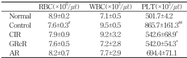
각 실험군의 혈액을 채취하여 적혈구, 백혈구 및 혈소 판의 수를 측정하였다(Table 1).
(1) 적혈구 수
각 실험군의 혈액을 채취하여 적혈구의 수를 측정한 결 과, 대조군에서는 정상군에 비하여 적혈구 수가 유의하게 (p<0.01) 감소하였다. 각 약물 투여군들에서는 대조군에 비하 여 약간 증가하는 경향이 있으나 유의성은 없었다(Fig. 1).
Table 1. Hematology Analysis
RBC(×106/㎕) WBC(×103/㎕) PLT(×103/㎕) Normal 8.9±0.2 7.1±0.5 501.7±4.2 Control 7.6±0.3# 9.5±0.5 865.7±161.3##
CIR 7.9±0.9 9.2±3.2 542.6±68.9*
GRcR 7.6±0.5 7.2±2.8 542.0±54.3*
AR 8.2±0.7 7.7±2.9 694.4±71.1
The rats were injected intraperitoneally (i.p) with AAPH for 7 days (once a day. 50 mg/kg) and orally administered with ClR, GRcR and AR decoction (1%) for 7 days (onc a day. 200 ul). The animals were anesthetized with ethyl ether and the blood samples were taken from the heart.
Normal : Normal SD rat.
Control : AAPH injection.
ClR : AAPH injection and treatment with ClR decoction (1%).
GRcR : AAPH injection and treatment with GRcR decoction (1%).
AR : AAPH injection and treatment with AR decoction (1%).
Values represent the means±SD of 8 rats.
* : p < 0.05, compared to control group.
# : p < 0.05, ## : p < 0.01 compared to normal group.
6.5 7 7.5 8 8.5 9 9.5
Normal Control ClR GRcR AR
A A PH RB C ( x 1 0
6/u l)
#
Fig. 1. Effects of ClR, GRcR and AR decoction on the number of RBC in rats injected with AAPH
The rats were injected intraperitoneally (i.p.) with AAPH for 7 days (once a day. 50 mg/kg) and orally administered with ClR, GRcR and AR decoction (1%) for 7 days (onc a day. 200 ㎕). The animals were anesthetized with ethyl ether and the blood samples were taken from the heart and the number of RBC was measured.
Normal : Normal SD rat.
Control : AAPH injection.
ClR : AAPH injection and treatment with ClR decoction (1%).
GRcR : AAPH injection and treatment with GRcR decoction (1%).
AR : AAPH injection and treatment with AR decoction (1%).
Values represent the mean±SD of 8 rats.
# : p< 0.05 compared to normal group.
(2) 백혈구 수
각 실험군의 혈액을 채취하여 백혈구의 수를 측정한 결과, 대조군은 정상군에 비하여 백혈구의 수가 증가하였 으나 유의성은 없었다. 각 약물 투여군들에서는 대조군에 비하여 백혈구 수가 감소했으나 유의성은 없었다(Fig. 2).
(3) 혈소판 수
각 실험군의 혈액을 채취하여 혈소판 수를 측정한 결 과, 대조군에서는 정상군에 비하여 혈소판의 수가 유의성 있게(p<0.01) 증가하였고, 羊乳根 투여군과 海防風 투여 군에서는 대조군에 비하여 유의성 있게(p<0.05) 감소하였 으며, 沙參 투여군에서도 대조군에 비하여 감소하였으나 유의성은 없었다(Fig. 3).
0 2 4 6 8 10 12 14
Normal Control ClR GRcR AR
A A PH
WBC ( x 10
3/u l)
Fig. 2. Effects of ClR, GRcR and AR decoction on the number of WBC in rats injected with AAPH
The rats were injected intraperitoneally (i.p.) with AAPH for 7 days (once a day. 50 mg/kg) and orally administered with ClR, GRcR and AR decoction (1%) for 7 days (onc a day. 200 ㎕). The animals were anesthetized with ethyl ether and the blood samples were taken from the heart and the number of WBC was measured.
Normal : Normal SD rat.
Control : AAPH injection.
ClR : AAPH injection and treatment with ClR decoction (1%).
GRcR : AAPH injection and treatment with GRcR decoction (1%).
AR : AAPH injection and treatment with AR decoction (1%).
Values represent the mean±SD of 8 rats.
0 200 400 600 800 1000 1200
Normal Control ClR GRcR AR
AAP H PLT (x103/ul)
* *
##
Fig. 3. Effects of ClR, GRcR and AR decoction on the number of PLT in rats injected with AAPH
The rats were injected intraperitoneally (i.p.) with AAPH for 7 days (once a day. 50 mg/kg) and orally administered with ClR, GRcR and AR decoction (1%) for 7 days (onc a day. 200 ㎕). The animals were anesthetized with ethyl ether and the blood samples were taken from the heart and the number of PLT was measured.
Normal : Normal SD rat.
Control : AAPH injection.
ClR : AAPH injection and treatment with ClR decoction (1%).
GRcR : AAPH injection and treatment with GRcR decoction (1%).
AR : AAPH injection and treatment with AR decoction (1%).
Values represent the mean±SD of 8 rats.
## : p< 0.01 compared to normal group.
* : p< 0.05 compared to control group.
2) Blood chemistry 분석
실험군에서 취한 혈청에서 albumin, total bilirubin, LDL cholesterol 및 glucose을 측정하였다(Table 2).
(1) Albumin
각 실험군에서 취한 혈청에서 albumin을 측정한 결과, 대 조군은 정상군에 비하여 albumin이 감소하였으나 유의성은 없었다. 각 약물 투여군들에서는 대조군에 비하여 albumin 이 증가 했으나 역시 유의성은 보이지 않았다(Fig. 4).
Table 2. Blood Chemistry Analysis
Albumin (g/㎗)
Total bilirubin
(㎎/㎗)
LDL cholesterol
(㎎/㎗)
Glucose (㎎/㎗) Normal 2.90±0.06 0.240±0.017 16.2±1.9 128.3±4.0 Control 2.61±0.14 0.153±0.015### 24.7±0.6## 57.3±4.0###
CIR 2.73±0.10 0.172±0.019 20.2±6.0* 77.6±11.1 GRcR 2.82±0.06 0.176±0.026 19.8±5.3* 66.2±19.7 AR 2.78±0.07 0.155±0.005 22.3±1.7* 78.0±10.9 The rats were injected intraperitoneally (i.p) with AAPH for 7 days (once a day. 50 mg/kg) and orally administered with ClR, GRcR and AR decoction (1%) for 7 days (onc a day. 200 ul). The animals were anesthetized with ethyl ether and the blood samples were taken from the heart.
Normal : Normal SD rat.
Control : AAPH injection.
ClR : AAPH injection and treatment with ClR decoction (1%).
GRcR : AAPH injection and treatment with GRcR decoction (1%).
AR : AAPH injection and treatment with AR decoction (1%).
Values represent the means±SD of 8 rats.
## : p< 0.01, ### : p< 0.001 compared to normal group.
2.4 2.5 2.6 2.7 2.8 2.9 3
Normal Control ClR GRcR AR
A A P H
Albumin(g/dl)
Fig. 4. Effects of ClR, GRcR and AR decoction on the level of albumin in rats injected with AAPH
The rats were injected intraperitoneally (i.p.) with AAPH for 7 days (once a day. 50 mg/kg) and orally administered with ClR, GRcR and AR decoction (1%) for 7 days (onc a day. 200 ㎕). The animals were anesthetized with ethyl ether and the blood samples were taken from the heart and the level of albumin in serum was measured.
Normal : Normal SD rat.
Control : AAPH injection.
ClR : AAPH injection and treatment with ClR decoction (1%).
GRcR : AAPH injection and treatment with GRcR decoction (1%).
AR : AAPH injection and treatment with AR decoction (1%).
Values represent the mean±SD of 8 rats.
(2) Total bilirubin
각 실험군에서 취한 혈청에서 total bilirubin을 측정한 결과, 대조군은 정상군에 비하여 total bilirubin이 유의성 있게 증가하였다. 각 약물 투여군들에서는 대조군에 비하 여 total bilirubin이 증가 했으나 유의성은 없었다(Fig. 5).
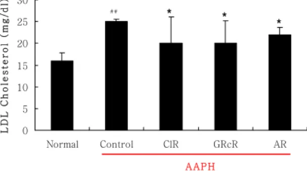
(3) Low-density lipoprotein(LDL) cholesterol 각 실험군에서 취한 혈청에서 LDL cholesterol의 생성 을 측정한 결과, 대조군에서는 정상군에 비하여 LDL cholesterol이 유의성 있게(p<0.01) 증가하였고, 각 약물 투여군에서는 대조군에 비하여 羊乳根, 海防風, 沙參 모 두 감소하였으나 통계적 유의성은 없었다(Fig. 6).
0 0.05 0.1 0.15 0.2 0.25 0.3
Normal Control ClR GRcR AR
A A PH
Total bilirubin (mg/dl)
###
Fig. 5. Effects of ClR, GRcR and AR decoction on total bilirubin in rats injected with AAPH
The rats were injected intraperitoneally (i.p.) with AAPH for 7 days (once a day. 50 mg/kg) and orally administered with ClR, GRcR and AR decoction (1%) for 7 days (onc a day. 200 ㎕). The animals were anesthetized with ethyl ether and the blood samples were taken from the heart and the level of total bilirubin in serum was measured.
Normal : Normal SD rat.
Control : AAPH injection.
ClR : AAPH injection and treatment with ClR decoction (1%).
GRcR : AAPH injection and treatment with GRcR decoction (1%).
AR : AAPH injection and treatment with AR decoction (1%).
Values represent the mean±SD of 8 rats.
### : p< 0.001 compared to normal group.
0 5 10 15 20 25 30
Normal Control ClR GRcR AR
AAPH
LDL C h ol es te r o l (m g/ dl )
## * *
*
Fig. 6. Effects of ClR, GRcR and AR decoction on LDL cholesterol in rats injected with AAPH
The rats were injected intraperitoneally (i.p) with AAPH for 7 days (once a day. 50 mg/kg) and orally administered with ClR, GRcR and AR decoction (1%) for 7 days (onc a day. 200 ul). The animals were anesthetized with ethyl ether and the blood samples were taken from the heart and the level of LDL cholesterol in serum was measured.
Normal : Normal SD rat.
Control : AAPH injection.
ClR : AAPH injection and treatment with ClR decoction (1%).
GRcR : AAPH injection and treatment with GRcR decoction (1%).
AR : AAPH injection and treatment with AR decoction (1%).
Values represent the means±SD of 8 rats.
* : p< 0.05 compared to normal group.
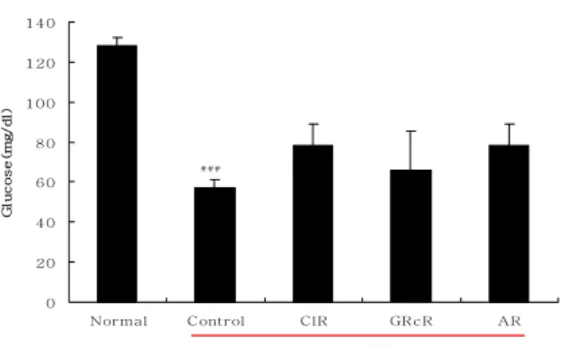
(4) Glucose
각 실험군에서 취한 혈청에서 glucose의 생성을 측정 한 결과, 대조군은 정상군에 비하여 glucose가 유의성 있게(p<0.001) 감소하였다. 각 약물 투여군들에서는 대 조군에 비하여 glucose가 증가했으나 유의성은 없었다 (Fig. 7).
0 20 40 60 80 100 120 140
Normal Control ClR GRcR AR
A A P H
Glucose(mg/dl)
###
Fig. 7. Effects of ClR, GRcR and AR decoction on the level of glucose in rats injected with AAPH
The rats were injected intraperitoneally (i.p.) with AAPH for 7 days (once a day. 50 mg/kg) and orally administered with ClR, GRcR and AR decoction (1%) for 7 days (onc a day. 200 ㎕). The animals were anesthetized with ethyl ether and the blood samples were taken from the heart and the level of glucose in serum was measured.
Normal : Normal SD rat.
Control : AAPH injection.
ClR : AAPH injection and treatment with ClR decoction (1%).
GRcR : AAPH injection and treatment with GRcR decoction (1%).
AR : AAPH injection and treatment with AR decoction (1%).
Values represent the mean±SD of 8 rats.
## : P< 0.01 compared to normal group.
* : p< 0.05. **: p<0.01 compared to control group.
3) 간 조직에서의 항산화 효과
흰쥐에서 적출한 간 조직을 분획하여 SOD의 활성, GSH의 함량, NO함량, MDA함량 및 catalase의 함량을 측정하였다(Table 3).
Table 3. Antioxidative Effects of ClR, GRcR and AR SOD
(%) GSH
(μmol/ℓ) NO
(μmol/ℓ) MDA
(μmol/㎖) Catalase (U/㎎) Normal 96.2±5.1 94.4±5.6 24.2±9.7 2.9±0.1 287.7±13.5 Control 75.8±11.0## 31.6±4.8### 111.9±9.4### 3.9±0.4 117.0±33.5###
CIR 85.8±5.8 71.8±9.5* 56.2±8.6* 3.4±0.4 195.7±6.1**
GRcR 95.8±5.0** 43.1±2.1* 72.7±7.5* 3.2±0.1 205.3±8.7* AR 101.6±2.6** 42.3±3.6 47.9±2.3* 3.5±0.5 191.3±11.2**
The rats were injected intraperitoneally (i.p) with AAPH for 7 days (once a day. 50 mg/kg) and orally administered with ClR, GRcR and AR decoction (1%) for 7 days (onc a day. 200 ul). After the animals were sacrificed, the liver was removed and the concentrations of SOD, glutathione, NO, MDA and catalase in liver were estimated.
Normal : Normal SD rat.
Control : AAPH injection.
ClR : AAPH injection and treatment with ClR decoction (1%).
GRcR : AAPH injection and treatment with GRcR decoction (1%).
AR : AAPH injection and treatment with AR decoction (1%).
Values represent the means±SD of 8 rats.
* : p< 0.05. ** : p< 0.01compared to control group.
## : p< 0.01. ### : p< 0.001 compared to normal group.
(1) SOD assay
흰쥐에서 적출한 간 조직을 분획하여 항산화 효소인
0 20 40 60 80 100 120
Normal Control ClR GRcR AR
A A PH
S O D a c ti vi ty ( % )
##** **
Fig. 8. Effects of ClR, GRcR and AR decoction on SOD activity in liver
The rats were injected intraperitoneally (i.p.) with AAPH for 7 days (once a day. 50 mg/kg) and orally administered with ClR, GRcR and AR decoction (1%) for 7 days (onc a day. 200 ㎕). After the animals were sacrificed, the liver was removed and SOD was estimated.
Normal : Normal SD rat.
Control : AAPH injection.
ClR : AAPH injection and treatment with ClR decoction (1%).
GRcR : AAPH injection and treatment with GRcR decoction (1%).
AR : AAPH injection and treatment with AR decoction (1%).
Values represent the mean±SD of 8 rats.
## : p< 0.01 compared to normal group.
** : p< 0.01 compared to control group.
SOD의 활성을 측정한 결과, 대조군은 정상군에 비하여 SOD의 활성이 유의성 있게(p<0.01) 감소하였다. 海防風 투여군과 沙參 투여군에서는 대조군에 비하여 유의성 있 게(p<0.01) 증가했으며, 羊乳根 투여군에서는 대조군에 비하여 증가했으나 유의성은 없었다(Fig. 8).
(2) GSH assay
흰쥐에서 적출한 간 조직을 분획하여 GSH의 함량을 측정한 결과, 대조군은 정상군에 비하여 GSH가 유의성 있게(p<0.001) 감소하였다. 羊乳根 투여군과 海防風 투여 군에서는 대조군에 비하여 유의성 있게(p<0.05) 증가했으 며, 沙參 투여군에서는 대조군에 비하여 증가했으나 유의 성은 없었다(Fig. 9).
(3) NO assay
흰쥐에서 적출한 간 조직을 분획하여 NO함량을 측정 한 결과, 대조군은 정상군에 비하여 NO의 함량이 유의성 있게(p<0.001) 증가하였다. 각 약물 투여군들에서는 대조 군에 비하여 羊乳根, 海防風, 沙參 모두 유의성 있게 (p<0.01) 감소하였다(Fig. 10).
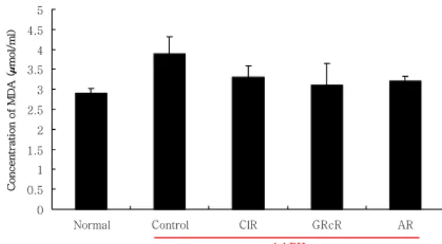
(4) Lipid peroxidation assay
흰쥐에서 적출한 간 조직을 분획하여 Malondialdehyde (MDA) 함량을 측정한 결과, 대조군은 정상군에 비하여 MDA의 함량이 증가하였으나 유의성은 없었다. 각 약물 투여군들에서는 대조군에 비하여 MDA가 감소했으나 유 의성은 없었다(Fig. 11).
0 10 20 30 40 50 60 70 80 90 100
Normal Control ClR GRcR AR
AAPH
Concentration of GSH (µmol/l)
###
*
*
Fig. 9. Effects of ClR, GRcR and AR decoction on the concentration of hepatic glutathione
The rats were injected intraperitoneally (i.p.) with AAPH for 7 days (once a day. 50 mg/kg) and orally administered with ClR, GRcR and AR decoction (1%) for 7 days (onc a day. 200 ㎕). After the animals were sacrificed, the liver was removed and glutathione level was estimated.
Normal : Normal SD rat.
Control : AAPH injection.
ClR : AAPH injection and treatment with ClR decoction (1%).
GRcR : AAPH injection and treatment with GRcR decoction (1%).
AR : AAPH injection and treatment with AR decoction (1%).
Values represent the mean±SD of 8 rats.
### : P< 0.001 compared to normal group.
* : p< 0.05 compared to control group.
0 20 40 60 80 100 120 140
Normal Control ClR GRcR AR
A A PH
Concentration of NO (µmol/l)
###
* * *
Fig. 10. Effects of ClR, GRcR and AR decoction on the concentration of hepatic nitric oxide
The rats were injected intraperitoneally (i.p.) with AAPH for 7 days (once a day. 50 mg/kg) and orally administered with ClR, GRcR and AR decoction (1%) for 7 days (onc a day. 200 ㎕). After the animals were sacrificed, the liver was removed and NO concentration was measured.
Normal : Normal SD rat.
Control : AAPH injection.
ClR : AAPH injection and treatment with ClR decoction (1%).
GRcR : AAPH injection and treatment with GRcR decoction (1%).
AR : AAPH injection and treatment with AR decoction (1%).
Values represent the mean±SD of 8 rats.
### : p< 0.001 compared to normal group.
* : p< 0.05 compared to control group.
(5) Catalase activity
흰쥐에서 적출한 간 조직을 분획하여 catalase의 함량 을 측정한 결과, 대조군은 정상군에 비하여 catalase가 유 의성 있게(p<0.001) 감소하였다. 각 약물 투여군들에서는 대조군에 비하여 羊乳根, 海防風, 沙參 모두 유의성 있게 (p<0.01) 증가하였다(Fig. 12).
0 0.5 1 1.5 2 2.5 3 3.5 4 4.5 5
Normal Control ClR GRcR AR
AAPH
Concentration of MDA (µmol/ml)
Fig. 11. Effects of ClR, GRcR and AR decoction on the concentration of hepatic MDA
The rats were injected intraperitoneally (i.p.) with AAPH for 7 days (once a day. 50 mg/kg) and orally administered with ClR, GRcR and AR decoction (1%) for 7 days (onc a day. 200 ㎕). After the animals were sacrificed, the liver was removed and the concentration of hepatic MDA was estimated.
Normal : Normal SD rat.
Control : AAPH injection.
ClR : AAPH injection and treatment with ClR decoction (1%).
GRcR : AAPH injection and treatment with GRcR decoction (1%).
AR : AAPH injection and treatment with AR decoction (1%).
Values represent the mean±SD of 8 rats.
0 50 100 150 200 250 300
Normal Control ClR GRcR AR
AAPH
Catalase activity(U/mg)
###
** ** **
Fig. 12. Effects of ClR, GRcR and AR decoction on catalase activity in liver
The rats were injected intraperitoneally (i.p.) with AAPH for 7 days (once a day. 50 mg/kg) and orally administered with ClR, GRcR and AR decoction (1%) for 7 days (onc a day. 200 ㎕). After the animals were sacrificed, the liver was removed and catalase activity was estimated.
Normal : Normal SD rat.
Control : AAPH injection.
ClR : AAPH injection and treatment with ClR decoction (1%).
GRcR : AAPH injection and treatment with GRcR decoction (1%).
AR : AAPH injection and treatment with AR decoction (1%).
Values represent the mean±SD of 8 rats.
### : p< 0.001 compared to normal group.
** : p< 0.01 compared to control group.
고 찰
沙參은 ≪神農本草經≫
1)
에 “味苦微寒, 主血積驚氣, 除 寒熱, 補中, 益肺氣, 久服利人, 一名知母, 生川谷”이라고 처음 수록되었고, ≪名醫別錄≫17)
에 “主治胃痹, 心腹痛, 結熱, 邪氣, 頭痛, 皮間邪熱, 安五臟, 補中”이라고 한 이래 味甘 性微寒하고 養陰淸肺, 祛痰止咳의 효능이 있어 肺熱燥咳, 虛勞久咳, 乾咳稠痰, 煩熱口乾, 咽乾喉痛 등의 증상 을 치료하는 데 상용되고 있다
2)
. 중국에서는 우리나라에 서 海防風으로 사용하는 갯방풍을 北沙參이라 하여 사용 하는데, 北沙參은 ≪本草滙言≫에 처음 수재되었는데9,18)
≪本草從新≫
19)
에 北沙參은 “甘苦微寒. 味淡體輕. 專補肺 陰, 淸肺火. 治久咳肺痿. 金收火刑者宜之. 寒客肺中作嗽者 勿服. 白實長大者良”이라 하였고, 南沙參은 “功同北沙參而 力稍遜. 色稍黃. 形稍瘦 小而短. 近有一種 味帶辣者不可 用”이라 하여 北沙參과 南沙參을 구별하여 수재하였는데, 南沙參은 北沙參과 효능은 비슷하나 매운 맛이 있는 것 은 사용하여서 안된다고 하였다.갯방풍은 우리나라에서는 性溫 味辛甘하고 解表袪風, 勝濕止痙의 효능이 있어 感冒頭痛, 風濕痺痛, 骨節痠痛, 風疹瘙痒, 四肢攣急, 破傷風등의 증상을 치료하는 防風으 로 사용되고 있으나
2)
, 중국에서는 味甘 性凉하고 養陰淸 肺, 益胃生津의 효능이 있어 肺虛有熱, 乾咳小痰, 熱病後 口乾 등의 증상을 치료하는 北沙參으로 사용하여9,18)
서로 다른 증을 치료하고 있다.羊乳根은 ≪名醫別錄≫
17)
에 “味甘, 溫, 無毒. 主治頭眩 痛, 益氣, 長肌肉, 一名 地黃. 三月採, 立夏後母死”라고 처 음 수재되었으며, 山海螺, 四葉參 등의 異名이 있고9,18)
, 味甘辛 性平하고 消腫, 解毒, 排膿, 祛痰, 催乳의 효능이 있어 肺癰, 乳癰, 腸癰, 腫毒, 瘰癧, 喉蛾, 乳少, 白帶 등의 증상을 치료하는 데 상용되고 있다18)
.이처럼 沙參, 羊乳根, 北沙參은 본초학적으로는 서로 性味와 쓰이는 바가 다른데 실제 임상에서는 잔대, 더덕, 갯방풍 등이 혼용되고 있어 이들의 효능을 검증할 필요 가 있다고 사료된다.
이들 식물에 대해서 Zhao 등
20)
은 Adenophora속과 Glehnia littoralis의 근연성을 DNA sequence를 통해 규 명하였으며, 성분연구로는 Asano 등21)
이 잔대Adenophora triphylla var. japonica에서 알카로이드 성분을 추출하였 고, Lee 등22)
이 더덕Codonopsis lanceolata에서 사포닌인 codonoposide를 분리하였고, Yuan 등23)
은 갯방풍G. littoralis 에서 lignan glycosides, neolignan glycoside, phenylpropanoid glycoside 를 분리하였다. 약리작용으로는 Lee 등24)
은 잔대A. triphylla가 in vitro에서 항암 효과가 있다고 하였 으며, Lee 등25)
은 더덕C. lanceolata에서 추출된 β-D- xylopyranosyl-(1-->3)--β-D-glucuronopyranosyl echinocystic acid가 급성 promyelocytic leukemia HL-60 cells을 개선 한다고 하였으며, Han 등10)
은 더덕이 늙은 쥐에서 항산 화 효과가 있다고 하였고, Liu 등11)
은 갯방풍의 항산화 효과를, Wang 등26)
은 갯방풍의 면역효과를 보고하였다.이처럼 항산화 효과, 면역조절 효과 등이 보고되었지만 잔대와 더덕, 갯방풍의 항산화 효과를 직접 비교한 연구 는 보고된 바가 없다.
이에 저자는 잔대(沙參), 더덕(羊乳根) 및 갯방풍(海防 風)의 항산화 효과를 비교 관찰하였다.
AAPH[2.2’-azobis (2-aminodinopropane) hydrochloride]는 수용성 azo 화합물의 일종으로서 열분해에 의해서 자유
기를 생성하는 것으로 알려져 있다. 복강내로 투여된 AAPH는 수분 내에 혈류를 타고 전신순환하며, AAPH가 산소분자와 반응하여 생성된 carbon radical은 peroxyl radical을 생성하여 여러 형태의 생물학적 분자와 결합반 응을 일으키면서 생체막의 구조를 붕괴시키는 것으로 생 각되고 있다
27)
.본 실험에서는 SD 흰쥐에 AAPH를 투여하여 실험적 급성 산화증을 유발시킨 후, 羊乳根, 海防風과 沙參의 1% 전탕액을 투여해 전반적인 생화학적 변화와 항산화 반응을 추적하였다.
AAPH에서 생성된 peroxyl radical은 생체막의 구조를 붕괴시킨다. 본 실험에서 대조군의 적혈구 수가 정상군에 비하여 통계적으로 유의하게 감소하여 산화에 의한 적혈 구 파괴를 추정할 수 있었다. 羊乳根, 海防風과 沙參 처리군 모두 적혈구의 수가 대조군에 비하여 증가하였 으나 유의성은 발견되지 않았다. 백혈구의 수는 대조군 에서도, 전탕액 투여군에서도 유의한 증가나 감소는 보 이지 않았다.
혈소판 수는 대조군에서 정상군에 비하여 유의하게 증 가하였고, 羊乳根과 海防風 투여군에서 대조군에 비해 유 의하게 감소하였다. 따라서 羊乳根과 海防風 전탕액은 혈 액성분 중 혈소판의 생성에 일정한 영향을 미치는 것으 로 사료되며, 뇌경색이나 혈전 등의 질환에 대한 임상적 활용이 기대된다.
AAPH로부터 생성된 과산화 자유기에 의해 endoplasmic reticulum에서 과산화반응이 일어나 비가역적 단백질 합 성 억제가 일어나고, 이는 혈중 albumin 농도의 감소로 이어진다. 또한 간세포에서의 bilirubin 처리에도 영향을 미치게 되므로 정상 흰쥐에 비해 그 농도가 뚜렷이 증가 하는 양상을 보이게 된다
28)
.본 실험에서 혈청 albumin 치는 대조군과 沙參, 羊乳 根, 海防風 투여군 모두 유의한 변화가 발견되지 않았다.
총 bilirubin 치는 대조군에서 정상군에 비하여 유의하게 감소하였으며, 沙參, 羊乳根 및 海防風을 투여한 군에서 는 대조군에 비해 증가하였으나 통계적 유의성은 보이지 않았다
정상적인 경우 혈중 LDL cholesterol은 세포 내에서 분해되지만, AAPH가 투여되어 산화반응에 의해 세포가 파괴되면 LDL은 분해되지 못하고 혈중에 분포하게 되고, 만일 산화적 스트레스를 받을 경우 축적되어 산화 LDL 을 형성하게 된다
29)
. 생성된 산화 LDL은 높은 세포 독성 이 있는 지질 과산화물을 가지고 있어 세포 조직에 확산 되어 독성을 나타내고, 내피세포에 염증을 일으켜 동맥경 화를 유발한다30)
.본 실험에서 LDL cholesterol은 대조군에 비해서 약물 투여군에서는 모두 감소하였으나 통계적 유의성은 없었다.
혈청 glucose는 노화에 따라 감소하는 것으로 알려져 있다. 본 실험 결과 대조군의 혈청 glucose는 정상군에 비하여 유의하게 감소하였고, 약물 투여군에서는 대조군 에 비하여 증가하였으나 통계적 유의성은 보이지 않았다.
항산화계는 크게 효소계, 비효소계, 소분자와 비효소계 로 나뉘는데, SOD와 catalase는 효소계 항산화계에 속한 다. 산소 유리기들을 선택적으로 차단하는 여러 다양한 외인성 소거제들이 있는 바, superoxide anion 생성은 SOD에 의해 억제되고, hydrogen peroxide는 catalase에 의해 억제된다. SOD는 O
2
가 H2
O2
와 O2
로 전환되는 것을 촉진하는 금속 함유 효소로 진핵세포의 세포질 내에 존 재하는 CuZu-SOD, 그리고 영장류의 세포질 내와 사립체 내강 내에 국한되어 존재하는 Mn-SOD의 두 종류가 있 다31)
. 세포질 내에 존재하는 catalase는 세포 소기관인 peroxizome 내에서 과산화수소를 물과 산소로 분해하는 효소로, cytochrome system을 가진 호기성 세포에 광범 위하게 분포되어 있는데, 포유동물의 간세포나 적혈구 내 에 고농도로 존재하여 과산화수소에 의한 산화적 손상으 로부터 세포를 보호하는 역할을 한다.본 실험 결과 항산화 효소 SOD의 활성은 대조군에서 정상군에 비하여 유의하게 감소하였고, 沙參과 海防風 투 여군에서는 대조군에 비하여 유의하게 증가하였으며, 羊乳根 투여군에서도 증가하였으나 유의성은 없었다.
Catalase는 대조군에서 정상군에 비하여 유의한 감소를 보인 반면, 세 약물 투여군 모두에서 대조군에 비하여 유 의한 증가를 보였다.
GSH는 비특이성 내인성 소거제로서, 내피세포 및 간 세포에 고농도로 존재한다. 이것은 hydrogen peroxidase 를 H
2
O로 환원시켜서 산화성 스트레스에 대하여 세포를 보호하는 작용이 있다32)
.본 실험에서 GSH 함량은 대조군에서 정상군에 비하 여 유의하게 감소하였고, 羊乳根과 海防風 투여군에서 대 조군에 비하여 유의한 증가를 보였다.
질소 중간대사물 중 하나인 NO (nitric oxide)는 면역 전달물질인 cytokine의 영향으로 면역세포로부터 생산되 어, 염증반응부위에 유리됨으로써 Fe-S를 함유하는 효소 의 작용을 억제시키거나 DNA에 손상을 미쳐 항미생물작 용이나 항암작용을 나타낸다고 알려졌다.
본 실험 결과 대조군에서는 정상군에 비하여 NO 함량 이 유의하게 증가한 반면, 세 약물 투여군 모두에서는 대 조군에 비하여 유의한 감소를 보였다.
MDA (malondialdehyde)는 세포막에서 다중불포화지 방산과 새로운 지질자유기가 반응하여 산소 radical 화합 물이 생성되고, 과잉산소가 존재할 때 과산화 radical이 지질과산화라고 하는 연쇄반응을 일으키게 되어 나오는 지질과 산화물의 지표로서 이용된다
33)
.본 실험의 대조군과 투여군 모두 간 조직의 MDA함량 에 유의한 변화를 보이지 않았다.
이상과 같이 沙參, 羊乳根 및 海防風 전탕액 투여가 실험적 산화 흰쥐의 肝內 NO, GSH, catalase, SOD, MDA 함량에 미치는 영향을 관찰한 결과, 羊乳根, 海防 風 및 沙參 모두 catalase 함량을 증가시키고, NO 함량을 감소시켰다. 또한 SOD의 활성은 沙參과 海防風 투여군 에서, GSH 함량은 羊乳根과 海防風 투여군에서 유의하
게 증가하였다. 페놀 함량은 海防風이 羊乳根 및 沙參에 비하여 높았으며, in vitro에서의 산화반응 억제 효과는 沙參이 羊乳根 및 海防風에 비하여 우수하였으나, 산화적 동물모델을 이용한 실험에서는 전반적으로 유사한 항산 화 효능을 보였으며, 그 중 海防風이 더 많은 항목에서 유의한 항산화 효능을 나타내었다.
본 실험결과를 종합할 때, 동물 모델을 이용한 항산화 실험에서 沙參, 羊乳根 그리고 海防風의 항산화 효능이 유사하였다. 그러므로 본초학적으로 氣味와 효능이 구별 되어 사용되는 沙參, 羊乳根 및 海防風이 항산화 효과에 있어서는 함께 사용될 수 있다고 사료된다.
결 론
沙參, 羊乳根 및 海防風의 AAPH로 유발된 실험적 산 화증에서 적혈구, 백혈구, 혈소판, 혈청 중 albumin, total bilirubin, LDL cholesterol, glucose 및 肝內 SOD, GSH, catalase, NO, MDA 등을 측정하여 다음과 같은 결론을 얻었다.
1. 산화적 스트레스에 의해 증가된 혈소판 수치는 羊 乳根 및 海防風 투여군에서 유의하게 감소하였다.
2. 산화적 스트레스에 의해 감소된 SOD의 활성은 海 防風과 沙參 투여군에서, GSH 함량은 羊乳根과 海 防風 투여군에서, catalase 함량은 세 약물 투여군 모두 대조군에 비하여 유의한 증가를 보였다 3. 산화적 스트레스에 의해 증가된 NO 함량은 沙參,
羊乳根 및 海防風 투여군 모두 대조군에 비하여 유 의하게 감소하였으며, MDA 함량은 세 약물군 모 두 유의한 변화가 없었다.
이상의 결과에서 본초학적으로 氣味와 효능이 다른 沙 參, 羊乳根 및 海防風 모두 항산화 효능이 인정되어 항산 화제로 개발이 가능하다고 사료된다.
감사의 글
이 논문은 2009년도 경원대학교 연구비 지원을 받았으 며 이에 감사드린다.
참고문헌
1. 孫星衍, 孫馮翼 輯. 神農本草經. 北京 : 科學技術出版 社. 1999 : 13.
2. 전국한의과대학 공동교재편찬위원회 편저. 본초학.
서울 : 영림사. 2004 : 644-5.
3. 식품의약품안전청 고시번호2007-90. 대한약전외한약 (생약)규격집. 2007 : 173.
4. 中華人民共和國 衛生部 藥典委員會 編. 中華人民共 和國 藥典 2005年版 1部. 北京 : 化學工業出版社.
2005 : 66, 170.
5. 조선민주주의인민공화국 보건부 약전위원회. 조선민 주주의인민공화국약전. 제5판. 평양 : 의학과학출판 사. 1996 : 232.
6. 行政院 衛生署 編. 中華民國中藥典範. 1985年版. 臺 北 : 達昌印刷有限公司. 1985 : 340.
7. 식품의약품안전청 고시 제2002-73. 대한약전. 제8개 정. 2002 : 1560.
8. 陰健, 郭力弓 主編. 中藥現代硏究與臨床應用. 北京 : 中醫古籍出版社. 1993 : 101-2, 249.
9. 國家中醫藥管理局 中華本草編委會. 中華本草. 上海 : 上海科學技術出版社. 1999 ; (5) : 955-7. (7) : 590-4, 601-3.
10. Han C, Li L, Piao K, Shen Y, Piao Y. Experimental study on anti-oxygen and promoting intelligence development of Codonopsis lanceolata in old mice.
Zhong Yao Cai. 1999 ; 22(3) : 136-8.
11. Liu F, Wang HX. The antioxidant effects of aqueous and organic extracts of Panax quinquefolium, Panax notoginseng, Codonopsis pilosula, Pseudostellaria heterophylla and Glehnia littoralis. J Ethnopharmacol.
2004 ; 93(2-3) : 285-8.
12. Evance CR, Halliwell B and Lunt GG. Free radicals and oxidative stress : environment, drugs and food additives. Portland Press. 1995 : 1-31.
13. Sozmen EY, Tanyakin T, Onat T, Kufay F, Erlacin S. Ethanol-induced oxidative stress and membrane injury in rat erythrocytes. Eur J Clin Chem Clin Biochem. 1994 ; 32 : 741-4.
14. Ichiyanagi T, Hatano Y, Matsugo S, Konishi T.
Kinetic comparisons of anthocyanin reactivities towards 2,2'-azobis(2-amidinopropane) (AAPH) radicals, hydrogen peroxide and tert-buthylhydroperoxide by capillary zone electrophoresis. Chem Pharm Bull. 2004 ; 52(4) : 434-8.
15. Bansal VS, Hattori H, Orihel D, Kanfer JN.
Distribution of selected phospholipid modifying enzymes in rat brain microsomal subfractions prepared by density gradient zonal rotor centrifugation.
Neurochem Res. 1985 ; 10(4) : 439-51.
16. Roos D, Weening RS, Wyss SR, Abei HE.
Protection of human neutrophils by endogenous catalase: studies with cells from catalase-deficient individuals. J Clin Invest. 1980 ; 65(6) : 1515-22.
17. 陶弘景 集, 尙志均 輯校. 名醫別錄. 北京 : 人民衛生 出版社. 1986 : 127, 162.
18. 江蘇新醫學院 編. 中藥大辭典. 上海 : 上海科學技術 出版社. 1977 : 195, 644, 1560.
19. 吳儀洛. 本草從新. 서울 : 행림서원. 1972 : 5.
20. Zhao KJ, Dong TT, Cui XM, Tu PF, Tsim KW.
Genetic distinction of radix adenophorae from its adulterants by the DNA sequence of 5S-rRNA spacer domains. Am J Chin Med. 2003 ; 31(6) : 919-26.
21. Asano N, Nishida M, Miyauchi M, Ikeda K, Yamamoto M, Kizu H, Kameda Y, Watson AA, Nash RJ, Fleet GW. Polyhydroxylated pyrrolidine and piperidine alkaloids from Adenophora triphylla var. japonica (Campanulaceae). Phytochemistry. 2000 ; 53(3) : 379-82.
22. Lee KT, Choi J, Jung WT, Nam JH, Jung HJ, Park HJ. Structure of a new echinocystic acid bisdesmoside isolated from Codonopsis lanceolata roots and the cytotoxic activity of prosapogenins.
J Agric Food Chem. 2002 ; 50(15) : 4190-3.
23. Yuan Z, Tezuka Y, Fan W, Kadota S, Li X.
Constituents of the underground parts of Glehnia littoralis. Chem Pharm Bull. 2002 ; 50(1) : 73-7.
24. Lee IS, Yang EJ, Kim HS, Chung SK, Furukawa F, Nishikawa A. Suppressive effects of Adenophora triphylla extracts on in vitro tumor cell growth and in vivo gastric epithelial proliferation. Anticancer Res. 2000 ; 20(5A) : 3227-31.
25. Lee KW, Jung HJ, Park HJ, Kim DG, Lee JY, Lee KT. Beta-D-xylopyranosyl-(1->3)-beta-D-glucuronopyranosyl echinocystic acid isolated from the roots of Codonopsis lanceolata induces caspase-dependent apoptosis in human acute promyelocytic leukemia HL-60 cells. Biol Pharm Bull. 2005 ; 28(5) : 854-9.
26. Wang SR, Guo ZQ, Liao JZ. Experimental study on effects of 18 kinds of Chinese herbal medicine for synthesis of thromboxane A2 and PGI2.
Zhongguo Zhong Xi Yi Jie He Za Zhi. 1993 ; 13(3) : 167-70.
27. Terao K, Niki E. Damage to biological tissues induced by radical initiator 2.2‘-azobis (2-aminodinopropane) hydrochloride and its inhibition by chain-breaking antioxidants. J Free Radical in Biol Med. 1986 ; 2 : 193-201.
28. Terao K. Liver injuries induced by free radical. J Toxicol Pathol. 1989 ; 2 : 11-8.
29. Esterbauer H, Gebicki J, Pohl H and Jurgens G.
The role of lipid peroxidation and antioxidants in oxidative modification of LDL. Free Radic Biol Med. 1992 ; 13 : 341-90.
30. Morel DW, Hessler JR and Chisolm GM. Low density lipoprotein cytotoxicity induced by free radical peroxidation of lipids. J Lipid Res. 1983 ; 24 : 1070-6.
31. Heflin AC and Brigham KL. Prevention by granulocyte depletion of increased vascular permeability of lung following endotoxemia. J Clin Invest. 1981 ; 68 : 1253-60.
32. Meister A. Glutathione deficiency produced by inhibition of its synthesis, and its reversal applications
in research and therapy. Pharmacol Ther. 1991 ; 51 : 155-94.
33. Sumida S, Tanka K, Kitao H and Nakadomo H.
Excercise induced lipid peroxidation and leakage of enzymes before and after Vitamin E supplementation.
Int J Biochem. 1989 ; 21 : 835-8.