산화동물 모델 흰쥐 간세포에 대한 枳椇子와 枳椇枝의 항산화 효과 연구
최정국, 한효상, 이영종*1)
경원대학교 한의과대학 본초학교실
Study on Antioxidant Effect of Hoveniae Semen cum Fructus and Hoveniae Ramulus on Liver Cells Isolated from Oxidatively Stressed Rat
Jeong-Kook Choi, Hyo-Sang Han, Young-Jong Lee*
Dept. of Herbology, College of Oriental Medicine, Kyungwon University
ABSTRACTS
Objectives : The Objective of this study is to compare the anti-oxidative effect of the Hoveniae Semen cum Fructus (HScF) to that of Hoveniae Ramulus (HR) in vivo.
Methods : The extracts of HScF and HR were tested in vivo for their anti-oxidative effects. SD Rats were injected with the AAPH (2.2‘-azobis (2-aminodinopropane) hydrochloride) to induce the acute oxidization stress, and the oxidized rats were administered with the HScF and HR decoction orally. The RBC, WBC and PLT populations in plasma, and the blood chemistry levels of the rats were measured to observe the overall changes in the blood. The anti-oxidative effects of the HScF and HR decoctions were investigated on the liver tissues of the oxidated rats.
Results : 1. Thrombocyte decreased significantly in the group medicated with HScF.
2. Blood glucose increased significantly in the group medicated with HR.
3. SOD activity increased significantly in both the group medicated with HScF and that with HR.
4. NO concentration decreased significantly in both the group medicated with HScF and that with HR.
5. The catalase content increased significantly in both the group medicated with HScF and that with HR.
Conclusions : The HScF and HR extracts have an anti-oxidative effect.
Key words : Hovenia dulcis Thunb., Hoveniae Semen cum Fructus, Hoveniae Ramulus, anti- oxidative effect
서 론
헛개나무Hovenia dulcis Thunb.는 갈매나무과(Rhamnaceae) 낙엽교목으로1), 우리나라에서는 경기, 강원 이남의 標高 70~900 m의 斜面이나 계곡 부위의 비옥한 林地에서 자 란다2).
≪대한약전외한약규격집≫3)에 헛개나무의 과병을 가진
열매 또는 씨를 枳椇子로 수재하고 있으며, ≪中華本草≫4) 에는 그 밖에도 잎을 枳椇葉, 줄기 껍질을 枳椇木皮, 수 액을 枳椇木汁, 뿌리를 枳椇根이라고 수재하고 있는데, 枳椇子는 味甘 性平하여 解酒毒, 止渴除煩, 止嘔, 利大小 의 효능이 있어 宿醉, 煩渴, 二便不利를 다스리고, 枳椇 木皮는 味甘 性溫하여 活血, 舒筋, 消食, 療痔의 효능이 있어 筋脈拘攣, 食積, 痔瘡을 다스리며, 枳椇木汁은 味甘
* 교신저자 : 이영종, 경기도 성남시 수정구 복정동 산 65 경원대학교 한의과대학 본초학교실
․ Tel : 031-750-5415 ․ E-mail : [email protected]
․ 접수:2009년 8월 25일 ․ 수정:2009년 9월 22일 ․ 채택:2009년 9월 23일
性平하여 辟穢除臭의 효능이 있어 암내를 없애며, 枳椇 根은 味甘澁 性溫하여 祛風活絡, 止血, 解酒의 효능이 있어 風濕筋骨痛, 勞傷咳嗽, 喀血, 小兒驚風, 宿醉를 다스 린다고 하였다4).
헛개나무의 생리활성 연구로 Yoshikawa5,6)는 hovenidulcioside가 histamine 유출을 저해하며, ampelopsin과 dihydroflavonol이 에 탄올 유도 근육이완을 저해한다 하였고, 김7)은 헛개나무 과병 추출물이 알코올분해능과 ADH 효소의 활성, GOS 활성을 증진시켰고, cathepsin 효소의 활성을 억제시켰다 고 보고하였으며, 최8)는 헛개나무 목부의 에탄올 추출물 이 높은 항균활성을 보였다고 보고하였다.
이와 같이 헛개나무의 생리활성에 대한 여러 연구가 진행되었으나, 枳椇子와 枳椇枝의 항산화 효과를 비교할 필요성이 있다. 이에 저자는 흰쥐의 간세포를 대상으로 하여 枳椇子와 枳椇枝의 산화반응 억제 효과 실험을 수 행하고 유의한 결과를 얻었기에 보고하는 바이다.
실 험 1. 재료
1) 약재
실험에 사용된 枳椇子(국산, Hoveniae Semen cum Fructus 이하 HScF로 표현)와 枳椇枝(국산, Hoveniae Ramulus 이하 HR로 표현)는 서울 경동시장에서 유통품 을 구입하였다.
2) 동물
동물은 수컷 6주령의 Sprague-Dawley Rat을 (주)샘타 코로부터 공급받아 실험당일까지 고형사료(抗生劑 無添 加, 삼양사료)와 물을 충분히 공급하고, 실온 22 ± 2℃를 유지하여 2주일간 실험실 환경에 적응시킨 후 실험에 사 용하였다.
3) 시약 및 기기 (1) 시약
시 약 명 제조사 제조국
Ethyl ether Samchun
Chemical Korea Sodium phosphate dibasic
Sodium phosphate monobasic Isopropanol
Sulfanilamide H2SO4
Potassium phosphate Xhantine
Sigma USA
Phosphoric acid Junsei Japan
10× reaction buffer, 2.5mM dNTP, 1.5mM MgCl2, 10pM primer
Bioneer Korea
Agarose FMC USA
SOD assay kit Dojondo Japan
Glutathione kit Nitric Oxide assay kit
Lipid peroxidation assay kit Pharmingen USA AccuPrep GMO DNA extraction kit Bionner Korea
(2) 기기
기 기 명 제조사 제조국
Pulverizer Rong tsong Taiwan
Rotary evaporator
Air compressor Eyela
Tamiya Japan
Homogenizer OMNi USA
Centrifuge Fume hood Clean bench
Hanil Hanil
Jeio thec Korea
Bio-freezer Sanyo Japan
Thermo aluminum bath
Vortex mixer Fine PCR
Vision Scientific Co Korea
Plate shaker Lab-Line USA
Primus 96 thermocycler system MWG Biotech Germany
ELISA Reader TECAN Austria
Electric chemical balance MC1 Germany
2. 방법 1) 약물 추출
枳椇子, 枳椇枝 100g씩에 각각 1 L의 증류수를 가하고 약탕기(웅진약탕기, 한국)를 이용하여 3시간 동안 끓인 다음 여과지로 여과한 후 강압증류장치를 이용하여 수분 을 제거하여 분말로 만들었다.
2) 산화 동물 모델에서의 항산화 효과 측정 (1) AAPH에 의한 산화적 스트레스 유발 및 약물 투여 SD Rat을 각 Normal군, Control군, 枳椇子 전탕액 투 여군(HScF), 枳椇枝 전탕액 투여군(HR)으로 실험하였다.
Control군은 50 mg/kg의 AAPH를 매일 1회씩 7일간 복 강에 투여 하였고, 치료군에는 AAPH 투여와 함께 각 1%의 각 약재 전탕액을 200 ㎕씩 7일간 매일 1회씩 Rat 존대를 이용하여 구강투여 하였다. 약물 투여 종료 후 12 시간 절식시킨 다음 간 조직 적출과 채혈을 실시하였다
(2) Hematology 분석
Rat은 실험 종료 24시간 후 ethyl ether를 이용해 마취 한 후 심장채혈법으로 채혈하였다. 채혈한 혈액은 CBC (complete blood count) bottle에 넣어 RBC WBC PLT를 (주) 이원임상검사센터(대전, 한국)에 의뢰하여 측정하였다.
(3) Blood chemistry 분석
Rat은 실험 종료 24시간 후 ethyl ether를 이용해 마취 한 후 심장 채혈 법으로 채혈하였다. 채혈한 혈액은 6,500 rpm 15분간 원심 분리하여 LDL cholesterol, total bilirubin, albumin, glucose를 (주) 이원임상검사센터(대 전, 한국)에 의뢰하여 측정하였다.
(4) 간장에서의 항산화 효과 측정
① 간 조직의 분획
간 조직의 일부를 적출한 후 Bansal 등9)의 방법에 의
해 4배의 150 mM KCl을 가하여 homogeniger를 이용하 여 균질화 하였다. 균질화 한 조직을 1차 원심분리(1,000 rpm 20분)한 후 균질화 되지 않은 조직 등을 제거하고 2 차 원심분리(4,000 rpm 30분)하여 상등액을 취하여 Nitric oxide (NO), Glutathione (GSH), catalase를 측정하였고, 나머지는 3차 원심분리(20,000 rpm 1시간) 후 상등액을 취하여 Superoxide dismutase (SOD). Malondialdehyde (MDA)를 측정하였다.
② SOD activity
SOD assay kit (Dojindo, Japan)을 이용하여 측정하였 고, 450 nm에서 흡광도를 측정한 후 SOD activity를 계 산 하였다.
③ Glutathione
Glutathione kit (Dojindo, Japan)을 이용하여 측정하였 고 405 nm에서 흡광도를 측정한 후 GSH의 농도를 계산 하였다.
④ NO assay
Nitric Oxide assay kit (Dojindo, Japan)을 이용하여 측정하였고, 450 nm에서 흡광도를 측정한 후 NO의 함량 을 계산하였다.
⑤ Lipid peroxidation
Lipid peroxidation assay kit (Oxford Biomedical Research, USA)을 이용하여 측정하였고 586 nm에서 흡 광도를 측정한 후 MDA를 계산하였다.
⑥ Catalase activity
Catalase 활성도 측정은 Aebi의 방법10)에 따라 3.0 mL cuvette에 130 nm phosphate buffer(pH 7,0) 500 ㎕, 간 분획물 40 ㎕와 증류수 660 ㎕를 혼합하여 기질인 15 mM H2O2 농도에 의한 흡광도의 감소율을 측정하였다.
효소의 활성도는 1분 동안 1 uM 의 H2O2를 분해시키는 효소의 량을 1 unit로 표시하였다.
3) 통계처리
본 실험에서 얻은 결과를 ANOVA test (JAVA, Bonferroni Ver 1.1)로 분석하여 p값을 구하였다. 각 실험 군을 control군 및 normal군과 비교하여 p<0.05일 때 유 의성이 있는 것으로 판정하였다.
성 적
1. 산화 동물 모델에서의 항산화 효과
1) Hematology 분석
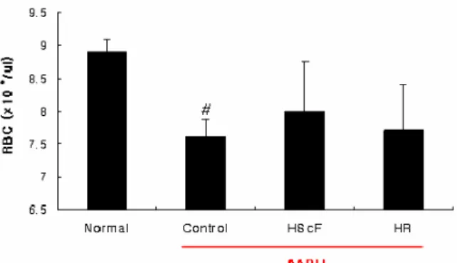
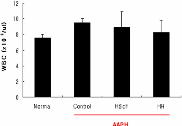
각 실험군의 혈액을 채취하여 적혈구, 백혈구 및 혈소 판의 수를 측정하였다(Table 1, Fig. 1~3).
Table 1. Hematology Analysis
RBC(×106/㎕) WBC(×103/㎕) PLT(×103/㎕) Normal 8.9±0.2 6.9±0.4 501.7±4.2 Control 7.6±0.3# 9.5±0.5 865.7±161.3##
HScF 8.0±0.8 9.0±2.1 602.6±126.2* HR 7.7±0.7 8.3±1.5 631.4±56.9 The rats were injected intraperitoneally (i.p) with AAPH for 7 days (once a day. 50 mg/kg) and orally administered with HScF and HR decoction (1%) for 7 days (onc a day. 200 ul). The animals were anesthetized with ethyl ether and the blood samples were taken from the heart.
Normal : Normal SD rat.
Control : AAPH injection.
HScF : AAPH injection and treatment with HScF extract.
HR : AAPH injection and treatment with HR extract.
Values represent the means±SD of 8 rats.
(1) RBC
각 실험군의 혈액을 채취하여 적혈구의 수를 측정한 결과, control군에서 normal군에 비하여 적혈구 수가 유 의성 있게 감소하였고, 약물 투여군에서는 枳椇子와 枳椇 枝 모두 control에 비하여 증가하였으나 유의성은 없었다.
枳椇子가 枳椇枝에 비하여 약간 더 증가하였다(Fig. 1).
Fig. 1. Effects of HScF and HR decoction extracts on RBC population in blood of rats injected with AAPH
The rats were injected intraperitoneally (i.p) with AAPH for 7 days (once a day. 50 mg/kg) and orally administered with HScF and HR decoction (1%) for 7 days (once a day. 200 ul). The animals were anesthetized with ethyl ether and the blood samples were taken from the heart and the number of RBC was measured.
Normal : Normal SD rat.
Control : AAPH injection.
HScF : AAPH injection and treatment with HScF extract.
HR : AAPH injection and treatment with HR extract.
Values represent the means±SD of 8 rats.
# : p< 0.05 compared with normal group.
(2) WBC
각 실험군의 혈액을 채취하여 백혈구의 수를 측정한 결과, control군은 normal군에 비하여 백혈구의 수가 증 가하였으나 유의성은 없었고, 약물 투여군에서는 control 군에 비하여 백혈구 수가 감소하였으며 枳椇子 투여군에 비하여 枳椇枝에서 더 감소하였으나 모두 유의성은 없었 다(Fig. 2).
Fig. 2. Effects of HScF and HR decoction extracts on WBC population in blood of rats injected with AAPH
The rats were injected intraperitoneally (i.p) with AAPH for 7 days (once a day. 50 mg/kg) and orally administered with HScF and HR decoction (1%) for 7 days (once a day. 200 ㎕). The animals were anesthetized with ethyl ether and the blood samples were taken from the heart and the number of WBC was measured.
Normal : Normal SD rat.
Control : AAPH injection.
HScF : AAPH injection and treatment with HScF extract.
HR : AAPH injection and treatment with HR extract.
Values represent the means±SD of 8 rats.
(3) PLT
각 실험군의 혈액을 채취하여 혈소판 수를 측정한 결 과, normal군에 비하여 control군에서 혈소판이 유의성 있게 증가하였고, 枳椇子 투여군에서는 control에 비하여 유의성 있게 감소하였으며, 枳椇枝 투여군에서는 control 군 보다 감소하였으나 유의성은 없었다(Fig. 3).
Fig. 3. Effects of the HScF and HR decoction extracts on PLT population in blood of rats injected with AAPH
The rats were injected intraperitoneally (i.p) with AAPH for 7 days (once a day. 50 mg/kg) and orally administered with HScF and HR decoction (1%) for 7 days (once a day. 200 ㎕). The animals were anesthetized with ethyl ether and the blood samples were taken from the heart and the number of PLT was measured.
Normal : Normal SD rat.
Control : AAPH injection.
HScF : AAPH injection and treatment with HScF extract.
HR : AAPH injection and treatment with HR extract.
Values represent the means±SD of 8 rats.
* : p< 0.05 compared with control group.
## : p< 0.01 compared with normal group.
2) Blood chemistry 분석
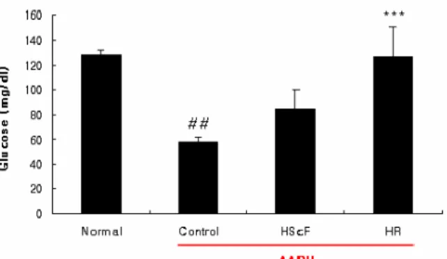
각 실험군에서 취한 혈청에서 albumin, total bilirubin, LDL cholesterol 및 glucose을 측정하였다(Table 2, Fig.
4~7).
Table 2. Blood Chemistry Analysis Albumin
(g/㎗) Total bilirubin
(㎎/㎗) LDL cholesterol
(㎎/㎗) Glucose (㎎/㎗) Normal 2.90±0.06 0.240±0.017 16.2±1.9 128.3±4.0 Control 2.61±0.14# 0.153±0.015### 24.7±0.6# 57.3±4.0##
HScF 2.76±0.08 0.168±0.013 21.8±5.7 84.6±15.2 HR 2.70±0.15 0.156±0.018 21.6±3.8 126.3±24.1***
The rats were injected intraperitoneally (i.p) with AAPH for 7 days (once a day. 50 mg/kg) and orally administered with HScF and HR decoction (1%) for 7 days (once a day. 200 ㎕). The animals were anesthetized with ethyl ether and the blood samples were taken from the heart.
Normal : Normal SD rat.
Control : AAPH injection.
HScF : AAPH injection and treatment with HScF extract.
HR : AAPH injection and treatment with HR extract.
Values represent the means±SD of 8 rats.
(1) Albumin
각 실험군에서 취한 혈청에서 albumin을 측정한 결과, control군은 normal군에 비하여 albumin이 유의성 있게 감소하였고, 약물 투여군에서는 control군에 비하여 증가 하였으며, 枳椇子 투여군에서 枳椇枝 투여군에 비하여 더 욱 크게 증가하였으나 모두 유의성은 없었다(Fig. 4).
Fig. 4. Effects of HScF and HR decoction extracts on serum albumin level in oxidated rats with AAPH
The rats were injected intraperitoneally (i.p) with AAPH for 7 days (once a day. 50 mg/kg) and orally administered with HScF and HR decoction (1%) for 7 days (once a day. 200 ㎕). The animals were anesthetized with ethyl ether and the blood samples were taken from the heart and the level of albumin in serum was measured.
Normal : Normal SD rat.
Control : AAPH injection.
HScF : AAPH injection and treatment with HScF extract.
HR : AAPH injection and treatment with HR extract.
Values represent the means±SD of 8 rats.
# : p< 0.05 compared with normal group.
(2) Total bilirubin
각 실험군에서 취한 혈청에서 total bilirubin을 측정한 결과, control군은 normal군에 비하여 유의성 있게 감소
하였고, 약물 투여군에서는 枳椇子 투여군과 枳椇枝 투여 군 모두 control군에 비하여 증가하였으나 유의성은 없었 다(Fig. 5).
Fig. 5. Effects of the HScF, HR decoction on the total bilirubin level in oxidated rat serum
The rats were injected intraperitoneally (i.p) with AAPH for 7 days (once a day. 50 mg/kg) and orally administered with HScF and HR decoction (1%) for 7 days (once a day. 200 ㎕). The animals were anesthetized with ethyl ether and the blood samples were taken from the heart and the level of total bilirubin in serum was measured.
Normal : Normal SD rat.
Control : AAPH injection.
HScF : AAPH injection and treatment with HScF extract.
HR : AAPH injection and treatment with HR extract.
Values represent the means±SD of 8 rats.
### : p< 0.001 compared with normal group.
(3) LDL cholesterol
각 실험군에서 취한 혈청에서 LDL cholesterol을 측정 한 결과, control군은 normal군에 비하여 유의성 있게 증 가하였고, 약물 투여군에서는 control군에 비하여 감소하 였으나 유의성은 없었다(Fig. 6).
Fig. 6. Effects of HScF and HR decoction on LDL cholesterol level in oxidated rat serum
The rats were injected intraperitoneally (i.p) with AAPH for 7 days (once a day. 50 mg/kg) and orally administered with HScF and HR decoction (1%) for 7 days (once a day. 200 ㎕). The animals were anesthetized with ethyl ether and the blood samples were taken from the heart and the level of LDL cholesterol in serum was measured.
Normal : Normal SD rat.
Control : AAPH injection.
HScF : AAPH injection and treatment with HScF extract.
HR : AAPH injection and treatment with HR extract.
Values represent the means±SD of 8 rats.
# : p< 0.05 compared with normal group.
(4) Glucose
각 실험군에서 취한 혈청에서 glucose의 생성을 측정 한 결과, control군은 normal군에 비하여 유의성 있게 감 소하였고, 枳椇枝 투여군에서는 control군에 비하여 유의성 있게(p<0.001) 증가하였으며, 枳椇子 투여군에서는 control 군에 비하여 약간 증가하였으나 유의성은 없었다(Fig. 7).
Fig. 7. Effects of HScF and HR decoction on serum glucose level in oxidated rats
The rats were injected intraperitoneally (i.p) with AAPH for 7 days (once a day. 50 mg/kg) and orally administered with HScF and HR decoction (1%) for 7 days (once a day. 200 ㎕). The animals were anesthetized with ethyl ether and the blood samples were taken from the heart and the level of glucose in serum was measured.
Normal : Normal SD rat.
Control : AAPH injection.
HScF : AAPH injection and treatment with HScF extract.
HR : AAPH injection and treatment with HR extract.
Values represent the means±SD of 8 rats.
*** : p< 0.001 compared with control group.
## : p< 0.01 compared with normal group.
2. 간장에서의 항산화 효과
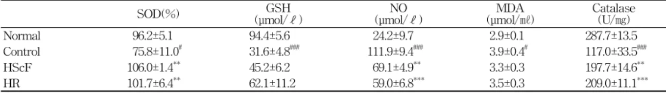
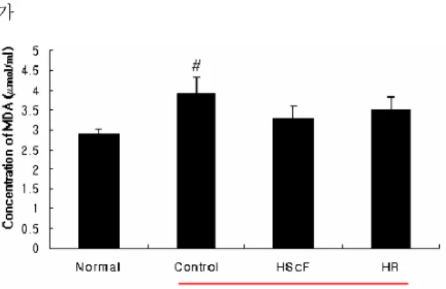
Rat에서 적출한 간 조직을 분획하여 SOD의 활성, glutathione의 함량, NO함량, MDA함량 및 catalase의 함 량을 측정하였다(Table 3, Fig. 8~12).
1) SOD assay
Rat에서 적출한 간 조직을 분획하여 SOD의 활성을 측정한 결과, control군은 normal군에 비하여 유의성 있 게 감소하였고, 약물 투여군에서는 枳椇子 투여군과 枳椇 枝 투여군 모두 SOD의 활성이 control군에 비하여 유의 성 있게(p<0.001) 증가하였다(Fig. 8).
2) Glutathione assay
Rat에서 적출한 간 조직을 분획하여 glutathione의 함 량을 측정한 결과, control군은 normal군에 비하여 유의 성 있게 감소하였고, 약물 투여군에서는 control군에 비하 여 증가하였으나 유의성은 없었다(Fig. 9).
3) Nitric oxide assay
Rat에서 적출한 간 조직을 분획하여 NO함량을 측정한
Table 3. Antioxidative Activity of HScF and HR
SOD(%) GSH
(μmol/ℓ) NO
(μmol/ℓ) MDA
(μmol/㎖) Catalase (U/㎎) Normal 96.2±5.1 94.4±5.6 24.2±9.7 2.9±0.1 287.7±13.5 Control 75.8±11.0# 31.6±4.8### 111.9±9.4### 3.9±0.4# 117.0±33.5###
HScF 106.0±1.4** 45.2±6.2 69.1±4.9** 3.3±0.3 197.7±14.6**
HR 101.7±6.4** 62.1±11.2 59.0±6.8*** 3.5±0.3 209.0±11.1***
The rats were injected intraperitoneally (i.p) with AAPH for 7 days (once a day. 50 mg/kg) and orally administered with HScF and HR decoction (1%) for 7 days (once a day. 200 ㎕). After the animals were sacrificed, the liver was removed and the concentrations of SOD, glutathione, NO, MDA and catalase in liver were estimated.
Normal : Normal SD rat.
Control : AAPH injection.
HScF : AAPH injection and treatment with HScF extract.
HR : AAPH injection and treatment with HR extract.
Values represent the means±SD of 8 rats.
Fig. 8. Effects of HScF and HR decoction on SOD activity in liver
The rats were injected intraperitoneally (i.p) with AAPH for 7 days (once a day. 50 mg/kg) and orally administered with HScF and HR decoction (1%) for 7 days (once a day. 200 ㎕). After the animals were sacrificed, the liver was removed and SOD was estimated.
Normal : Normal SD rat.
Control : AAPH injection.
HScF : AAPH injection and treatment with HScF extract.
HR : AAPH injection and treatment with HR extract.
Values represent the means±SD of 8 rats.
** : p< 0.01 compared with control group.
# : p< 0.01 compared with normal group.
Fig. 9. Effects of HScF and HR decoction on the concentration of hepatic glutathione
The rats were injected intraperitoneally (i.p) with AAPH for 7 days (once a day. 50 mg/kg) and orally administered with HScF and HR decoction (1%) for 7 days (once a day. 200 ㎕). After the animals were sacrificed, the liver was removed and GSH concentration was measured.
Normal : Normal SD rat.
Control : AAPH injection.
HScF : AAPH injection and treatment with HScF extract.
HR : AAPH injection and treatment with HR extract.
Values represent the means±SD of 8 rats.
### : p< 0.001 compared with normal group.
Fig. 10. Effects of HScF and HR decoction on the concentration of hepatic niric oxide
The rats were injected intraperitoneally (i.p) with AAPH for 7 days (once a day. 50 mg/kg) and orally administered with HScF and HR decoction (1%) for 7 days (once a day. 200 ㎕). After the animals were sacrificed, the liver was removed and NO concentration was measured.
Normal : Normal SD rat.
Control : AAPH injection.
HScF : AAPH injection and treatment with HScF extract.
HR : AAPH injection and treatment with HR extract.
Values represent the means±SD of 8 rats.
*** : p< 0.001, ** : p< 0.01 compared with control group, ### : p< 0.001 compared with normal group.
결과, control군은 normal군에 비하여 유의성 있게 증가 하였고, 약물 투여군에서는 枳椇子 투여군과 枳椇枝 투여 군 모두 control군에 비하여 유의성 있게 감소하였으며, 枳椇枝 투여군에서 枳椇子 투여군보다 NO함량이 더욱 감소하였다(Fig. 10).
4) Lipid peroxidation assay
Rat에서 적출한 간 조직을 분획하여 MDA함량을 측정 한 결과, control군은 normal군에 비하여 유의성 있게 증 가하였고, 약물 투여군에서는 control군에 비하여 감소하 였으나 유의성은 없었다(Fig. 11).
5) Catalase activity
Rat에서 적출한 간 조직을 분획하여 catalase의 함량 을 측정한 결과, control군은 normal군에 비하여 유의성 있게 증가하였고, 약물 투여군에서는 枳椇子 투여군과 枳椇枝 투여군 모두 control군에 비하여 유의성 있게 증
가
Fig. 11. Effects of HScF and HR decoction on the concentration of hepatic MDA
The rats were injected intraperitoneally (i.p) with AAPH for 7 days (once a day. 50 mg/kg) and orally administered with HScF and HR decoction (1%) for 7 days (once a day. 200 ㎕). After the animals were sacrificed, the liver was removed and the concentration of hepatic MDA was estimated.
Normal : Normal SD rat.
Control : AAPH injection.
HScF : AAPH injection and treatment with HScF extract.
HR : AAPH injection and treatment with HR extract.
Values represent the means±SD of 8 rats.
# : p< 0.05 compared with normal group.
Fig. 12. Effects of HScF and HR decoction on catalase activity in liver
The rats were injected intraperitoneally (i.p) with AAPH for 7 days (once a day. 50 mg/kg) and orally administered with HScF and HR decoction (1%) for 7 days (once a day. 200 ㎕). After the animals were sacrificed, the liver was removed and catalase activity was estimated.
Normal : Normal SD rat.
Control : AAPH injection.
HScF : AAPH injection and treatment with HScF extract.
HR : AAPH injection and treatment with HR extract.
Values represent the means±SD of 8 rats.
*** : p< 0.001, **: p< 0.01 compared with control group.
### : p< 0.001 compared with normal group.
하였으며, 枳椇子 투여군보다 枳椇枝 투여군에서 더욱 증 가하였다(Fig. 12).
고 찰
枳椇子는 ≪新修本草≫11)에 “枳椇, 味甘平, 無毒. 主頭
風 少腹拘急. 陸機云 一名 木蜜. 其木皮 溫 無毒. 主五痔 和五臟. 以木爲屋, 屋中酒則味薄, 此亦奇物”이라고 처음 수재된 이후 현재 임상에서는 解酒毒, 止渴除煩, 止嘔, 利 大小便의 효능이 있어 宿醉, 煩渴, 二便不利 등의 증을 치료하는 데 사용되고 있다4).
≪대한약전외한약규격집≫3)에는 헛개나무Hovenia dulcis Thunb.(갈매나무과 Rhamnaceae)의 과병을 가진 열매 또 는 씨를 枳椇子로 수재하고 있는데, ≪本草綱目≫12)에는 열매 이외에도 木汁과 木皮가, ≪中華本草≫4)에는 잎을 枳椇葉, 줄기 껍질을 枳椇木皮, 수액을 枳椇木汁, 뿌리를 枳椇根이라고 수재하고 있는데, 枳椇木皮는 味甘 性溫하 여 活血, 舒筋, 消食, 療痔의 효능이 있어 筋脈拘攣, 食積, 痔瘡을 다스리며, 枳椇木汁은 味甘 性平하여 辟穢除臭의 효능이 있어 암내를 없애며, 枳椇根은 味甘澁 性溫하여 祛風活絡, 止血, 解酒의 효능이 있어 風濕筋骨痛, 勞傷咳 嗽, 喀血, 小兒驚風, 宿醉를 다스린다고 하였다.
헛개나무 식물에 대한 성분과 생리활성 연구로는 헛개 나무 잎에서 Yoshikawa K.13)가 hoduloside Ⅰ, Ⅱ, Ⅲ,
Ⅳ, Ⅴ 등을 분리하였으며, 헛개나무의 종자와 과경으로 부터 Yoshikawa M.5,6)이 hovenidulcioside와 hovenitin 등 을 분리하였고, Ding14)이 dihydrokaemferol, quercetin 등 을 분리하였으며, Hase15)가 ampelopsin을 분리하였고, 헛 개나무의 어린 잎과 줄기로부터 Cho16)는 vanillic acid와 ferulic acid를 분리하였다. 헛개나무 잎에서 Yoshikawa K.17)는 dammarane glycosides가 anti-sweet activity 물 질이라고 하였으며, Yoshikawa M.5,6)은 hovenidulcioside가 histamine 유출 저해 효과가 있다고 하였고, ampelopsin과 dihydroflavono는 에탄올로 유도된 근육이완을 저해한다 고 하였다. 헛개나무의 과병에서 Hase15)는 ampelopsin이 간 손상에 대한 hepatoprotective activity를 갖는다고 보 고하였다. 그리고 안18)은 枳椇子가, 이19)는 枳椇枝가 간 보호와 항섬유화 효과가 우수하다고 보고하였다.
항산화제는 물질의 변질방지, 인체의 노화방지 및 성 인병 예방 등의 기능을 할 수 있는 물질이며20,21), 현재 사용되는 항산화제로는 BHA (butylated hydroxy anisole), BHT (butylated hydroxy toluene), PG (propyl gallate) 등과 같은 합성 항산화제와 토코페롤과 같은 천연 항산화 제가 개발되어 이용되고 있다. 그러나 BHA, BHT는 우 수한 효과가 있지만 독성이 문제가 되고 있으며, 토코페 롤은 가격이 높은 단점을 가지고 있기 때문에22), 이들을 대체할 수 있는 효과적이고도 안전한 항산화제의 개발이 요구되고 있다.
이에 저자는 숙취해소와 간장질환에 사용되어 항산화 효과가 있을 것으로 사료되는 枳椇子와 枳椇枝의 항산화 효과를 실험을 통하여 비교 관찰하고자 하였다.
AAPH (2.2’-azobis (2-aminodinopropane) hydrochloride) 는 수용성 azo 화합물의 일종으로서 열분해에 의해서 자 유기를 생성하는데, 복강 내로 투여되면 수분 내에 혈류를 타고 전신순환하여 산소분자와 반응하여 peroxyl radical을 생성하고, 여러 형태의 생물학적 분자와 결합반응을 일
으키면서 생체막의 구조를 붕괴시키는 것으로 생각되고 있다23).
본 실험에서는 AAPH (2.2’-azobis (2-aminodinopropane) hydrochloride)를 SD Rat에 주사하여 급성적 산화 스트 레스를 유발한 다음, 枳椇子 및 枳椇枝 전탕액을 투여하 고 hematology 분석, blood chemistry 분석, 간 조직 내 SOD 활성, glutathione 함량, catalase 활성, NO 함량, MDA 함량 등을 측정하였다.
RBC 수는 枳椇子와 枳椇枝 전탕액 투여군 모두 control군에 비해 증가하였으나 유의성은 없었다. WBC 수는 control군, 약물 투여군 모두 유의한 변화를 보이지 않았다. 혈소판은 枳椇子 투여군에서 control군에 비하여 유의하게 감소하였다.
AAPH로부터 생성된 과산화자유기에 의해 endoplasmic reticulum에서 과산화반응이 일어나 비가역적 단백질 합 성 억제가 일어나 혈중 albumin 농도는 감소하게 된다24). 본 실험에서 albumin은 control에서 normal에 비하여 유 의하게 감소하였으며, 枳椇子 및 枳椇枝 투여군에서는 control에 비하여 증가하였으나 유의성은 없었다.
Total bilirubin은 control군에서 normal군에 비하여 유 의하게 감소하였으며, 枳椇子 및 枳椇枝 투여군에서는 control군에 비하여 증가하였으나 유의성은 없었다.
AAPH로 free radical이 생성되면서 그로 인해 세포가 파괴되면, LDL cholesterol이 분해되지 못하고 혈중에 분 포하게 된다. 본 실험 결과 LDL cholesterol은 control군 에서 normal군에 비해 증가했으며, 枳椇子와 枳椇枝 투여 군 모두 control군에 비해 감소했으나 유의성은 없었다.
혈중 glucose는 control군에서 normal군에 비하여 유의 하게 감소하였으며, 枳椇枝 투여군에서 control군에 비하 여 유의성 있게 증가하였다.
SOD는 superoxide anion을 과산화수소로 전환시키는 효소로서 진핵세포에서는 세포질의 copper-and zinc- superoxide dismutase (Cu,Zn-SOD)와 사립체의 maganese- superoxide dismutase (Mn-SOD), 원핵세포에서는 세포 질의 Mn-SOD와 원형질막 외강의 iron-superoxide dismutase (Fe-SOD) 등 여러 가지 동종효소가 알려져 있다25).
본 실험에서 간 조직 내 SOD 활성을 측정한 결과, control군에서 normal군에 비하여 SOD 활성이 유의하게 감소한 반면, 枳椇子 및 枳椇枝 투여군에서는 control군에 비하여 유의하게 증가하였다.
GSH (Glutathione)는 hydrogen peroxidase를 H2O로 환원시켜서 산화성 스트레스에 대하여 세포를 보호하는 작용이 있다(Meister A.). 본 실험에서 GSH농도는 control군 에서 normal군에 비하여 유의하게 감소하였고, 枳椇子 및 枳椇枝 투여군에서는 control군에 비하여 증가하였으나 유의성은 없었다.
AAPH에 의한 세포손상과 관련하여, NO는 맥관계 (vascular system)에서 내피 유래 확장인자(endothelium- derived relaxation factor)로서 작용하며26), 혈소판응집 (platelet aggregation)과정에 관여한다27). 본 실험 결과
NO 농도는 control군에서 normal군에 비하여 유의하게 증가하였으나, 枳椇子 및 枳椇枝 투여군에서 유의하게 감 소하였다.
MDA는 식이성 불포화 지방산의 주된 과산화 생성물 형태로서 지질 과산화물의 지표로 널리 이용되고 있다28). 본 실험 결과 MDA 함량은 control군에서 normal군에 비하여 유의하게 증가하였으며, 枳椇子 및 枳椇枝 투여 군에서는 control군에 비하여 감소하였으나 유의성은 없었다.
Catalase는 세포 소기관인 peroxizome 내에서 과산화 수소를 물과 산소로 분해하는 효소로서, 과산화수소에 의 한 산화적 손상으로부터 세포를 보호하는 역할을 한다29). 본 실험 결과 catalase 함량은 control군에서 normal군에 비하여 유의하게 감소한 반면, 枳椇子 및 枳椇枝 투여군 에서는 control군에 비하여 유의하게 증가하였다.
따라서 枳椇子 및 枳椇枝는 SOD 활성과 catalase 활 성을 증가시키고, NO를 억제함으로써 항산화 효능을 나 타냄을 알 수 있었다.
이상의 결과를 종합하면 枳椇子와 枳椇枝의 총 페놀 함량은 거의 유사하며, 枳椇子와 枳椇枝의 양자 간의 항 산화 효능은 추출방법, 분획, 농도 등에 따라 다소의 차 이는 있었으나 통계적 유의성은 나타나지 않았다. 그러나 枳椇子와 枳椇枝 모두 항산화 효능이 인정되어 항산화제 로 개발할 수 있다고 사료된다.
결 론
枳椇子와 枳椇枝의 항산화 효과를 동물실험을 통하여 비교 분석한 결과 아래와 같은 결론을 얻었다.
1. 혈소판은 枳椇子 투여군에서 유의하게 감소하였다.
2. 혈중 glucose는 枳椇枝 투여군에서 유의성 있게 증 가하였다.
3. SOD 활성은 枳椇子 및 枳椇枝 투여군 모두에서 유 의하게 증가하였다.
4. NO 농도는 枳椇子 및 枳椇枝 투여군 모두에서 유 의하게 감소하였다.
5. Catalase 함량은 枳椇子 및 枳椇枝 투여군 모두 유 의하게 증가하였다.
이상의 실험결과 枳椇子와 枳椇枝의 항산화 효과는 거 의 유사하며, 추출방법, 분획, 농도 등에 따라 다소의 차 이는 있으나, 枳椇子와 枳椇枝 모두 항산화 효과가 인정 되므로 항산화 치료제로 개발할 수 있다고 사료된다.
감사의 글
이 논문은 2009년도 경원대학교 연구비 지원을 받았으 며 이에 감사드린다.
참고문헌
1. 이창복 . 대한식물도감. 서울 : 향문사. 2003 : 714.
2. 김세현, 나천수, 김만조, 정남철. 헛개나무의 분포와 증식 특성. 한국임학회. 2002 ; 1(1) : 161-82.
3. 식품의약품안전청 고시번호2007-90. 대한약전외한약 (생약)규격집. 2007 : 323.
4. 國家中醫藥管理局 中華本草編委會. 中華本草. 上海 : 上海科學技術出版社. 1999 : (5) : 238-41.
5. Yoshikawa M, Murakami T, Ueda T, Muraoka H, Yamahara J, Murakami N. Bioactive saponins and glycosides. Ⅳ Four methylmigrated 16, 17-seco- damarane Triterpene Glycosides from Chinese natural medicine, Hovenia semen Seu Fructus, the Seeds and Fruit of Hovenia dulcis Thunb.
Absolute Stereo Structures and inhibitory activity on Histamine release of Hovenidulciosides A1 and A2, B1, and B2. Chem Pharm Bull. 1996 ; 44(9) : 1736-43.
6. Yoshikawa M, Murakami T, Ueda T, Yoshizumi s, Ninomiya K, Yanahara J. Bioactive constituents of Chinese natural medicine. Ⅲ. Absolute Stereo Structures of new dihydroflavonols, Hovenitins Ⅰ,
Ⅱ, and Ⅲ, isolated from Hovenia semen Seu Fructus, the seeds and fruit of Hovenia dulcis Thunb. (rhamnaceae) : Inhbitory effect on a alcohol-induced muscular relaxation and hepatoprotective activity. Vakugaku Zasshi. 1997 ; 117(2) : 108-18.
7. 김영길, 이상호, 권혁일, 황백, 이현용, 안상욱, 김민 희, 이병익. 헛개나무와 오리나무 추출물의 간해독작 용 및 체내 알콜 분해능 비교. 한국약용작물학회지.
1997 ; 7(4) : 263.
8. 최윤정, 최돈하, 이학주, 이성숙, 수목추출물의 생리 활성에 관한 연구(10) - 헛개나무 목부의 향균활성 물질. 목재공학. 2003 ; 31(1) : 1.
9. Bansal VS, Hattori H, Orihel D, Kanfer JN.
Distribution of selected phospholipid modifying enzymes in rat brain microsomal subfractions prepared by density gradient zonal rotor centrifugation.
Neurochem Res. 1985 Apr ; 10(4) : 439-51.
10. Roos D, Weening RS, Wyss SR, Aebi HE.
Protection of human neutrophils by endogenous catalase: studies with cells from catalase-deficient individuals. J Clin Invest. 1980 Jun ; 65(6) : 1515-22.
11. 蘇敬 等 撰, 尙志鈞 輯校. 新修本草(輯復本). 合肥 : 安徽科學技術出版社. 1981 : 356.
12. 李時珍. 本草綱目. 北京 : 人民衛生出版社. 1982 : 1845, 1846.
13. Yoshikawa K, Tumura S, Yamada K and Arihara
S. Antisweet Natural products : Ⅶ. Hodulosides
Ⅰ, Ⅱ, Ⅲ, Ⅳ, and Ⅴ from the leaves of Hovenia dulcis. Thunb Chem Pharm Bull. 1992 ; 40(9) : 2287-91.
14. Ding LS, Liang QL, Teng YF. Study on flavonoids in seeds of Hovenia dulcis Thunb. Acta Pharmaceutics Sinica. 1997 ; 32(8) : 600-2.
15. Hase K, Ohsugi M, Xiong QB, BASNET B, Kadota S. Hepatoprotective effect of Hovenia dulcis Thunb. On experimental liver injuries induced by carbon tetrachloride or D-galactosamin/
lopopolysaccharide. Biological and Pharmaceutical Bulletin. 1997 ; 20(4) : 381-5.
16. Cho JY, Moon JH. Isolation and identification of 3-methoxy-4hidroxy benzoic acid and 3-methoxy- 4hydroxy cinamic acid from hot water extract of Hovenia dulcis Thunb. and confimation of their antioxidative and antimicrobial activity. Korean J Food Sci Tecnol. 2000 ; 32(6) : 1403-8.
17. Yoshikawa K, Nagai Y, Yoshida M, Arihara S.
Antisweet Natural products : Ⅷ. Structures of hodulosides Ⅵ X from Hovenia dulcis Thunb. var.
tomentella. Makino Chem Pharm Bull. 1993 ; 41(10) : 1722-5.
18. 안민수. 枳梖子의 간보호 및 항섬유화 효과에 관한 연구. 서울 : 경희대학교 대학원. 2005.
19. 이종안. 枳椇枝 물추출물의 간보호 및 항섬유화 효 과에 관한 연구. 경희대학교 대학원. 2005.
20. Farag RS, Badei AZMA, Hewedi FM, El-Baroty GSA. Antioxidant activity of some spice essential oils on linoleic acid oxidation in aqueous media.
JAOCS. 1989 ; 66 : 792-9.
21. Frei B. Natural antioxidants in human health and disease. San Diego : Academic Press. 1994 : 20- 55.
22. Choe SY, Yang KH. Toxicological studies of antioxidants, butylated hydroxy toluene (BHT), and butylated hydroxy anisole (BHA). Iorean J Food Sci Technol. 1982 ; 14 : 283-8.
23. Terao K., Niki E. Damage to biological tissues induced by radical initiator 2.2‘-azobis (2-aminodinopropane) hydrochloride and its inhibition by chain-breaking antioxidants. J Free Radical in Biol Med. 1986 ; 2 ; 193-201.
24. Terao K. Liver injuries induced by free radical. J Toxicol Pathol. 1989 ; 2 ; 11-8.
25. Proctor PH. Free radicals and human disease. In CRC handbook of free radical and antioxidants in biomedicine. vol I. Miquel J, Quintanilha AT and Weber H eds. Florida : CRC Press. 1989 : 209-22.
26. Teo S, Pohl L, Halpert J. Production of superoxide anion radicals during the oxidative metabolism of amino-chloramphenicol. Biochem Pharmacol. 1986 Dec 15 ; 35(24) : 4584-6.
27. Radamski MW, Palmer RM, Moncada S. An.
L-arginine/nitric oxide pathway present in human platelets regulates aggregation. Proc Natl Acad Sci USA. 1990 Jul ; 87(13) : 5193-7.
28. Piche LA, Draper HH, Cole PD. Malondialdehyde excretion by subjects consuming cod liver oil vs.
a concentrate n-3 fatty acid. Lipids. 23 : 370, 371.
29. Deisseroth A and Dounce AL. Catalase physical and chemical properties, Mechanism of catalysis, and physiological role. Physiological reviews. 1970 ; 50 : 319-75.