이 논문은 2012년 1월 27일 접수하여 2012년 3월 9일 채택되었음.
책임저자:배성수, 서울아산병원 방사선종양학과 Tel: 02)3010-4419, Fax: 02)3010-6905 E-mail: [email protected]
방광암 환자의 영상유도 방사선치료에 관한 고찰
서울아산병원 방사선종양학과
배성수ㆍ배선명ㆍ김진산ㆍ강태영ㆍ백금문ㆍ권경태
목 적: 현재 본원에서 방광암 환자의 영상유도 방사선치료는 재현성을 높이기 위하여 환자의 상태에 따라 알맞은 양의 생리 식염수를 주입하고 영상유도 시스템(On-Board Imager system, OBI, VARIAN, USA)의 Cone–Beam CT (CBCT)로 3차원 정합 (3D-3D matching)을 하여 치료를 한다. 본 연구에서는 방광암 환자의 치료 시 획득한 CBCT 영상의 분석을 통해 뼈를 기준으 로 한 정합과 방광을 기준으로 한 정합의 차이를 알아보고, 생리 식염수를 주입한 방광의 체적 변화를 알아보고 방광암 환자 의 치료 시 더욱 적절한 영상정합방법을 평가하고 고찰하고자 한다.
대상 및 방법: 본원에서 2009년 1월에서 2010년 4월까지 방사선치료를 위해 내원한 방광암 환자 7명을 대상으로 Folly catheter를 이용하여 방광 내 잔류 소변을 제거한 뒤 환자 개개인에 맞게 정해진 양 만큼의 생리식염수를 주입하고 CT-Sim 후 치료계획을 설계하였다. 그 뒤 OBI system을 이용하여 치료 전 자세 확인을 위해 CBCT를 찍었고, 담당 주치의가 모든 대 상 환자의 영상 정합을 진행하였다. 총 45개 CBCT 영상을 이용하여 뼈를 기준으로한 영상정합과 방광을 기준으로 한 영상정 합의 차이를 분석하였다. 또, 방광의 체적 변화를 Eclipse (version 8.0, VARIAN, USA)를 통해 얻어냈다.
결 과: 뼈를 기준으로 한 영상정합을 한 후 다시 방광을 기준으로 한 정합의 차이는 X축으로 평균 3±2 mm, Y축으로 1.8±1.3 mm, Z축으로 2.3±1.7 mm이고 전체 이동거리는 4.8±2.0 mm로 나타났다. 또 방광의 체적은 기준 대비 4.03±3.97%의 차이를 나타냈다.
결 론: 방광의 특성상 해부학적 위치 및 내부의 움직임으로 인해 뼈를 이용한 영상정합 후에도 방광의 위치 차이가 발생하였 다. 또, 생리식염수를 채운 방광의 체적은 4.03%의 차이를 나타냈으나 영상 정합 시 모두 계획한 볼륨 안에 포함되는 것을 확인 할 수 있었다. 따라서 생리식염수를 주입한 뒤 방광을 기준으로 영상 정합을 실시함으로써 더욱 정확한 치료를 실시 할 수 있을 것으로 사료된다.
핵심용어: 영상유도 방사선치료, bladder filling, OBI, 2D 영상정합, 3D 영상정합
서 론
방사선치료를 시행할 경우 모의치료 및 치료계획을 수립 할 때와 동일한 환자 자세가 유지되어야 하는 set-up의 정확 성이 전제 되어야 한다. 환자 set-up을 위해 전통적인 방법으 로는 환자 피부 표면에 잉크로 레이저와 일치되는 선을 표시 하거나 문신을 하는 방법이 이용 되어 왔으며 보조기구 및 고정 장치를 이용하기도 하였다. 최근에 세기변조 방사선치 료(Intensity-Modulated Radiation Therapy, IMRT)가 보편화 되면서 정확한 set-up의 중요성이 더욱 커지게 되었으며, 그 로인해 영상 유도 방사선 치료(Image-Guided Radiation Therapy, IGRT)의 적용으로 치료부위의 정확성을 향상 시킬 수 있게 되었다.1) 환자의 치료 계획용 표적체적(Planning
Target Volume, PTV)을 직접적으로 확인하기가 어려운 경우 가 많기 때문에 뼈의 구조를 기준으로 환자 set-up 확인과 보 정이 이루어져 왔다.
현재 본원에서 방광암의 초기(Initial) 치료는 영상유도 시 스템(On-Board Imager system, OBI, VARIAN, USA)을 이용 하여 골반 내 전체 방광을 포함한 영역에 대하여 뼈를 기준 으로 정합을 한 뒤 치료를 시행하고 있다(Fig. 1). 이때 CBCT를 통해 환자의 재현성이 확인되면 주 1회는 CBCT영 상을 얻어 뼈를 기준으로 3D 정합을, 나머지 4회는 KV를 이 용한 2D 정합을 하고 있다.
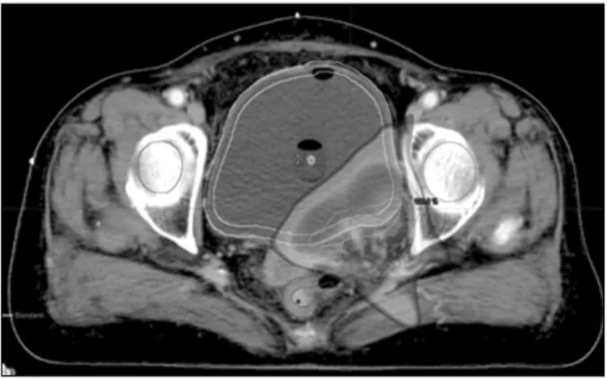
방사선 치료범위를 축소한(Reduce field) 치료 시에는 육안 적 종양체적(Gross Tumor Volume, GTV) 영역에 일정한 여 유 폭(Margin)을 두고 치료 계획용 표적체적을 결정하여 방 광의 일부분에만 치료를 시행하고 있다(Fig. 2).2) 이때 CBCT 를 통해 영상을 얻어 뼈를 기준으로 정합을 한 결과 방광 및 직장의 상태에 따라 방광의 위치 변화가 있었다(Fig. 3).3-5)
Fig. 1. Initial treatment for bladder cancer.
Fig. 2. Reduce field treatment for bladder cancer.
Fig. 3. When bone match changes in the bladder volume (-
Ref. CT bladder volume, ㆍCBCT bladder volume).
Fig. 4. Without injection of saline volume change of the bladder.
또한 방광의 해부학적 특성으로 인해 CT-simulation 촬영 시의 방광 체적과 치료 시 CBCT로 얻은 방광 체적이 차이가 나타났다(Fig. 4).6-8) 따라서 재현성을 높이기 위하여 치료 전 환자의 방광에 CT-simulation 당시의 동일한 양의 생리식염 수를 주입하고 매일 CBCT를 찍어 방광을 기준으로 정합을 실시한 뒤 치료를 진행하고 있다.
이와 같은 원인으로 인해 다음과 같은 목적으로 본 연구를 시행하였다.
첫 번째로, 방광암의 방사선치료 범위를 축소한(Reduce field) 치료 시 내부의 움직임을 고려하여 치료 계획용 표적 체적에 대해 보다 정확히 치료하기 위한 영상정합 방법을 알 아보기 위해 뼈를 기준으로 한 정합과 방광을 기준으로 한 정합을 비교하여 방광의 위치 변화 정도를 평가하였다.
두 번째는, 생리식염수를 주입한 방광의 체적 변화를 분석 함으로써 재현성 및 유용성을 평가하고 더욱 정확한 영상정 합 방법을 제시하여 궁극적으로 치료의 정확도를 향상 하는 것이 본 연구의 목적이다.
대상 및 방법
본 연구의 대상으로는 2009년 1월부터 2010년 4월까지 방 광의 영상유도 방사선 치료 환자 7명의 Reduce field치료 대 한 CBCT 영상 총 45개의 Data를 분석하였다. GE사의 Light speed RT 16 chanel CT Simulator를 사용하여 scan을 하고 치 료용적, 주변정상장기 등을 CT 영상에 그려 넣은 후 전산화 치료계획을 수립하였다. CT simulation 및 치료 전 처치로 환 자의 방광에 생리식염수 주입을 위하여 먼저 소변을 보게 한 후 주사기를 통하여 잔류 소변을 제거한다. 다음은 Folly catheter set를 이용하여 환자의 상태에 따른 알맞은 양의 생 리 식염수를 주입하고 시간에 따른 방광체적 변화 및 환자의 고통을 최소화하기 위하여 전 처치 직후에 환자 set-up을 시 행한다(Fig. 5). CT simulation 시와 동일한 자세로 치료실에 서는 피부에 표시된 기준선과 치료실 레이져를 이용해 환자 를 set-up 한 후 본원의 선형가속기(21-ix, Varian, USA)에 부 착된 OBI를 이용하여 CBCT 영상을 획득하였다.
환자 자세는 앙와위(Supine)로 하였고, 자세 고정을 위해 Ankle pillow를 사용하였다.
먼저 뼈를 기준으로 한 정합과 방광을 기준으로 한 정합의
Fig. 6. Switching the position vector of values of the bladder (unit mm).
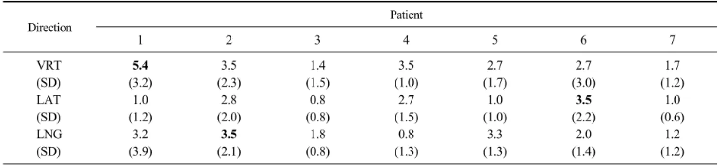
Table 1. The difference of shift value between bone match and volume match (unit mm)
Direction Patient
1 2 3 4 5 6 7
VRT 5.4 3.5 1.4 3.5 2.7 2.7 1.7
(SD) (3.2) (2.3) (1.5) (1.0) (1.7) (3.0) (1.2)
LAT 1.0 2.8 0.8 2.7 1.0 3.5 1.0
(SD) (1.2) (2.0) (0.8) (1.5) (1.0) (2.2) (0.6)
LNG 3.2 3.5 1.8 0.8 3.3 2.0 1.2
(SD) (3.9) (2.1) (0.8) (1.3) (1.3) (1.4) (1.2)
Fig. 5. Folly catheter set.
차이로 방광의 위치변화를 알아보기 위해 CBCT 데이터를 획득한 후 뼈를 기준으로 정합을 하여 set-up 오류를 보정 하 였다. 그 상태에서 다시 reference CT의 방광과 CBCT의 방 광을 정합 하여 방광의 위치 변화를 측정 하였다.9)
다음은 방광 체적 변화를 비교하여 재현성 및 생리식염수 의 유용성 평가를 위하여 Eclipse를 이용해 CBCT의 data를 불러와 방광의 윤곽(contour)을 그린 뒤에 체적을 얻어내고 CT simulation 당시 방광과의 체적을 정량적으로 비교 평가 하였다. 이때 모든 과정에서 정합의 보정은 한명의 담당 주 치의가 시행하였고 CBCT상의 방광 윤곽을 그리는 것(Volume cotouring)은 환자 1명 당 3회씩 그려서 평균값을 이용하였다.
결 과
1. 뼈를 기준으로 한 정합 후 방광을 기준으로 한 정 합 분석
첫 번째, 7명의 환자를 대상으로 환자 set-up 후 OBI를 이 용하여 CBCT를 촬영 한 뒤 뼈를 기준으로 정합을 하여 set-up 오차를 제거한 후 방광을 기준으로 정합을 시행 하여
그 차이를 통해 뼈와 방광의 위치변화를 분석한 값은 다음과 같다(Table 1). 가장 큰 변화를 보인 Vertical 방향은 최대 5.4 mm 최소 1.4 mm Lateral 방향으로 최대 3.5 mm 최소 0.8 mm Longitudinal 방향으로 최대 3.5 mm 최소 0.8 mm의 차 이를 나타내었다.
이를 vector 값으로 환산하였을 때 7명의 환자 중 최대 7 mm 최소 2.6 mm의 차이를 나타냈다(Fig. 6).
2. 방광 체적 분석
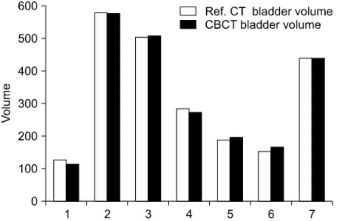
두 번째로, 생리식염수를 주입한 방광 체적을 Eclipse를 통 해 얻은 결과값의 비교이다. 가장 큰 차이를 나타내는 환자 의 경우 12 cc의 차이를 나타내었고 대부분의 환자의 경우 10 cc 미만의 차이를 나타내었다. 평균 4% 미만의 방광 체적 차이를 나타내었다(Fig. 7).
고안 및 결론
위의 첫 번째 실험결과로부터 뼈를 기준으로 정합을 시행 하였을 경우 방광의 해부학적 특성 및 직장의 상태에 따라
Fig. 7. Changes in bladder volume compared to Ref. CT and CBCT.
방광의 위치 변화가 있음을 알 수 있었다. 위치 변화 값이 가 장 크게 나타났을 경우에는 치료계획 시 설정한 여유 폭 5 mm 이상의 차이가 나타나는 경우도 볼 수 있었다. 또, 위치 변화의 평균값보다 표준 편차 값이 크게 나타나는 경우도 나 타났는데 이는 매번 치료 시 마다 방광의 위치 변화 및 방향 성이 일정하기 보다는 무작위적임을 알 수 있었다. 이러한 방광의 위치변화는 주변장기인 Femoral head 및 직장에 원하 지 않는 선량이 전달 될 수 있다. 이로 인해 방사선 치료범위 를 축소한(Reduce field) 경우 자칫 표적이 PTV를 벗어날 수 있는 확률을 증가 시킬 수 있으며, 이는 결국 PTV가 증가하 는 요인이 된다.10-13) 때문에 뼈를 기준으로 하는 정합보다는 방광을 기준으로 정합을 함으로써 PTV 여유 폭 감소 및 인 접장기 합병증(complication) 감소의 효과를 기대할 수 있었 다. 또한, 정합을 시행 한 후 방광의 위치 변화를 분석한 결 과 vertical 방향으로 가장 크게 나타났는데 이는 환자의 허 리 들림 및 직장의 상태 변화로 인한 현상으로 판단된다.
두 번째로, 방사선 치료범위를 축소한(Reduce field)치료 시 방광에 생리식염수를 주입함으로써 Reference CT 방광 체적과 CBCT상 방광 체적의 재현성을 4% 이내로 높일 수 있었고 GTV가 모두 계획한 용적 안에 포함됨을 알 수 있었 다. 그러나 요도를 통해 생리식염수를 넣는 과정이 환자들로 하여금 많은 고통을 감수해야 하기 때문 환자 상태에 따라 주입여부를 결정해야 한다. 또 주입 후에도 고통 경감 및 방 광의 체적변화 최소화를 위해 신속하게 치료를 진행해야 한 다. 본 연구를 통해 방광암의 방사선 치료범위를 축소한 (Reduce field)치료 시에는 방광의 특성상 계획된 PTV 용적 에서 표적이 빠질 염려가 있으므로 치료 전 방광에 CT-simu- lation 당시의 동일한 양의 생리식염수를 주입하여 매일 CBCT촬영을 통해 방광을 기준으로 정합을 함으로써 더욱
정확한 치료를 실시 할 수 있을 것으로 사료된다.
참고문헌
1. Meijer GJ, Rasch C, Remeijer P, et al.: Three-dimensional analysis of delineation errors, setup errors, and organ motion during radiotherapy of bladder cancer. Int J Radiat Oncol Biol Phys 2003;55:1277-1287
2. Miralbell R, Nouet P, Rouzaud M, et al.: Radiotherapy of bladder cancer: Relevance of bladder volume changes in planning boost treatment. Int J Radiat Oncol Biol Phys 1998;41:741-746
3. Lars Fokdal Henriette Honor: Impact of changes in bladder and rectal filling volume on organ motion and dose distri- bution of the bladder in radiotherapy for urinary bladder cancer. Int J Radiation Oncology Biol Phys 2004;59:436-444 4. Harris SJ, Buchanan RB: An audit and evaluation of bladder
movements during radical radiotherapy. Clin Oncol (R CollRadiol) 1998;10:262-264
5. Turner SL, Swindell R, Bowl N, et al.: Bladder movement duringradiation therapy for bladder cancer: Implications for treatment planning. Int J Radiat Oncol Biol Phys 1997;39:
355-360
6. Pos FJ, Koedooder K, Hulshof MC, et al.: Influence of blad- der and rectal volume on spatial variability of a bladder tu- mor during radical radiotherapy. Int J Radiat Oncol Biol Phys 2003;55:835-841
7. Sur RK, Clinkard J, Jones WG, et al.: Changes in target vol- ume during radiotherapy treatment of invasive bladder carci- noma. Clin Oncol (R Coll Radiol) 1993;5:30-33
8. Don Y, Matthew P: Cone beam CT imaging analysis of in- ternational variations in bladder volume and position during radiotherapy for bladder cancer. Int J Radiation Oncology Biol Phys 2010;76:1045-1053
9. Ma CM, Paskalev K: In-room CT techniques for image- guidedradiation therapy. Med Dosim 2006;31:30-39
10. Sur RK, Clinkard J, Jones WG, et al.: Changes in target vol- ume during radiotherapy treatment of invasive bladder carci- noma. Clin Oncol 1993;5:30-33
11. Muren LP, Smaaland R, Dahl O: Organ motion, set-up var- iation and treatment margins in radical radiotherapy of uri- nary bladder cancer. Radiother Oncol 2003;69:291-304 12. Meijer GJ, Rasch C, Remeijer P, Lebesque JV: Three-di-
mensional analysis of delineation errors, setup errors, and or- gan motion during radiotherapy of bladder cancer. Int J Radiat Oncol Biol Phys 2003;55:1277-1287
13. Fokdal L, Honore H, Hoyer M, Meldgaard P, Fode K, von derMaase H: Impact of changes in bladder and rectal filling volume on organ motion and dose distribution of the bladder in radiotherapy for urinary bladder cancer. Int J Radiat Oncol Biol Phys 2004;59:436-444
Abstract
The Investigation Image-guided Radiation Therapy of Bladder Cancer Patients
Seong Soo Bae, Sun Myoung Bae, Jin San Kim, Tae Young Kang, Geum Mun Back, Kyung Tae Kwon Department of Radiation Oncology, Asan Medical Center, Seoul, Korea
Purpose: In hospital image-guided radiation therapy in patients with bladder cancer to enhance the reproducibility of the appropriate amount, depending on the patient’s condition, and image-guided injection of saline system (On-Board Imager system, OBI, VARIAN, USA) three of the Cone-Beam CT dimensional matching (3D-3D matching) to be the treatment. In this study, the treatment of patients with bladder cancer at Cone-Beam CT image obtained through the analysis of the bones based matching and matching based on the bladder to learn about the differences, the bladder’s volume change injected saline solution by looking at the bladder for the treatment of patients with a more appropriate image matching is to assess how the discussion.
Materials and Methods: At our hospital from January 2009 to April 2010 admitted for radiation therapy patients, 7 patients with bladder cancer using a Folly catheter of residual urine in the bladder after removing the amount determined according to individual patient enough to inject saline CT-Sim was designed after the treatment plan.
After that, using OBI before treatment to confirm position with Cone-Beam CT scan was physician in charge of matching was performed in all patients. CBCT images using a total of 45 bones, bladder, based on image matching and image matching based on the difference were analyzed. In addition, changes in bladder volume of Eclipse (version 8.0, VARIAN, USA) persuaded through.
Results: Bones, one based image matching based on the bladder and re-matching the X axis is the difference between the average 3±2 mm, Y axis, 1.8±1.3 mm, Z-axis travel distance is 2.3±1.7 mm and the overall 4.8±2.0 mm, respectively. The volume of the bladder compared to the baseline showed a difference of 4.03±3.97%.
Conclusion: Anatomical location and nature of the bladder due to internal movement of the bones, even after mat- ching with the image of the bladder occurred in different locations. In addition, the volume of saline-filled bladder showed up the difference between the 4.03 percent, but matched in both images to be included in the planned volumes were able to confirm. Thus, after injection of saline into the bladder base by providing a more accurate image matching will be able to conduct therapy.
Key words: image-guided radiation therapy, bladder filling, OBI, 2D match, 3D match