氣血疏通丸이 Monosodium iodoacetate로 骨關節炎을 誘導한 랫드에 미치는 影響
허문강1#, 김순근1, 최학주2, 김동희1,3*
1 : 대전대학교 한의과대학 병리학교실, 2 : 건강나라 자연의학 연구소 3 : 대전대학교 난치성 면역질환의 동서생명의학연구 지역혁신센터
Effects of gihyeolsotong-hwan on monosodium iodoacetate-induced osteoarthritis in rats
Wen-Qiang Xu1#, Soon-Keun Kim2, Hak-Joo Choi3, Dong-Hee Kim1,3*
1 : Department of Pathology, College of Oriental Medicine, Daejeon University 2 : Health country and Natural medicine laboratory
3 : Traditional and Biomedical Research Center(TBRC-RIC), Daejeon University
ABSTRACT
Objectives : In cases of osteoarthritis, the hypofunction of the cartilage and joint leads to a limited range of joint motion, swelling, and pain, which is generally treated using pharmaceutical drugs (e.g., anti-inflammatory agents, cartilage protectants, and nonsteroidal anti-inflammatory drugs) or replacement arthroplasty. However, long-term drug treatment is associated with adverse effects on the gastrointestinal systems. The present study aimed to evaluate the ability of Giheolsotong-hwan to treat of osteoarthritis symptoms in the MIA-induced rat model based on histological analysis, and factors that are associated with inflammation and bone mineral metabolism.
Methods : Giheolsotong-hwan was administered orally at doses of 200 ㎎/㎏/day or 400 ㎎/㎏/day for 2 weeks before direct injection of monosodium iodoacetate (3 ㎎/50㎕ of 0.9% saline) into the intra-articular space of the rats’ right knee. The rats subsequently received the same doses of oral Giheolsotong-hwan for another 4 weeks. We evaluated the treatment effects based on serum biomarkers and histopathological analysis of the knee joints.
Results : Compared to those in control rats, the Giheolsotong-hwan treatments significantly decreased the serum concentration of inflammation factors (i.e., IL-1β , IL-6, TNF-α , PGE
2, and LTB
4), and bone degrade factors (i.e., MMP-9, CTX-Ⅱ, and COMP). In addition, the Giheolsotong-hwan treatments significantly increased the concentration of glycosaminoglycans of bone defence factors, but no chage the TIMP-1. Furthermore, the Giheolsotong-hwan treatments effectively preserved the knee cartilage and proteoglycan.
Conclusion : The results indicate that Giheolsotong-hwan treated osteoarthritis symptoms. Thus, Giheolsotong- hwan may be a novel oriental therapeutic option for the management of osteoarthritis.
1)
Key words : bone metabolism, Giheolsotong-hwan, inflammation factors, oriental therapeutic, osteoarthritis
*Corresponding author : Dong-Hee Kim. Daejeon University, Korea.
·Tel : +82-42-280-2636 ·E-mail : [email protected]
#First author : Wen-Qiang Xu. Daejeon University, Korea.
·Tel : +82-42-280-2827 ·E-mail : [email protected]
·Received : 17 April 2018 ·Revised : 9 May 2018 ·Accepted : 25 May 2018
Ⅰ. 서 론
골관절염 (Osteoarthritis)은 '퇴행성관절염'이라고 지칭되 며, 중년 또는 노년기에 흔히 발생하는 임상적 질환으로 관절 연골의 국소적인 퇴행성 변화, 연골하골의 비대, 주변 골연부의 과잉 골형성 및 관절의 변형에 따른 반복적인 동통, 관절 강 직감과 점진적인 운동장애 등을 초래한다
1,2).
건강보험심사평가원 자료
3)에 의하면 골관절염은 50대 이 상의 중노년층에서 91% 이상의 유병률을 차지하고 있으며, 2015년 약 449만 명에서 2016년 약 463만 명으로 여전히 증가 추세이다.
현재 골관절염의 치료는 통증과 염증을 완화시키는 NSAIDs (Nonsteroidal Anti-inflammatory Drugs), 코르티코스테 로이드 호르몬 (corticosteroid hormone, 코데인 (codeine), 옥시코돈 (oxycodone) 등의 약물 요법과 더불어 동통 제거, 기능회복, 연골파손 억제를 위한 유리체의 제거, 관절 치환술, 활막 절제술, 관절 유합술 등의 수술요법이 활용되고 있다
4).
평균 수명의 증가로 의료계 해결 과제로 인식되고 있는 골 관절염 치료를 위해 다양한 약물과 수술방법 등이 상기한 바와 같이 사용되어 유의적인 성과를 거두고 있지만, 수술요법은 인체에 대한 손상이 크고 재수술과 같은 문제점이 초래되고 있으며, 약물은 장기간 복용할 경우 위장 장애 등 다양한 부 작용이 보고되고 있는 실정이다
5).
한의학에서 골관절염은 '痺證' 범위에 속하며 발병 초기에는 風寒濕邪 침입으로 體虛邪實 또는 虛實夾雜의 병기가 발생하고, 점진적으로 熱, 痰 및 瘀血 등의 복잡한 병리적 변화가 나타 남으로써 만성적이고 고질적인 증상을 유발한다
6).
이에 대한 치료법으로는 辨證論治에 준하여 祛風濕, 補肝腎, 强筋骨, 淸熱化痰, 活血祛瘀 등의 방법이 사용되고 있으며, 침, 약침, 국부 마사지, 운동요법 등이 병용되고 있다
1,7).
Monosodium iodoacetate (MIA)으로 골관절염을 유도한 동물모델은 연골의 손상과 기능 장애, 통증 등의 증상이 실제 골관절염 질환과 유사하여 골관절염의 효능 평가에 활발히 이용 되고 있는데, 桂枝附子湯
8), 健步丹
9), 附子瀉心湯
10), 麻黃附子 細辛湯
11), 疎風活血湯
12)등이 본 병태 모델을 통하여 유의성이 증명된 바가 있다.
본 실험에 사용한 기혈소통환(氣血疏通丸)의 구성 약재는 祛風解表, 勝濕止痛 등에 대한 효능을 가진 羌活, 防風, 獨活, 秦艽 등과 散寒止痛, 補血行氣 등에 효능을 갖는 當歸, 白芷, 杜仲, 白芍藥, 薑黃. 散熱燥濕 효능의 蔓荊子, 仙茅, 甘菊, 薄荷, 黃柏 등과 化痰消腫 효능의 白芥子로 구성되어 현재 관절염에 대한 임상방이다
13,14).
이에 본 연구는 현재 임상에서 활용하는 氣血疏通丸이 골 관절염 질환에 대한 객관적인 효능을 실험적으로 규명하여 EBM (Evidence-Based Medicine)을 구축하고자, MIA 골 관절염 병태 모델 제작 2주 전부터 경구 투여하고 병태 모델 제작 후 4주간 총 6주를 경구 투여한 결과, 혈액 바이오마커 및 조직병리학적 검사 등을 통해 관절염 유관 인자와 형태학적 관찰에 있어 유의성 있는 결과가 도출되었기에 보고하는 바이 다.
Ⅱ. 재료 및 방법
1. 약재
본 실험에 사용한 氣血疏通丸(이하, GSH)의 구성 약재들은
㈜옴니허브에서 防風, 蔓荊子, 仙茅, 蓁艽, 黃柏, 芥子, 甘草는 중국산, 薑黃은 미얀마산이었으며, 나머지 약재는 국산으로 구매하였다. 구매한 약재는 대전대학교 TBRC-RIC에서 정선 후 사용하였고, 그 내용과 분량 (1첩)은 다음과 같다(Table 1).
Herbal
medicine name Abbreviation Weight
(g)
羌 活 Osterici Radix 3
防 風 Saposhnikoviae Radix 3
當 歸 Angelicae Gigantis Radix 3
蔓 荊 子 Vitex negundo L. 3
香 附 子 Cyperi Rhizoma 2
黃 芪 Astragali Radix 2
白 芷 Angelicae dahuricae Radix 2
甘 菊 Chrysanthemi Flos 2
薄 荷 Mentha haplocalyx Briq. 2
仙 茅 Curculigo orchioides Gaertn. 2 獨 活 Angelicae Pubescentis Radix 2
杜 仲 Eucommiae Cortex 2
蓁 艽 Gentianae Macrophyllae Radix 2
白 芍 藥 Paeoniae Radix Alba 2
薑 黃 Curcumae longae Rhizoma 2
黃 柏 Phellodendron chinese Schneid. 2
芥 子 Sinapis Semen 1
甘 草 Glycyrrhizae Radix 1
Total amount 38
Table 1. The Prescription of GSH
2. 동물 및 사료
실험동물인 수컷 7주령의 SD-Rat (200∼220 g)는 ㈜샘 타코BIOKOREA (Korea)에서 공급받아 실험 당일까지 일반 고형사료 (2018S, ENVIGO Co., U.K)와 물을 충분히 공급 하고 온도 22 ± 2℃, 습도 55 ± 15%, 12시간-12시간 (light-dark cycle)의 환경에서 2주간 적응시킨 후 실험에 사용 하였다. 본 실험은 대전대학교 동물실험윤리 위원회의 승인 (동물사용 윤리위원회 승인 번호–DJUARB 2017-020)을 받아 동물 윤리준칙에 의거하여 실험하였다.
3. 시약 및 기기
사용된 시약은 Monosodium iodoacetate (MIA ; Sigma
Co., U.S.A.), Rat cytokine milliplex map immunoassay kit
(Millipore Co., U.S.A.), PGE2 (Prostaglandin E2) ELISA
Kit (Elabscience, U.S.A.), LTB
4ELISA Kit (Elabscience, U.S.A.), Rat MMP-9 (Matrix Metalloproteinase 9/
Gelatinase B) ELISA Kit (Elabscience, U.S.A.), Rat CTX-Ⅱ (Cross Linked C-telopeptide of Type Ⅱ Collagen) ELISA Kit (Elabscience, U.S.A.), Rat COMP (cartilage oligomeric matrix protein) ELISA kit (MyBioSource Co., U.S.A.), Rat TIMP-1 (Tissue Inhibitors of Metalloproteinase 1) ELISA Kit (Elabscience, U.S.A.), Rat Glycosaminoglycans ELISA kit (MyBioSource Co., U.S.A.) 등을 사용하였으며, 기기는 rotary vacuum evaporator (Büchi B-480 Co., Switzerland), freeze dryer (EYELA FDU-540 Co., Japan), centrifuge (Sigma Co., U.S.A.), Luminex (Millipore Co., U.S.A.), ELISA reader (Molecular Devices Co., U.S.A.), Light Microscope (Carl Zeiss, Co., Germany) 등을 사용하였다.
4. 시료 제조
GSH 20첩 (760 g) 기준으로 증류수 10 L를 넣고 4시간 동안 100℃에서 끓인 후 여과액을 얻었으며, 여과액의 500
㎖을 rotary vacuum evaporator에서 감압 농축하였다. 농 축된 용액을 freeze dryer로 동결 건조하여 분말 16.23 g(500 ㎖ 대비 수득율 3.24%)을 얻었으며, 얻어진 분말은 초 저온 냉동고 (-80℃)에서 보관하며 사용하였다.
5. 시료 처리 및 골관절염 병태 모델 제작
실험그룹은 골관절염을 유발하지 않은 정상군 (이하, Normal), 골관절염 유발 전·후에 증류수만을 경구 투여한 음성대조군 (이하, Control) 및 골관절염 유발 전부터 GSH을 200, 400 ㎎/㎏의 농도로 투여한 실험군으로 나누어 매일 같은 시각에 경구 투여를 진행하였다.
골관절염 병태 모델을 제작하기 위해 마취제 (럼푼 0.1 ㎖ + 케타민 0.4 ㎖)를 복강 내에 주입하여 마취된 Rat의 오른쪽 무릎관절 주변을 깨끗이 제모한 후, 골 관절염 유발물질인 MIA를 당뇨주사기 (BD insulin syringe)를 사용하여 무릎 관절강 내에 60 ㎎/㎏의 농도로 50 ㎕씩 투여하였으며, MIA 희석에는 0.9% Saline을 사용하였다.
6. 혈액 채취 및 혈청 분리
실험 종료 후 ether로 마취한 상태에서 심장 천자법을 이용 하여 채혈한 다음 3,000 rpm에서 20분간 원심 분리하여 혈 청을 분리하였다. 분리된 혈청은 혈액 바이오마커 측정을 진 행하기 위해 측정 항목별로 분주하여 초저온 냉동고 (-80℃)에 넣어 보관하였다.
7. 혈청 내 관절염 관련 바이오마커 측정 1) Cytokine 생성량 측정
Cytokine (IL-1β , IL-6, TNF-α ) 생성량은 merck
millipore사의 mouse cytokine milliplex map immunoassay kit을 사용하여 제조사의 프로토콜에 맞춰 절차를 수행한 후 Luminex 기기로 측정하였다.
2) PGE2 및 LTB4 생성량 측정
PGE
2및 LTB
4생성량은 Elabscience사의 ELISA kit을 사용하여 제조사의 프로토콜에 맞춰 절차를 수행한 후 ELISA reader 기기로 측정하였다.
3) MMP-9, CTX-Ⅱ, COMP, TIMP-1, GAGs 생성량 측정
MMP-9 및 CTX-Ⅱ, COMP, TIMP-1, GAGs 생성량은 Mybiosource사의 ELISA kit을 사용하여 제조사의 프로토콜에 맞춰 절차를 수행한 후 ELISA reader 기기로 측정하였다.
8. 조직병리학 검사
조직병리학 검사는 혈액 채취 후 경추 탈구 시킨 동물의 우측 대퇴부위와 경골 부위를 절단한 뒤 근육조직을 제거하고 10%
포르말린에 고정시켜 검사전문 업체인 KPNT (Korea)에 의뢰 하여 Hematoxylin and Eosin (H&E) 염색과 Safranin-O 염색을 진행하였다. 염색되어 온 조직슬라이드는 광학현미경을 통해 200배 배율에서 연골 주변과 proteoglycan 등을 확인 후 사진 촬영하였다.
9. 통계처리
본 연구의 실험 결과는 평균값±표준 편차 (mean±S.D.)로 표시하였다. 각 처리군의 비교는 one-way analysis of variance (ANOVA)로 분석한 후, student’s t-test를 사용하여 통계적 유의성을 검증하였으며, 정상군과 비교한 대조군의 결 과는
###p<0.001,
##p<0.01,
#p<0.05로 나타내었으며, 대조군 과 비교한 실험군의 결과는
***p<0.001,
**p<0.01,
*p<0.05로 표기하였다.
Ⅲ. 결 과
1. 혈청 내 염증 관련 바이오마커 1) Cytokine 생성량
혈청 내 사이토카인 생성량을 측정한 결과, IL-1β는 정상 군이 21.6±3.3%, 대조군이 100.0±4.4%, 200 ㎎/㎏ 투여 군이 38.1±3.0%, 400 ㎎/㎏ 투여군이 61.4±2.5%로 나타 났으며, IL-6 생성량은 정상군이 67.3±7.0%, 대조군이 100.0±7.8%, 200 ㎎/㎏ 투여군이 70.2±5.5%, 400 ㎎/㎏
투여군이 74.2±6.6%로 나타났다. 또한, TNF-α 생성량은 정상군이 50.3±7.5%, 대조군이 100.0±5.9%, 200 ㎎/㎏
투여군이 78.7±9.5%, 400 ㎎/㎏ 투여군이 83.9±10.0%로
나타나, GSH를 200 ㎎/㎏과 400 ㎎/㎏으로 경구 투여한 실
험군은 대조군과 대비하여 IL-1β, IL-6, TNF-ɑ 등의 염
증성 사이토카인 생성을 유의성 있게 (p<0.001, p<0.01) 감소시켰다(Table 2).
Cytokine level (% of
control)
Groups
Normal Control GSH
200 400
IL-1β 21.6±3.3 100.0±
4.4###
38.1±
3.0***
61.4±
2.5***
IL-6 67.3±7.0 100.0±
7.8###
70.2±
5.5***
74.2±
6.6***
TNF-α 50.3±7.5 100.0±
5.9###
78.7±
9.5***
83.9±
10.0**
Luminex assays was used to measure serum levels of interleukin-1 beta (IL-1β), interleukin-6 (IL-6), and tumor necrosis factor-alpha (TNF-α). The measured levels were expressed as percent of the control value (mean ± standard deviation, n = 6/group) (###p<0.001 indicates a significant difference from the normal group,
***p<0.001, or **p<0.01 indicates a significant difference from the osteoarthritis control group).
Table 2. Effects of GSH on serum pro-inflammatory cytokine in a monosodidum iodoacetate-induced rat model of osteoarthritis
2) PGE2 및 LTB4 생성량
혈청 내 PGE
2및 LTB
4생성량을 측정한 결과, PGE
2생성 량은 정상군이 734.9±78.4 ng/㎖, 대조군이 3913.0±209.1 ng/㎖, 200 ㎎/㎏ 투여군이 1085.0±151.5 ng/㎖, 400 ㎎/㎏
투여군이 988.0±142.2 ng/㎖로 나타났으며(Fig. 1A), LTB
4생성량은 정상군이 3797.1±192.3 pg/㎖, 대조군이 5464.5±377.4 pg/㎖, 200 ㎎/㎏ 투여군이 2926.4±208.1 pg/㎖, 400 ㎎/㎏ 투여군이 3131.7±430.5 pg/㎖로 나타 나(Fig. 1B), GSH를 200 ㎎/㎏과 400 ㎎/㎏으로 경구 투여한 실험군은 대조군과 대비하여 PGE
2및 LTB
4등의 염증 관련 바이오마커 생성을 유의성 있게 (p<0.001) 감소시켰다(Fig. 1).
2. 혈청 내 골 파괴 관련 바이오마커
혈청 내 MMP-9 및 CTX-Ⅱ, COMP 생성량을 측정한 결과, MMP-9 생성량은 정상군이 67.3±7.2%, 대조군이 100.0±
1.1%, 200 ㎎/㎏ 투여군이 91.0±3.6%, 400 ㎎/㎏ 투여군이 87.9±5.3%로 나타났으며, CTX-Ⅱ 생성량은 정상군이 50.7±
6.9%, 대조군이 100.0±5.6%, 200 ㎎/㎏ 투여군이 54.4±
5.5%, 400 ㎎/㎏ 투여군이 63.7±5.3%로 나타났다. 또한, COMP 생성량은 정상군이 270.7±12.9 ng/㎖, 대조군이 221.3±14.0 ng/㎖, 200 ㎎/㎏ 투여군이 179.1±17.5 ng/㎖, 400 ㎎/㎏ 투여군이 178.4±8.8 ng/㎖로 나타나, GSH를 200 ㎎/㎏과 400 ㎎/㎏으로 경구 투여한 실험군은 대조군과 대비하여 MMP-9, CTX-Ⅱ, COMP 등의 연골 파괴 관련 바 이오마커 생성을 유의성 있게 (p<0.001, p<0.01) 감소시켰 다(Table 3).
Fig. 1. Effects of GSH on serum inflammatory factors in a monosodium iodoacetate-induced rat model of osteoarthritis.
Enzyme-linked immunosorbent assays was used to measure serum levels of prostaglandin E2 (PGE2) (A), and leukotriene B4 (LTB4) (B). The measured levels were expressed as percent of the control value (mean ± standard deviation, n = 6/group) (###p<0.001 indicates a significant difference from the normal group, ***p<0.001 indicates a significant difference from the osteoarthritis control group).
Cytokine level (% of
control)
Groups
Normal Control GSH
200 400
MMP-9 67.3±7.2 100.0±
1.1###
91.0±
3.6*
87.9±
5.3*
CTX-Ⅱ 50.7±6.9 100.0±
5.6###
54.4±
5.5***
63.7±
5.3***
COMP 270.7±12.9 100.0±
6.3###
80.9±
7.9***
80.6±
4.0***
Enzyme-linked immunosorbent was used to measure serum levels of Matrix Metalloproteinase-9 (MMP-9), C-Telopeptide of Type II Collagen (CTX-Ⅱ), and cartilage oligomeric matrix protein (COMP). The measured levels were expressed as percent of the control value (mean ± standard deviation, n = 6/group) (###p<0.001 indicates a significant difference from the normal group, ***p<0.001, or *p<0.05 indicates a significant difference from the osteoarthritis control group).
Table 3. Effects of GSH on serum bone degrade factors in a monosodidum iodoacetate-induced rat model of osteoarthritis
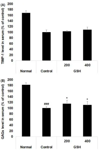
3. 혈청 내 골 파괴 방어 관련 바이오마커
혈청 내 TIMP-1 및 GAGs 생성량을 측정한 결과, TIMP-1 생성량은 정상군이 168.1±7.6%, 대조군이 100±
7.9%, 200 ㎎/㎏ 투여군이 103.3±2.7%, 400 ㎎/㎏ 투여 군이 108.7±10.1%로 나타났으며(Fig. 2A), GAGs 생성량은 정상군이 182.0±6.8%, 대조군이 100±5.0%, 200 ㎎/㎏
투여군이 115.8±12.2%, 400 ㎎/㎏ 투여군이 111.2±
12.2%로 나타나(Fig. 2B), GSH를 200 ㎎/㎏과 400 ㎎/㎏
으로 경구 투여한 실험군은 대조군과 대비하여 연골 파괴 관련 바이오마커인 GAGs 생성을 유의성 있게 (p<0.001, p<0.01) 증가시켰으나 TIMP-1 생성은 차이가 나타나지 않았다(Fig. 2).
Fig. 2. Effects of GSH on serum bone defence factors in a monosodidum iodoacetate-induced rat model of osteoarthritis.
Enzyme-linked immunosorbent was used to measure serum levels of Tissue inhibitor of matrix metalloproteinases 1 (TIMP-1), and glycosaminoglycans (GAGs). The measured levels were expressed as percent of the control value (mean ± standard deviation, n = 6/group) (###p<0.001 indicates a significant difference from the normal group, *p<0.05 indicates a significant difference from the osteoarthritis control group).
4. 조직변화에 미치는 영향
무릎 연골 조직 변화를 H&E 및 safranin-O 염색을 통해 확인한 결과, 정상군은 대조군과 GSH 투여군과 대비하여 연골 (Fig. 3A-동그라미)과 연골 주변으로 proteoglycan의 보존 (Fig. 3B-동그라미)이 잘 되어 있는 것이 관찰되었다. 상대
적으로 GSH 투여군은 대조군과 대비하여 정상군에 가까운 연골 및 proteoglycan 손실이 적은 것으로 관찰되었다(Fig. 3)
Fig. 3. Histopathological effects of GSH on knee joints in a monosodium iodoacetate (MIA)-induced rat model of osteoarthritis.
Representative histological images of knee joint tissues were stained using hematoxylin and eosin (A) or safranin-O (B) (magnification, 200×).
Ⅳ. 고 찰
골관절염은 주로 고관절, 손가락, 무릎 등 장기간 지속적인 사용을 하는 관절 부위에 발생하며, 연골뿐만 아니라 인대, 근육, 활액 등 관절을 구성하는 요소들로 인해 더욱 심화되어 심한 통증을 수반한 채 관절의 운동범위를 제한하게 된다
15,16).
이러한 골관절염은 손상된 연골세포에서 IL-1β , IL-6, TNF-α 등의 염증성 cytokine들이 다량으로 생성되어 염증 매개물질의 생성 촉진 및 MMPs의 생성을 유의하게 증가시키는 것으로 알려져 있다
16).
본 시료인 氣血疏通丸은 祛風濕止痛, 强筋骨 약물을 중심 으로 처방이 구성되어 현재 골관절염에 활용되고 있는 임상방 이다. 따라서 본 연구는 새로운 치료제 개발이 아닌 현재 활용 되고 있는 처방의 객관적인 효능과 치료기전을 규명하여 EBM (Evidence-Based Medicine)을 구축하는데 목적이 있다.
氣血疏通丸의 구성 약재 중 蔓荊子
17), 薄荷
18), 秦艽
19), 白芥 子
20)黃芪
21), 當歸
22)등은 항염증 혹은 항산화 효능이 규명된 바가 있고, 또한 羌活
23), 獨活
23), 香附子
24), 白芷
25), 甘菊
26), 仙茅
27), 防風
28), 杜仲
29), 白芍藥
30), 薑黃
31), 黃柏
32)등도 단일 약재, 약침, 처방 등으로 관절염에 대한 유효성이 입증된 바가 있다.
본 연구에서는 MIA로 골관절염을 유도한 랫 동물 병태 모 델을 활용하여 GSH를 경구 투여하여 골관절염 유관 인자 및 형태학적 관찰을 통해 염증, 골 파괴 및 골 파괴 방어 등에 대한 효능을 혈액 바이오마커 및 조직병리학적 검사를 통해 확인하 였다.
골관절염의 주된 원인으로 알려진 염증에 의한 발생은
TNF-α , IL-1β, IL-6와 같은 염증성 cytokine이 다량으로
생성되어 연골세포, 대식세포, 골아세포 및 활막 조직에 작용
함으로써 관절 파괴에 주된 역할을 한다
33-35). 특히, IL-1β 와
TNF-α 는 골관절염에서 중요한 역할을 하고 있는데 이들의
과량 생성은 염증 매개 인자인 PGE
2와 LTB
4등의 합성을 촉진
하여 proteoglycan을 소실시키고 재합성을 억제하며, MMPs의
활성화 및 생성을 증가시킴으로써 관절염증을 증폭시킴과 동
시에, 연골 기질파괴를 일으킨다. 또한, IL-6의 분비를 촉진 하여 골 회복 및 생성을 방해하는 것으로 알려져 있다
35,36). 본 연구에서 GSH 투여군은 대조군과 대비하여 염증성 cytokine IL-1β , IL-6 및 TNF-α의 생성을 유의성 있게 감소시켜 골 관절염의 주된 증상인 염증 반응을 억제하였다(Table 2), .
또한, 염증 반응의 중요한 매개물질인 PGE
2는 손상 부위나 조직에서 통증과 발열 전달에 관여하는 것으로 알려져 있으며
37,38)
, LTB
4는 lipoxygenase의 활성화에 따른 주요 산물로서
염증 반응에서 호산구 및 호중구에 대하여 강력한 유주성 특 성을 갖는 성분으로 알려져 있으며 세포의 화학주성에 필요한 여러 인자들과 세포부착인자의 발현에 영향을 주어 치주염, 류마티스관절염 및 다수의 염증성 질환을 일으켜 골 손실 질 환에 핵심적 기능을 하는 인자로 알려져 있다
8,39). 본 연구에서 GSH 투여군은 대조군과 대비하여 PGE
2와 LTB
4의 생성량을 유의성 있게 감소시켜 앞선 염증성 cytokine 결과인 염증에 의한 골관절염 질환을 개선하는 효능이 있음을 뒷받침하고 있 다(Fig. 1A, B).
GSH의 투여가 염증 반응을 억제하여 골관절염 치료에 대한 가능성이 입증됨에 따라 골 파괴 및 골 파괴 방어 인자 등을 다각도로 검증하여 골관절염에 대한 효능을 증명하고자 다양한 인자를 추가로 확인하였다.
연골의 파괴는 우선 MMPs 합성 및 활성화에 의해 파괴되 는데 이러한 현상은 염증성 cytokine에 의해 유발된다. 연골 세포는 sulfated proteoglycan, type Ⅱ collgen 등의 연골 특이적 단백질로 합성되어 조직을 이루게 되는데, 관절조직의 파괴는 주로 연골 내 콜라겐의 소실로 인해 발생한다
40-44). 또한 골관절염이 지속되면 연골에 포함된 단백질 성분인 COMP의 발현량이 증가하여 혈액 검사를 통해 연골마모 정도, 손상 및 진행 여부 등을 예측할 수 있는 아주 중요한 바이오마커로 알 려져 있다
45,46). 본 연구에서 GSH 투여군은 대조군과 대비하여 MMP-9 및 CTX-Ⅱ, COMP 등의 생성량을 유의성 있게 감소 시켜 본 시료가 항염증 작용과 더불어 연골 파괴 억제 효능이 있음을 나타내고 있다(Table 3).
TIMP-1은 세포의 기질을 재건하는 MMPs의 내적인 길항 제로서 골 파괴를 지연하거나 형성하는 것으로 알려져 있다.
IL-1β , IL-6는 내피세포에서 TIMP 생산을 억제하는 반면 활액 및 연골세포에서는 TIMP의 발현을 증가시키고 상위 염증 매개 인자인 TNF-α 는 TIMP의 발현을 억제하는 반면 MMP의 발현을 증가시킨다. 이러한 MMPs와 TIMP의 활동 사이의 불 균형이 골관절염의 병리적 메커니즘으로 이해되고 있다
47). 그 러나 본 연구에서 TIMP-1 생성량은 대조군과 GSH 경구 투 여군 사이의 차이가 나타나지 않았는데, 이는 GSH가 TIMP 자극을 통하지 않고 MMPs의 발현을 직접적으로 조절한다는 것을 나타내고 있어 GSH가 MMPs 생산을 감소시켜 조직 재생 및 연골, 뼈 파괴에 결정적인 역할을 할 수 있음을 시사한다 (Fig. 2A).
GAGs는 관절연골에서 주로 탄력성과 관련되어 콜라겐을 보호하는 작용을 하며, 또한 관절 연골의 주요 성분인 proteoglycan을 생성하는 것으로 알려져 있다
48,49). 본 연구 에서 GSH 투여군은 대조군과 대비하여 GAGs 생성량을 유의성 있게 증가시켜 연골 회복에 대한 효능이 확인되었다(Fig. 2B).
이와 같은 혈액 바이오마커 결과를 통해 GSH의 경구 투여가
염증 반응, 골 파괴 및 방어 등에 효능이 확인되어 이를 더욱 자세히 증명하고자 H&E, Safranin-O 등의 염색을 통해 조 직병리학적 검사를 진행하였다. 조직병리학 소견 결과, 정상 군은 대퇴골, 경골, 연골 및 활막 조직 등의 손실이 나타나지 않았으나 대조군과 GSH 투여군은 연골과 연골 주변의 proteoglycan의 손실이 관찰되었다. 그러나 상대적으로 GSH 투여군은 대조군 대비 연골마모와 proteoglycan의 소실이 적은 것으로 나타나 혈액 바이오마커 결과와 부합되었다.
이상의 결과를 종합해 볼 때, GSH은 골관절염 병태 동물 모델을 통해 대표적인 원인이라 할 수 있는 염증 반응 (염증성 cytokine 및 염증 매개 인자)의 생성량 감소와 동시에 골 파 괴를 일으키는 대표적 인자 역시도 감소시키며 동시에 연골 파괴를 방어할 수 있는 효능이 혈액 및 조직병리학 결과를 통해 입증되었다. 현재 임상에서 활용되고 있는 GSH에 대한 객관 적인 본 연구결과는 추후 치험 사례연구가 보완 된다면 골관 절염에 대한 경제/사회적으로 손실을 보완해 줄 한의학 치료 제로써 상용화가 가능할 것으로 판단된다.
Ⅴ. 결 론
본 연구에서는 氣血疏通丸 (GSH)이 골관절염에 대한 효과를 객관적으로 증명하고자 관절염을 유발한 동물모델을 통해 혈액 및 조직학적 평가를 진행한 결과, 다음과 같은 결론을 얻었다.
1. GSH 투여군은 대조군과 대비하여 염증성 cytokine (IL-1β , IL-6, TNF-α ) 및 염증 매개 인자인 PGE
2, LTB
4등의 생성량을 유의성 있게 감소시켰다.
2. GSH 투여군은 대조군과 대비하여 골 파괴 관련 인자인 MMP-9, CTX-Ⅱ, COMP 등의 생성량을 유의성 있게 감소시켰다.
3. GSH 투여군은 대조군과 대비하여 골 파괴 방어 관련 인자인 TIMP-1 생성량은 차이가 나타나지 않았으나, GAGs 생성량을 유의성 있게 증가시켰다.
4. GSH 투여군은 대조군과 대비하여 H&E 및 safranin-O 염색을 통해 연골의 파괴가 적고 proteoglycan 손실이 적게 나타났다.
이상의 결과들로 보아 GSH 투여는 염증 및 골 관련 대사 질환에 유의적인 영향을 주어 골관절염을 치료할 수 있는 임상 방임이 실험적으로 규명되었다. 따라서 현재 임상에서 활용되고 있는 GSH가 EBM (Evidence-Based Medicine)을 구축에 일조할 것으로 사료된다.
감사의 글
본 연구는 산업통상자원부 지정 대전대학교 난치성 면역질
환의 동서생명의학연구 지역혁신센터의 지원에 의한 것입니다.
References