558 Copyright © 2012 The Korean Society of Cardiology Korean Circulation Journal
Introduction
Coronary artery aneurysms (CAAs) are relatively rare but have been diagnosed recently with increasing frequency by the use of coronary angiography. The use of percutaneous coronary interven- tion (PCI) for lesions containing small coronary aneurysms is under controversy. Here, we report a rare case of a patient with rupture and spontaneous sealing of a coronary aneurysm that occurred within an atherosclerotic stenotic lesion after deployment of a drug- eluting stent (DES).
Case
A 78-year-old man visited the cardiology outpatient department at our hospital for exertion-related chest pain that had persisted for 4 years. His only risk factor for coronary artery disease was hy- pertension. An electrocardiogram showed sinus rhythm and no ST-T
Case Report
http://dx.doi.org/10.4070/kcj.2012.42.8.558
Print ISSN 1738-5520 • On-line ISSN 1738-5555
Rupture and Spontaneous Sealing of a Coronary Aneurysm After Deployment of Drug-Eluting Stent
Tae Jung Kwon, MD, Jin Yong Hwang, PhD, Choong Hwan Kwak, PhD, Young-Hoon Jeong, PhD, Yong Whi Park, PhD, Seok-Jae Hwang, PhD, Jeong Rang Park, PhD, Jong-Hwa Ahn, MD, and Ji Hyun Min, MD
Department of Internal Medicine, Gyeongsang National University Hospital, Jinju, Korea
Lesions with coronary artery aneurysm (CAA) can become complicated during percutaneous coronary intervention. Here, we report a case of a 78-year-old man who developed a rupture, and spontaneous sealing of the CAA occurred after stent implantation, as shown by computed tomography coronary angiography. (Korean Circ J 2012;42:558-561)
KEY WORDS: Coronary aneurysm; Drug-eluting stents.
Received: September 6, 2011
Revision Received: November 22, 2011 Accepted: January 16, 2012
Correspondence: Jin Yong Hwang, PhD, Department of Internal Medicine, Gyeongsang National University Hospital, 79 Gangnam-ro, Jinju 660-702, Korea
Tel: 82-55-750-8064, Fax: 82-55-758-9122 E-mail: [email protected]
• The authors have no financial conflicts of interest.
This is an Open Access article distributed under the terms of the Creative Commons Attribution Non-Commercial License (http://creativecommons.
org/licenses/by-nc/3.0) which permits unrestricted non-commercial use, distribution, and reproduction in any medium, provided the original work is properly cited.
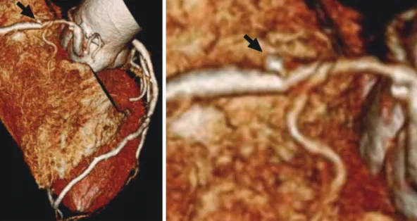
changes. Chest radiography showed unremarkable findings. Mild diastolic dysfunction and no regional wall motion abnormalities were noted in the echocardiogram. The plasma levels of both tropo- nin I and creatine kinase-MB were within the normal range. A 2.5- mm saccular coronary aneurysm and tubular stenosis (60-70%) was detected on the proximal right coronary artery (RCA) by using a computed tomography coronary angiography (Fig. 1).
The patient was pre-loaded with clopidogrel (600 mg) and aspi- rin (300 mg) 1 day before the coronary angiography. His coronary an- giography showed the presence of a small saccular CAA with dif- fuse, tubular stenosis in the proximal portion of the RCA (Fig. 2A).
The periprocedural antithrombotic regimen consisted of a 3500- unit bolus of unfractionated heparin. A 6 Fr JR4 guiding catheter was used to engage the RCA. The lesion was pre-dilated with a 3.0× 20 mm Pantera balloon (Biotronik, Berlin, Germany) at 10 atm. We then performed intravascular ultrasonography (IVUS), which showed a luminal stenosis with fibrous fatty plaque, a reference diameter of 4.5 mm, minimal lumen diameter of 1.0 mm, a post percentage dia- meter stenosis of 60%, and 2.5×2.5 mm sized small aneurysms in the proximal RCA (Fig. 3A). A 4.0×38 mm Endeavor® stent (zotaro- limus, Medtronic, Santa Rosa, CA, USA) was implanted in the prox- imal RCA. After stenting, the coronary aneurysm was no longer de- tected, and 1 side branch of the proximal RCA was jailed. Angio- graphy showed no luminal defect (Fig. 2B). Subsequently, adjunctive balloon dilatation using a 4.5×12 mm, non-compliant balloon was performed. Following this, acute stent thrombosis developed (Fig.
2C). We performed IVUS, which showed a thrombus in the stent, but
an aneurysm was not seen (Fig. 3B). A 500 µg bolus of tirofiban was
administered, followed by intravenous infusion of 0.10 µg/(kg · min).