204
책임저자:김대철, 부산시 서구 동대신동 3가 1번지
602-715, 동아대학교 의과대학 병리학교실 Tel: 051-240-2878, Fax: 051-980-0585 E-mail: [email protected]
접수일:2007년 4월 2일, 게재승인일:2007년 6월 13일
이 논문은 2005학년도 동아대학교 학술연구비(신진과제)에 의하여 연구되었음.
침윤성 유방암에서 Topoisomerase IIα와 Ki-67단백 발현의 임상병리학적 의의
동아대학교 의과대학 외과학교실, 1병리학교실
오상민ㆍ김대철1ㆍ조세헌
Expression of Topoisomerase II α and Ki-67 in Invasive Mammary Carcinoma and Their Clinico- pathologic Implication
Sang-Min Oh, M.D., Dae-Cheol Kim, M.D.1 and Se Heon Cho, M.D.
Purpose: Various prognostic indicators have been identified for mammary carcinomas, but the issue of their significance remains unsettled. The prognostic impact of c-erb B2, Ki-67 and topoisomerase II α expression was investigated in rela- tion to prognostic factors for carcinomas of the breast and to the tumor cell growth fraction.
Methods: One hundred eighteen cases of invasive mammary carcinoma were investigated by immunohistochemical stain- ing for c-erb B2, topoisomerase II α, and Ki-67. Clinicopa- thologic parameters were compared with the expression pat- tern and incidence of c-erb B2, topoisomerase II α and Ki- 67 in invasive mammary carcinoma.
Results: C-erb B2 showed significant correlation with top- oisomerase II α (P<0.05), but others were not significant.
Topoisomerase II α and Ki-67 index closely paralleled each other, indicating that both reflect the proliferate activity of tu- mor cells and were associated with high nuclear and histo- logical grade, ER and PR expression (P<0.05).
Conclusion: These results indicate that ki-67 and top- oisomerase II α proteins might play a role in tumor pro- gression of breast carcinoma. The Ki-67 and topoisomerase II α index may be proliferate factors of breast cancer. In addition, the increase expression of Ki-67 and topoisomerase II α and hormone receptor were closely correlated each oth- er, and could be used as factors suggesting poor prognosis in breast carcinoma. (J Korean Surg Soc 2007;73:204-209)
Key Words: Breast cancer, c-erb B2, Topoisomerase IIα, Ki-67
중심 단어:유방암, c-erb B2, Topoisomerase IIα, Ki-67
Departments of Surgery and 1Pathology, Dong-A Univer- sity College of Medicine, Busan, Korea
서 론
유방암은 여성에서 발생하는 가장 흔한 악성종양 중 하나 이지만, 많은 연구에도 불구하고 유방암을 유발하는 분자 생물학적 기전은 아직 확실히 규명되어 있지 않다. 유방암 에서 가장 흔히 발견되는 유전자의 이상은 p53 유전자의 변 이와 HER-2/neu 종양 유전자의 증폭 등으로 알려져 있다.(1) HER-2/neu는 염색체 17q21에 존재하는 종양유전자로 유 방암, 폐암, 난소암 및 위암 등에서 발현하고 구조적으로 표 피성장인자의 수용체와 유사하다. HER-2/neu 단백질의 발 현은 HER-2/neu 유전자의 증폭을 반영하는 것으로 알려져 있고, 유방암에서 HER-2/neu 유전자의 증폭은 나쁜 예후 인 자로 알려져 있다. 이 외에도 유방암의 예후를 추정할 수 있는 인자들에 관한 많은 연구가 이루어져 있는데, 현재까 지 림프절 전이상태, 조직학적 유형, 종양크기, 핵 등급, 조 직등급, 에스트로겐 수용체, 프로게스테론 수용체 그리고 세포 증식력 측정 등이 알려져 있다. 이 중 암세포의 증식능 력은 암세포의 특징 중 하나로 병의 진행을 결정짓는 중요 한 인자로 알려져 있다. Ki-67 발현은 세포의 증식능력을 측정하는 대표적 인자로 알려져 있고, 최근 세포의 증식능 력을 측정하는 새로운 표지자로 DNA topoisomerase IIα (topo IIα)가 있다.(2) DNA topoisomerase는 DNA 합성, 전사, 염색체 분리와 재결합과 같은 DNA 복제의 중요 단계에서 DNA 형태를 변화시키는 효소 중 하나이다. 이 중 DNA topo IIα는 세포증식에 있어서 변화가 민감하여 빠른 증식 을 할 때 높은 수준을 유지한다고 알려져 있으며, 또한 S기 에 있는 정상과 형질 전환된 모든 세포에서 검출되며 비증
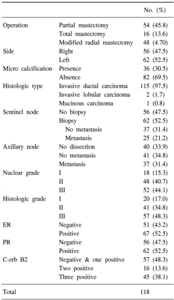
Table 1. Summary of clinicopathologic and immunohistochmical data in breast carcinomas
No. (%)
Operation Partial mastectomy 54 (45.8)
Total mastectomy 16 (13.6) Modified radial mastectomy 48 (4.70)
Side Right 56 (47.5)
Left 62 (52.5)
Micro calcification Presence 36 (30.5)
Absence 82 (69.5)
Histologic type Invasive ductal carcinoma 115 (97.5) Invasive lobular carcinoma 2 (1.7) Mucinous carcinoma 1 (0.8)
Sentinel node No biopsy 56 (47.5)
Biopsy 62 (52.5)
No metastasis 37 (31.4)
Metastasis 25 (21.2)
Axillary node No dissection 40 (33.9)
No metastasis 41 (34.8)
Metastasis 37 (31.4)
Nuclear grade I 18 (15.3)
II 48 (40.7)
III 52 (44.1)
Histologic grade I 20 (17.0)
II 41 (34.8)
III 57 (48.3)
ER Negative 51 (43.2)
Positive 67 (52.5)
PR Negative 56 (47.5)
Positive 62 (52.5)
C-erb B2 Negative & one positive 57 (48.3)
Two positive 16 (13.6)
Three positive 45 (38.1)
Total 118
식성 세포에서는 검출되지 않는다고 한다.(3) 또한 이는 an- thracycline와 epipodophyllotoxins과 같은 항암제의 치료 목 표로서, 유방암 조직 내 topo IIα의 발현이 높으면 anthracy- cline 항암 치료에 잘 반응하는 것으로 알려져 있고 HER-2/
neu의 과발현과 종종 동반하기 때문에,(4) HER-2/neu를 과 발현하는 유방암이 anthracycline 항암 치료에 잘 반응하는 이유 중의 하나로 설명하기도 한다.
이에 본 저자는 유방암 조직에서, Ki-67, topo IIα, HER-2/
neu의 발현강도와 유방암의 임상적 특징 및 예후 인자와의 관계를 알아보고 향후 항암치료에 대한 기본 자료로 활용 하고자 한다.
방 법 1) 연구대상
2004년 1월부터 2004년 12월까지 동아대학교 의료원 유 방센터에서 유방암으로 수술을 받은 118명의 여성 유방암 환자를 대상으로 의무기록과 병리조직 검사 보고서를 바탕 으로 자료를 수집하였다.
2) 연구방법
(1) 병리학적 분류: 종양의 조직학적 악성도는 Bloom과 Richardson(5,6)의 등급체계에 근거하여 각 세 등급으로 분 류하였고 핵 등급은 Nottingham/Tenovus Breast Cancer Study 에서 사용한 등급기준에 따라 분류하였다.(7) 병기는 AJCC 의 분류에 따랐다.
(2) 면역조직화학염색: 본 실험에 사용한 일차항체는 mo- noclonal mouse anti-rat Ki-67 (MIB-5, 1:100, Dako, Denmark), monoclonal mouse anti-human topoisomerase IIα (SWT3D1, 1:200, Dako, Denmark)와 anti-human c-erb B2 (1:400, Neomark)를 사용하였다.
파라핀 포매조직을 4μm의 두께로 절편하여 슬라이드에 부착하였다. 크실렌에서 파라핀을 제거한 후 100%, 90%, 80% 및 70% 에탄올에서 함수시키고 증류수로 씻어내었다.
내인성 과산화효소를 제거하기 위해 3% 과산화수소를 조 직절편 위에 떨어뜨려 30분간 실온에 방치한 후, 인산염완 충식염수로 세척한 후 구연산완충(pH 6.0)용액에 저주파방 법으로 전처리 하였다. 희석한 1차 항체와 실온에서 2시간 반응시킨 후 PBS로 항체를 제거하고 2차 항체와 avidin-bio- tin-peroxidase complex를 반응시킨 후에 Mayers Hematox- ylin으로 대조 염색 후 검경하였다.
(3) 면역조직화학염색 결과 판정: c-erb B2는 DAKO의 HercepTest for Immunoenzymatic Staining (Dako Corp, Car- pinteria, CA)(8)에 따라 0점(negative)과 1점(one positive)은 음성(0), 2점(two positive)은 중등도 양성(1), 3점(three pos- itive)은 강 양성(2)으로 하여 2점과 3점을 양성으로 판독하 였으며 병리전문의 1인이 판독하였다. Topo IIα는 가장 염
색강도가 높은 부분을 찾아 4군데의 고배율 시야(×400)에 서 핵에 진한 갈색으로 염색되는 강양성의 발현을 나타내 는 세포의 수를 세어 백분율을 구하고 topo IIα index로 표 기하였다. Ki-67도 topo IIα와 동일한 방법을 사용하여 백분율 을 구했고, Ki-67 index로 표기하였다. Topo IIα index와 Ki-67 index는 표준화 기준치(cut-off value)가 아직까지 없기 때문에 어떤 특정 수치를 기준값으로 정하여 비교하지 않았고, 각 집 단에서 나타난 값의 크기에 따라 나누어 비교하였다.
(4) 통계학적 분석: 환자의 나이, 종양의 크기, 조직학적 등급, 핵 등급, 림프절 전이유무, ER과 PR의 발현 여부와 HER-2/neu 발현강도, Ki-67 및 topo IIα index와의 관련성을 윈도우용 SPSS 10.0K을 이용하여 분석했다. 각 인자 간의 상관성은 spearman’s Rho test를 하였고 P값이 0.05 이하일 때 통계학적으로 유의성이 있는 것으로 인정하였다.
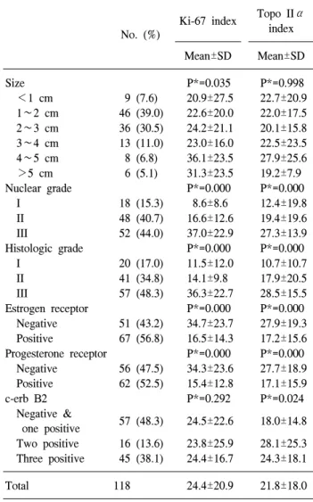
Table 2. Relationship between Ki-67 index and topoisomerase II α index and clinicopathologic data in breast carcinoma
No. (%)
Ki-67 index Topo IIα index
Mean±SD Mean±SD
Size P*=0.035 P*=0.998
<1 cm 9 (7.6) 20.9±27.5 22.7±20.9 1∼2 cm 46 (39.0) 22.6±20.0 22.0±17.5 2∼3 cm 36 (30.5) 24.2±21.1 20.1±15.8 3∼4 cm 13 (11.0) 23.0±16.0 22.5±23.5 4∼5 cm 8 (6.8) 36.1±23.5 27.9±25.6
>5 cm 6 (5.1) 31.3±23.5 19.2±7.9
Nuclear grade P*=0.000 P*=0.000
I 18 (15.3) 8.6±8.6 12.4±19.8
II 48 (40.7) 16.6±12.6 19.4±19.6
III 52 (44.0) 37.0±22.9 27.3±13.9
Histologic grade P*=0.000 P*=0.000
I 20 (17.0) 11.5±12.0 10.7±10.7
II 41 (34.8) 14.1±9.8 17.9±20.5
III 57 (48.3) 36.3±22.7 28.5±15.5
Estrogen receptor P*=0.000 P*=0.000
Negative 51 (43.2) 34.7±23.7 27.9±19.3 Positive 67 (56.8) 16.5±14.3 17.2±15.6
Progesterone receptor P*=0.000 P*=0.000
Negative 56 (47.5) 34.3±23.6 27.7±18.9 Positive 62 (52.5) 15.4±12.8 17.1±15.9
c-erb B2 P*=0.292 P*=0.024
Negative &
57 (48.3) 24.5±22.6 18.0±14.8 one positive
Two positive 16 (13.6) 23.8±25.9 28.1±25.3 Three positive 45 (38.1) 24.4±16.7 24.3±18.1
Total 118 24.4±20.9 21.8±18.0
*Spearman's Rho test.
Fig. 1. Immunohistochemical stain in breast carcinoma. (A) Ki-67 expression in breast carcinoma (×400), (B) c-erb B2 expression in breast carcinoma (×400), (C) topoisomerase IIα expression in breast carcinoma (×400).
결 과 1) 임상자료의 분석
총 118예의 유방암 환자의 평균연령은 49.9세였으며 오 른쪽 유방에서 56예(47.5%), 왼쪽 유방에서 62예(52.5%)였 고, 54예(45.8%)가 유방보존수술을 받았고, 16예(13.6%)가 유방전절제술을, 나머지 48예(40.7%)가 변형근치유방절제 술을 받아 유방보존수술의 빈도가 전체 환자의 절반에 가 까운 수치를 나타냈다.
감시림프절 생검은 전체 환자의 62예(52.5%)에서 감시림 프절 생검을 받았고, 생검예 중 25예(21.2%)에서 전이소견 이 있었다. 액와부림프절 곽청술은 전체 환자의 78예 (66.1%)에서 실시하였고, 그중 37예(31.4%)에서 전이가 있 었다(Table 1).
2) 병리조직학적 소견
종양의 크기는 평균 2.3 cm로 2 cm 이하의 종양이 반 이 상을 차지하였다. 미세석회화는 36예(30.5%)에서 동반한 것 으로 나타났다.
종양의 병리조직학적 형태는 침윤성 관암종이 115예 (97.5%)로 가장 많았다. 종양의 핵등급은 I등급이 18예(15.3%), II등급이 48예(40.7%) 그리고 III등급이 37예(31.4%)였고 조 직학적 등급은 I등급이 20예(17.0%), II등급이 41예(34.8%) 그리고 III등급이 57예(48.3%)였다(Table 1).
3) 면역조직화학염색 결과
118예 중 ER 양성은 67예(56.8%), PR 양성은 62예(52.5%) 였고, c-erb B2는 음성(negative and one positive)이 57예 (48.3%), 강양성(three positive)이 45예(38.1%) 그리고 중등도 양성(two positive)이 16예(13.6%)였다. 중등도 양성을 보인
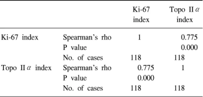
Table 3. Correlation expression of Ki-67 and topoisomerase IIα in breast carcinoma
Ki-67 Topo IIα
index index
Ki-67 index Spearman’s rho 1 0.775
P value 0.000
No. of cases 118 118
Topo IIα index Spearman’s rho 0.775 1
P value 0.000
No. of cases 118 118
16예에서는 HER-2/neu에 대한 형광제자리 부합검사는 실 시하지 않았다. Ki-67에 대한 면역조직화학염색에서 Ki-67 index는 최저 0점에서 최대 88.75점으로 평균 24.4점이었고, topo IIα는 최저 0점에서 최대 93.3점으로 평균 21.8점으로 나타났다(Table 2, Fig. 1A).
4) 면역조직화학검사 소견과 임상병리학적 소견과의 비교
Ki-67 index는 종양의 크기를 각 1 cm 간격으로 군을 나누 었을 때 종양의 크기가 증가함에 따라 Ki-67 index의 평균값 도 함께 증가하였고, 통계적으로 유의하였으나(P=0.035), topo IIα index는 종양의 크기가 증가함에도 불구하고 그 평 균값이 유사하여, 통계적으로 유의하지 않았다(P=0.998), (Table 2, Fig. 1B, C). 반면 종양의 크기를 2 cm 이하, 2 cm에 서 5 cm사이, 그리고 5 cm 이상의 군으로 나누었을 때 Ki-67 index와 topo IIα index 둘 다 통계적 의미를 찾을 수 없었다.
핵 등급이 증가함에 따라 Ki-67 index는 I등급에서 8.6±8.6, II등급에서 16.6±12.6, III등급에서 37.0±22.9로 증가하였고 topo IIα index도 핵등급이 증가함에 따라 index의 수치가 통계적으로 의미 있게 증가하였다(P=0.000) (Table 2, Fig.
1B, C). 조직학적 등급에서도 조직학적 등급이 높을수록 topo IIα index와 Ki-67 index가 c-erb B2는 Ki-67 index에서 음성군과 비교해서 양성군에서 현저하게 수치가 증가하지 않았지만, topo IIα index는 음성군보다 양성군에서 index의 수치가 의미 있게 증가하였다(P=0.024) (Table 2, Fig. 1B, C).
호르몬 수용체인 ER는 양성에서보다는 음성에서 topo IIα index와 Ki-67 index가 각각 27.9±19.3과 23.7±23.7으로 통계적 으로 의미 있게 높았고, PR에서도 topo IIα index와 Ki-67 in- dex가 각각 27.7±18.923.6과 34.3±23.6로 호르몬 수용체 음성 에서 통계적으로 의미 있게 높았다(P=0.000) (Table 2).
Ki-67과 topo IIα 발현의 상호 연관성에서 Ki-67 index가 증가할수록 topo IIα index가 증가하였고, 통계적으로 유의 하였다(P=0.000) (Table 3).
고 찰
HER-2/neu (c-erb B2) 종양유전자는 정상세포에서 단일 복제 형태로 있고, p185로 알려진 185 kD의 세포막 당 단백 이 암호화되어 있다. 이 p185의 세포 내 성분은 티로신 활성 효소를 가지고 있기 때문에 세포 내 성분의 40%와 세포 외 성분의 85%에서 표피성자인자 수용체와 구조적 상동성을 가진다.(9) 이러한 HER-2/neu 유전자의 증폭과 단백질의 과 발현은 유방암에서 현재 중요한 예후인자로 간주하고 있으 며, 최근 연구들에서 HER-2/neu 종양유전자의 증폭은 호르 몬이나 화학요법의 반응을 예측할 수 있는 유용한 인자로 알려져 있다.(10) 임상적으로 HER-2/neu 종양유전자가 중요 한 이유는 HER-2/neu 유전자 단백질의 세포 외 부분에 대한 단일 항체인 trastuzumab (HerceptinⓇ)이 새로운 항암제로 사 용이 증가할 것이 예측되며, 이 HerceptinⓇ은 HER-2/neu 종 양유전자의 증폭이나 과발현 시에만 효과가 있기 때문이 다.(11) 현재 유방암에서 HER-2/neu 종양유전자의 분석은 호르몬 수용체와 종양의 증식능과 함께 중요성이 강조되고 있다. HER-2/neu 유전자의 증폭이나 과발현은 14.6%에서부 터 42%까지 다양하게 보고되고 있으며,(12,13) 예후 인자로 서의 가치에 대한 보고 또한 다양하다.(14) 또 하나의 예후 를 예측할 수 있는 방법 중에 하나가 종양에서 세포의 증식 능을 평가하는 방법이다. 여러 가지 방법 중에서 PCNA나 MIB-1에 대한 면역조직화학적 염색이 널리 이용되고 있다.
MIB-1은 종양의 성장 분획을 측정하는 데 이용되는 Ki-67 의 단일클론항체이며, 이 Ki-67은 395 kDa와 354 kDa의 분 자량을 갖는 인간 핵의 비히스톤 단백과 반응하고,(15) 세 포주기의 G0기를 제외한 G1, S, G2와 M기에서 모두 발현된 다.(16) Ki-67을 이용한 세포 증식능은 많은 종양을 통하여 진단과 예후 인자로서의 가치가 있는 것으로 보고되어 있 다. 본 연구에서 Ki-67 index는 종양의 크기와 핵 등급 그리 고 조직학적 등급이 높을수록 Ki-67 index가 증가하였고, 통 계적으로 유의하였다. 이는 종양의 크기가 클수록, 병리조 직학적 등급이 나쁠수록 Ki-67 index 수치가 높아 예후가 나쁠 것으로 예측할 수 있을 것이다.
Ki-67에 대한 유용성에 대한 보고는 다양하며, 그 의미에 대한 분석 또한 저자마다 차이가 있었다. Wrba 등(17)은 Ki-67 index와 종양의 크기와 상관관계가 있다고 한 반면, Dettmar 등(18)은 종양의 크기와는 관계 없다는 보고를 하 였다. 또한 본 실험에서 각각 ER 음성, PR 음성일 때 Ki-67 index가 의미 있게 높게 나타났지만 이에 대한 보고도 저자 마다 차이가 나서, 상관관계에 대한 논란은 아직 남아 있는 것으로 볼 수 있다. c-erb B2의 경우에는 세포의 증식력과 상관관계가 있다는 논문도 있지만(19), 고 등(20)은 유의한
차이를 보이지 않았다고 보고하였고 본 실험에서도 c-erb B2는 Ki-67 index와 연관성이 없었다.
한편 새로운 증식표지자로 연구되고 있는 Topoisomerase 는 DNA 합성, 전사, 염색체 분리와 재결합과 같은 DNA 대 사의 중요 단계에서 DNA 형태를 변화시키는 효소로 알려 져 있으며,(21) 여기에는 크게 기능이 다른 topoisomerase I 과 II의 두 종류가 있다. Topoisomerase I은 DNA의 한 가닥 을 자르는 반면 topoisomerase II는 동시에 두 가닥을 자르며 그 기능의 수행을 위해 ATP를 필요로 한다. 포유류에는 top- oisomerase II의 두 isoform인 170 kDa의 α-isoform과 180 kDa 의 β-isoform이 존재하는데, 이들은 각각 염색체 17q12-21 와 3q24에 위치한다.(22) 이들은 서로 높은 상동성을 가짐에 도 불구하고 서로 독립적으로 조절되며 다른 성상과 기능 을 가진다고 알려져 있는데, 이 중 α-isoform은 anthracycline 계 항암제의 표적으로 작용하여 항암 치료에 있어 중요한 역할을 담당한다. Topoisomerase II 억제제에 대한 민감도는 표적세포에서 topo IIα의 표현 정도에 따라 달라지는데, topo IIα단백질의 농도가 낮을수록 topoisomerase II 억제제 에 대한 표적효소가 적어서 항암제에 대한 민감도가 낮다 고 한다.(23) 이러한 topo IIα는 정상세포와 조직에서 세포 증식의 표지자로 알려져 있으며,(24) 유방암종,(25,26) 대장 암종,(27) 요로상피암종,(28) 등에서의 연구 결과가 보고되 어 있다. 본 실험에서 topo IIα는 종양의 크기가 5 cm 이상 일 때 60.83으로 급격히 증가하였으나 그 예가 작아 통계적 인 의의는 없었다. 핵 등급은 고등급으로 갈수록 index 수치 가 증가하였고, 조직등급에서도 1등급은 10.67, 2등급은 17.93 그리고 3등급은 28.50으로 증가하여 통계적으로 유의 하였고 호르몬 수용체도 수용체의 발현이 없을 때 topo IIα index가 높게 나타나 통계적으로 유의하였다. Ki-67과는 달 리 topo IIα index는 c-erb B2의 발현과도 통계적으로 유의 하였다.
Ki-67과 topo IIα 발현의 상호 연관성에서, Ki-67 index가 증가할수록 topo IIα index가 증가하여 Ki-67의 세포 증식능 과 상호 연관성을 보였다. Nakopoulou 등(26)은 유방암종에 서 topo IIα의 발현이 핵 등급, 호르몬 수용체 상태, Ki-67 염색지수, p53 발현, HER-2/neu 단백질의 과발현과 연관성 이 있지만, 종양의 병기나 림프절 전이 등과는 연관성이 없 다고 보고하였다. 반면 Depowski 등(25)은 topo IIα의 증가 와 생존율, 병기, 림프절 전이, HER-2/neu의 증폭과 연관성 이 있으며, 종양의 크기, 등급, 호르몬 수용체 상태와는 연 관성이 없다고 하였다. 비록 본 실험에서 임상 및 병리조직 학적 예후 인자들간의 관련성을 통계적 데이터를 근거로 긍정적인 평가를 내릴 수 있겠지만, topo IIα 및 Ki-67이 각 예후 인자와의 관련성에 대해 주요 쟁점은, 현재 각 저자 마다 사용하고 있는 염색지수(index value)가 실험 방법마다 상이하며, 그 기준치 또한 광범위하게 협의된 바가 없기 때 문일 것으로 추측해 볼 수 있겠다. Topo IIα의 면역조직화
학 염색을 실시한 저자들 중 Nakopoulou 등(26)은 전체 종양 세포 중에서 10% 이상 염색이 되면 양성으로 판정하였고, Depowski 등(25)은 1% 이상을 양성으로 판정하였다. 본 실 험에서는 그 기준치를 정하지 않아 양성과 음성에 대한 각 예후 인자간에 관련성에 대한 자료는 만들지 않았다.
결 론
동아대학교 의료원 유방센터에서는 118명의 유방암 환자 를 대상으로 Topo IIα와 Ki-67의 단백질 발현을 분석한 결 과, Ki-67 index는 종양의 크기가 증가함에 따라 증가하였고 핵 등급과 조직학적 등급이 높을수록 Ki-67 index와 topo II α index도 의미 있게 증가하였다. c-erb B2는 topo IIα index 의 음성군보다 양성군에서 index가 증가하였다. 호르몬 수 용체인 ER/PR에서는 음성에서 topo IIα index와 Ki-67 index 가 높았고 Ki-67과 topo IIα 발현은 Ki-67 index가 증가할수 록 topo IIα index가 증가하였다. 즉 Ki-67 index와 topo IIα index는 종양의 크기가 클수록, ER/PR 음성일 때 의미 있게 증가하여 예후가 좋지 않음을 예측할 수 있을 것으로 보고, topo IIα index는 c-erb B2의 과발현과 연관성이 있는 것으 로 보아 항암제의 감수성을 예측해 볼 수 있을 것이다. 향후 면역염색과 topo IIα유전자에 대한 형광제자리부합법 등을 이용하여 topo IIα index와의 관계를 비교 분석해서, index 의 기준치를 표준화할 필요가 있을 것으로 생각하며, 이를 근거로 항암제 치료의 반응을 예측하는 데 기준이 될 수 있을 것으로 기대해 본다.
REFERENCES
1) Been ken SW, Grizzle WE, Crowe DR, Conner MG, Weiss HL, Sellers MT, et al. Mo55lecular biomarkers for breast can- cer prognosis: co expression of c-erbB-2 and p.53. Ann Surg 2001;233:630-8.
2) Lynch BJ, Guinee DG Jr, Holden JA. Human DNA top- oisomerase II-alpha: a new marker of cell proliferation in in- vasive breast cancer. Hum Pathol 1997;28:1180-8.
3) Hirabayashi S. Immunohistochemical detection of DNA top- oisomerase type II alpha and Ki-67 in adenoid cystic carcino- ma and pleomorphic adenoma of the salivary gland. J Oral Pathol Med 1999;28:131-6.
4) Harris LN, Yang L, Liotcheva V, Pauli S, Iglehart JD, Colvin OM, et al. Induction of topoisomerase II activity after ErbB2 activation is associated with a differential response to breast cancer chemotherapy. Clin Cancer Res 2001;7:1497-504.
5) Le Doussal V, Tubiana-Hulin M, Friedman S, Hacene K, Spyratos F, Brunet M. Prognostic value of histologic grade nu- clear components of Scarff-Bloom-Richardson (SBR). An im- proved score modification based on a multivariate analysis of 1262 invasive ductal breast carcinomas. Cancer 1989;64:1914-
21.
6) Elston CW, Ellis IO. Pathological prognostic factors in breast cancer. I. The value of histological grade in breast cancer: ex- perience from a large study with long-term follow-up.
Histopathology 1991;19:403-10.
7) Frierson HF Jr, Wolber RA, Berean KW, Franquemont DW, Gaffey MJ, Boyd JC, et al. Interobserver reproducibility of the Nottingham modification of the Bloom and Richardson histo- logic grading scheme for infiltrating ductal carcinoma. Am J Clin Pathol 1995;103:195-8.
8) Graziano C. HER-2 breast assay, linked to Herceptin, wins FDA's okay. CAP Today 1998;12:14-6.
9) Dean GS, Pusztai L, Xu FJ, O'Briant K, DeSombre K, Cona- way M, et al. Cell surface density of p185 (c-erbB-2) determi- nes susceptibility to anti-p185 (c-erbB-2)-ricin A chain (RTA) immunotoxin therapy alone and in combination with anti-p170 (EGFR)-RTA in ovarian cancer cells. Clin Cancer Res 1998;
4:2545-50.
10) Ross JS, Fletcher JA, Linette GP, Stec J, Clark E, Ayers M, et al. The Her-2/neu gene and protein in breast cancer 2003:
biomarker and target of therapy. Oncologist 2003;8:307-25.
11) Shak S. Overview of the trastuzumab (Herceptin) anti-HER2 monoclonal antibody clinical program in HER2-overexpressing metastatic breast cancer. Herceptin Multinational Investigator Study Group. Semin Oncol 1999;26:71-7.
12) Sutterlin MW, Haller A, Gassel AM, Peters K, Caffier H, Dietl J. The correlation of c-erbB-2 oncoprotein and estab- lished prognostic factors in human breast cancer. Anticancer Res 2000;20:5083-8.
13) Pinto AE, Andre S, Pereira T, Nobrega S, Soares J. C-erbB-2 oncoprotein overexpression identifies a subgroup of estrogen receptor positive (ER+) breast cancer patients with poor prognosis. Ann Oncol 2001;12:525-33.
14) Yamauchi H, Stearns V, Hayes DF. When is a tumor marker ready for prime time? A case study of c-erbB-2 as a predictive factor in breast cancer. J Clin Oncol 2001;19:2334-56.
15) Gerdes J, Li L, Schlueter C, Duchrow M, Wohlenberg C, Gerlach C, et al. Immunobiochemical and molecular biologic characterization of the cell proliferation-associated nuclear an- tigen that is defined by monoclonal antibody Ki-67. Am J Pathol 1991;138:867-73.
16) Popov Z, Hoznek A, Colombel M, Bastuji-Garin S, Lefrere- Belda MA, Bellot J, et al. The prognostic value of p53 nuclear overexpression and MIB-1 as a proliferative marker in transi- tional cell carcinoma of the bladder. Cancer 1997;80:1472-81.
17) Wrba F, Chott A, Reiner A, Reiner G, Markis-Ritzinger E, Holzner JH. Ki-67 immunoreactivity in breast carcinomas in relation to transferrin receptor expression, estrogen receptor status and morphological criteria. An immunohistochemical study. Oncology 1989;46:255-9.
18) Dettmar P, Harbeck N, Thomssen C, Pache L, Ziffer P, Fizi K, et al. Prognostic impact of proliferation-associated factors MIBI (Ki-67) and S-phase in node-negative breast cancer. Br J Cancer 1997;75:1525-33.
19) Barnes DM, Meyer JS, Gonzalez JG, Gullick WJ, Millis RR.
Relationship between c-erbB-2 immunoreactivity and thymi- dine labelling index in breast carcinoma in situ. Breast Cancer Res Treat 1991;18:11-7.
20) Ko CD, Kang HJ, Kim SW, Youn YK, Oh SK, Choe KJ, et al. Assessment of MIBI (Ki-67) labeling index and correlation with other well established prognostic factors in breast cancer.
J Korean Surg Soc 2001;60:361-7.
21) Berger JM, Gamblin SJ, Harrison SC, Wang JC. Structure and mechanism of DNA topoisomerase II. Nature 1996;379:225-32.
22) Drake FH, Hofmann GA, Bartus HF, Mattern MR, Crooke ST, Mirabelli CK. Biochemical and pharmacological properties of p170 and p180 forms of topoisomerase II. Biochemistry 1989;
28:8154-60.
23) Smith PJ, Soues S, Gottlieb T, Falk SJ, Watson JV, Osborne RJ, et al. Etoposide-induced cell cycle delay and arrest-de- pendent modulation of DNA topoisomerase II in small-cell lung cancer cells. Br J Cancer 1994;70:914-21.
24) Heck MM, Earnshaw WC. Topoisomerase II: a specific mark- er for cell proliferation. J Cell Biol 1986;103:2569-81.
25) Depowski PL, Rosenthal SI, Brien TP, Stylos S, Johnson RL, Ross JS. Topoisomerase II alpha expression in breast cancer:
correlation with outcome variables. Mod Pathol 2000;13:
542-7.
26) Nakopoulou L, Lazaris AC, Kavantzas N, Alexandrou P, Athanassiadou P, Keramopoulos A, et al. DNA topoisomerase II-alpha immunoreactivity as a marker of tumor aggressiveness in invasive breast cancer. Pathobiology 2000;68:137-43.
27) Staley BE, Samowitz WS, Bronstein IB, Holden JA. Expre- ssion of DNA topoisomerase I and DNA topoisomerase II-al- pha in carcinoma of the colon. Mod Pathol 1999;12:356-61.
28) Monnin KA, Bronstein IB, Gaffney DK, Holden JA. Eleva- tions of DNA topoisomerase I in transitional cell carcinoma of the urinary bladder: correlation with DNA topoisomerase II-alpha and p53 expression. Hum Pathol 1999;30:384-91.