구순구개열Vol. 5, No. 2 2002
A New Method of Synchronous Distraction Osteogene
이
s of the Maxilla and IVksndiblePill-Hoon Choung, Young-HoKang, Byoung-Moo Seo
Department of Oral and Maxillofacial Surgery, Maxillofacial Deformity Clinic, C이lege of Dentistry, Seoul National University, Seoul, Korea
상하악 동시 골 신장술 정필훈, 강영호, 서병무
서울대학교
치과대학 구강악안면외과학 교실,
악안면기형특수클리닉,
BI21
악안면
기형재건조직공학팀,
ERC편측 왜소증의 얼굴을 상하악과 하악 모두 신장할 필요가 있는 경우 하악을 신장하여 상악도 신장시키는 방법을 최근 소개한 바 있으나 본 논문에서는 오히려 상악을 신장시킬 때 하악도 같이 신장시키는 방법을 개발하여 좋은 결과를 얻었 다. 두 명의 편측안면 왜소증 환자에서 양악 동시에 골 신장술을 시행하였다. Ortiz Monasteries 방법과 달리 상악은 구내 르포트씨 제일 골절단술을 시행하였고, 하악은 구내 시상분할 골 절단술을 이용하여 골신장시 저항을 최소한으로 줄이려 하였다. 편방향 구내 골 신장기와 스플린트를 이용하여 양악 골신장술을 시행하였다. 5일의 잠재기 후 매일 1mm의 골신 장을 시행하였고 6주에서 8주간의 경화기를 둔 후, 골 신장기와 스플린트를 제거 하였다. 골신장 길이는 각각 13mm와 15mm이었고, 특이할 만한 부작용은 관찰되지 않았다. 이에 본 교실에서는 상악신장을 바탕으로 양악을 신장시키는 새로 운 술식을 보고하는 바이 다.
Keywords:distraction osteogenesis,maxilla,mandible
* This study was supportedbyagrantof theKoreaHealth21 R&D project, Ministryof Health &Welfare, Republic of Korea(00- PJ1-PG1-CH11-0004)
Introduction
In the field of osteodistraction of the craniofacial skeleton, Synder et al (1973) were the first to introduce elongation of the mandible in a dog experimental model using external device. McCarthy (1992) et al introduced unilateral mandibular elongation in patients with hemifacial microsomia which is reported or mandibular hypoplasia. Molina
구순구개 5:113~116, 2002
and Ortiz (1995) reported a series of cases of mandibular elongation using the corticotomy of the external surface of the mandible. Recently, Cohen (1999) introduced an intraoral distraction device to avoid external scarring. In hemifacial microsomia, the mandibular length elongation produces inevitable changes in dental occlusion. These ooclusal chages can be quickly corrected orthodontically in children, but not in adults. To solve this problem, Ortiz
113
Pill-Hoon Choung, Young-Ho Kang*, Byoimg-M。。Seo
Monasterio et al (1997) introduced simultaneous distraction of the mandible and maxilla using an incomplete LeFort I osteotomy on the maxilla.
In contrast to the Ortiz Monasterio* s technique, we performed a complete Le Fort I osteotomy and Intraoral Wrtico-Sagittal Ramus Osteotomy
Patients and Methods
Two patients with hemifacial microsomia were
treated by bimaxillary distraction osteogenesis in the Department of Oral and Maxillofacial Surgery of Seoul National University Hospital in 2002. A 7-year-
이d female with left hemifacial microsomia and 5-year- old male with right hemifacial microsomia. Ceph P- A/Lat, a panoramic view were taken preoperatively and postoperatively.
Distance from the horizontal plane at the level of the frontozygomatic suture, the zygomatic arch to the occlusal plane and length of the both ramus length
Hg. 1 Complete Le Fort I osteotomy and distractiondeviceapplication
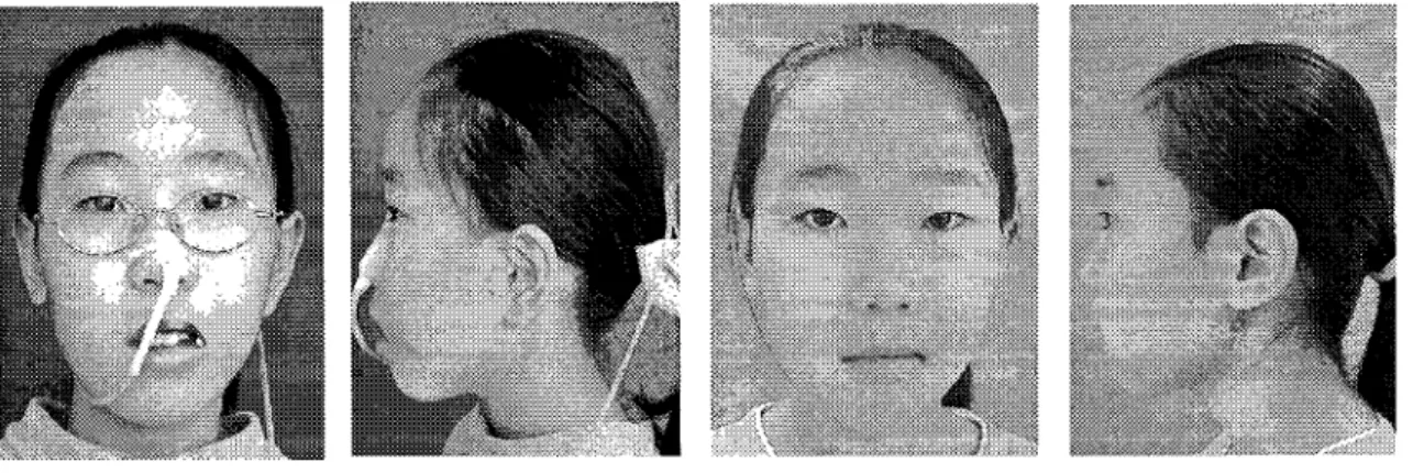
Fig.2 Case 1:7-yearoldgirl with left hemifacial microsomia
114
구순구개열 V이.5, No. 2 2002
Fig 3Case 1
Right pictures are pre-op and left arepost-op M-phto
Fig.4Case 2: 5-year oldboy with right hemifacialmicrosomia
Fig. 5 Case 2
Right pictures are pre-op and left are post-op M-phto
115
미l-Ho이] 아)oung, Young-Ho Kang*, Byoung-Moo Seo
and mi이 ine of maxilla and mandible were measured and recorded on the ceph PA. With a reciprocating saw, a complete horizontal osteotomy was made between the piriform aperture and the maxillary tuberosity and with curved chisel, both pterygomaxillary junction were freed. At mandible, Intraoral Vfertico-Sagittal Ramus Osteotomy was done at affected area.
Intraoral unidirectional distraction device (Martin Co.) and surgical wafer were used to perform bimaxillary distraction. After a latent period of 5days, distraction was performed at a rate 1mm per day.
After a consolidation period of 6-8 weeks , the wafer and distraction device were removed. The distraction distance(13 and 15mm) were obtained using one intraoral distraction device without complication.
Discussion
All the patients showed no complication, there was no infection, also any reoperation.
The patients with allogenic cranial bone grafting shows good initial stability, decrease of donor site morbidity and relapse tendency like autogenous bone grafting : The mean advancement was 4.97mm (SD 1.26) in group 1 and 5.03mm (SD 0.77) in group 2, as meas나!ed on the SN perpendicular line by IS. The mean relapse was 0.63mm (SD 0.43) in group 1 and 0.65mm (SD 0.35) in group 2 respectively. There was no statistical difference between both allogenic and
autogenic group by Mann-Whitney U test (p = 0.907).
And more relapse in Le Fort I and II combined surgery was observed, which was estimated to relapse of Le Fort I osteotomy.
References
1. Louis PJ, Waite PD, Austin RB. Long term skelet시 stability after rigid fixation of Le Fort I osteotomy with advancement. International Journal of Oral and Maxillofacial Surgery. 22:8- 86.1993
2. Baker DL, Stoelinga PJ, Blijdorp PA, Brouns JJ.
Long term stability after inferior maxillary repositioning by miniplate fixation. International Journal of Oral and Maxillofacial Surgery.
21:320-326.1992
3. de M이 van Otterloo JJ, Tuinzing DB, Kostense P Inferior positioning of the maxilla by Le Fort I osteotomy : a review of 25 patients with vertical maxillary deficiency. Journal of Cranio-Maxillo Facial Surgery. 24:69-77.1996
4. Emil WS. Variation of the Le Fort U osteotomies for Correction of Midfacial Deformities. Journal of Maxillofacial Surgery. 8:258-265.1980 5. Luyk NH, Ward-Booth RP. The stability of Le
Fort I advancement osteotomy using bone plates without bone grafts. Journal of Maxillofacial Surgery. 13:250-253.1985
저자 연락처
서을시 종로구 연건동28번지 서을대학교 치과대학 구강악안면외과 정필훈 우편번호)110-744 전화:02-760-3477 E-mail: [email protected]
116