• 검색 결과가 없습니다.

Geometrical design characteristics of orthodontic mini-implants predicting maximum insertion torque

N/A
N/A
Protected

Academic year: 2022

Share "Geometrical design characteristics of orthodontic mini-implants predicting maximum insertion torque"

Copied!
7
0
0

로드 중.... (전체 텍스트 보기)

전체 글

(1)

Geometrical design characteristics of orthodontic mini-implants predicting maximum insertion torque

Objective: To determine the unique contribution of geometrical design cha-

rac teristics of orthodontic mini-implants on maximum insertion torque while controlling for the influence of cortical bone thickness.

Methods: Total

num ber of 100 cylindrical orthodontic mini-implants was used. Geo met- rical design characteristics of ten specimens of ten types of cylindrical self- drilling orthodontic mini-implants (Ortho Easy®, Aarhus, and Dual Top

TM

) with diameters ranging from 1.4 to 2.0 mm and lengths of 6 and 8 mm were measured. Maximum insertion torque was recorded during manual insertion of mini-implants into bone samples. Cortical bone thickness was measured.

Retrieved data were analyzed in a multiple regression model. Results: Significant predictors for higher maximum insertion torque included larger outer diameter of implant, higher lead angle of thread, and thicker cortical bone, and their unique contribution to maximum insertion torque was 12.3%, 10.7%, and 24.7%, respectively. Conclusions: The maximum insertion torque values are best controlled by choosing an implant diameter and lead angle according to the assessed thickness of cortical bone.

[Korean J Orthod 2014;44(4):177-183]

Key words: Orthodontic mini-implant, Mini-implant stability, Mini-implant

orthodontic anchorage, Implant insertion torque

Višnja Katića Ervin Kamenarb David Blaževićb Stjepan Špalja

aDepartment of Pediatric Dentistry and Orthodontics, School of Medicine, University of Rijeka, Rijeka, Croatia

bDepartment of Mechanical Engineering Design, Faculty of Engineering, University of Rijeka, Rijeka, Croatia

Received July 19, 2013; Revised October 14, 2013; Accepted November 15, 2013.

Corresponding author: Višnja Katić.

Research Assistant, Department of Orthodontics, School of Medi cine, University of Rijeka, Kresimirova 40, HR-51000 Rijeka, Croatia.

Tel +385-51-345-636 e-mail [email protected]

*The affiliation of Višnja Katić and Stjepan Špalj was recently changed. Present affiliation is “Dapartment of Orthodontics, School of Medicine, University of Rijeka, Rijeka, Croatia".

©

2014 The Korean Association of Orthodontists.

The authors report no commercial, proprietary, or financial interest in the products or companies described in this article.

This is an Open Access article distributed under the terms of the Creative Commons Attribution Non-Commercial License (http://creativecommons.org/licenses/by-nc/3.0) which permits unrestricted non-commercial use, distribution, and reproduction in any medium, provided the original work is properly cited.

http://dx.doi.org/10.4041/kjod.2014.44.4.177

(2)

INTRODUCTION

Versatile temporary anchorage device (TAD) systems offer fixed anchorage to improve the efficiency of orthodontic treatment. Orthodontic mini-implants are proven to be very helpful as temporary skeletal anchorage, particularly in patients with low compliance or insufficient dental structures.

1,2

Miniscrew-type implants are useful because they are easy to insert and to remove in a wide range of areas, their surgical implantation is less traumatic, they can be used for various purposes, immediate orthodontic loading is possible, and their associated cost is low.

3,4

Successful orthodontic treatment greatly depends on appropriate, stable anchorage. It was reported that mini-implants have a lower success rate (80–85%) than osseointegrated implants (91.00% and 97.81%, for maxilla and mandible, respectively).

5

The displacement of a screw can occur due to inflammation of surrounding tissue or as a result of a poor bone-screw interface.

Suggested factors that cause degeneration of the bone at the implant-tissue interface include excessive insertion tor que, heat at the border between the screw and bone, and mechanical injury.

6

Therefore, the recommended optimal maximum insertion torque (MIT) values between 50−100 Nmm help clinicians improve clinical results.

7

The initial implant stability is important for clinical success if TADs are to be loaded immediately.

6

In cases where primary stability is not achieved during insertion, the mini-implant cannot be engaged in therapy for the next 6−8 weeks, which prolongs orthodontic treatment.

Many studies have been conducted to analyze MIT with respect to the diameter and length of the mini- screw.

8-10

Manufacturers offer their TADs mainly with handheld screwdrivers (Figure 1), which widens their applicability. The intentions of this study are to obtain data in conditions similar to those of routine in-office procedures and to determine critical factors of the screw design that could be controlled to avoid failure of TADs in a manual insertion procedure. Our goal is to determine the unique contribution of individual geo-

metrical design characteristics of orthodontic mini- implants—thread length and outer diameter, depth/

pitch ratio (thread shape factor [TSF]), and lead angle of thread—on MIT while controlling for the influence of cortical bone thickness.

We hypothesize that diameter, TSF, and lead angle of the thread are significant predictors of MIT.

MATERIALS AND METHODS

The following ten cylindrical self-drilling orthodontic mini-implant types with a bracket-like head (all made of titanium alloy grade 5, Ti-6Al-4V) were investigated in this study (Figure 2): Ortho Easy® (FORESTADENT®, Pforzheim, Germany), 1.7 × 6 mm and 1.7 × 8 mm;

Aarhus Anchorage System (MEDICON eG, Tuttlingen, Germany), 1.5 × 6 mm and 1.5 × 8 mm; and Jeil Dual Top

TM

Anchor System (Jeil Medical Corp., Seoul, Korea), 1.4 × 6 mm, 1.6 × 6 mm, 2.0 × 6 mm, 1.4 × 8 mm, 1.6

× 8 mm, and 2.0 × 8 mm.

Ten specimens of each mini-implant type were tested.

Precise measurement of all orthodontic miniscrews was conducted using the Olympus SZX16® optical stereo- microscope (Olympus Corporation, Tokyo, Japan) with 8× and 20× magnifications. Each microscopic sample was photographed alongside a calibrated chart, and the measurement was read on photographs. Length, outer diameter, depth, pitch, and lead angle of the screw thread were the geometrical design characteristics mea- sured in this study (Figure 3). All implants were found to have an asymmetric overall thread shape type. TSF was calculated as the ratio of thread depth to pitch for each miniscrew.

One hundred orthodontic mini-implants were manually inserted perpendicular to the bone surface using the handheld screwdriver of the respective mini-implant system without pre-drilling of a pilot hole. Each mini- implant was screwed into one bone sample. The inser-

Figure 1. Orthodontic mini-implant with handheld screwdriver.

Figure 2. Tested mini-implants (from left to right): Dual

Top™ (Jeil Medical Corp., Seoul, Korea) 1.4 × 6 mm, and

1.4 × 8 mm; Aarhus (MEDICON eG, Tuttlingen, Germany)

1.5 × 6 mm, and 1.5 × 8 mm; Dual Top™ 1.6 × 6 mm, and

1.6 × 8 mm; Ortho Easy® (FORESTADENT®, Pforzheim,

Germany) 1.7 × 6 mm, and 1.7 × 8 mm; Dual Top™ 2.0 ×

6 mm, and 2.0 × 8 mm.

(3)

tion torque was measured for the duration of the inser- tion up to the implant collar, and measured values of MIT (expressed as Nmm) were digitalized and saved using PicoLog® 5.14.6 (Pico® Technology Ltd., St. Neots, UK) data acquisition software. The torque-measuring device (Institut IGH, Zagreb, Croatia) consisted of a fixed body with four sensors connected to a high resolution PicoScope® oscilloscope (Pico® Technology Ltd.) and a custom made docking unit for the bone samples (Figure 4).

Each bone sample measuring 2 × 1.5 cm in size was prepared from the spinal aspect of swine ribs. Fresh swine ribs were deperiosted, cut into bone samples with a water-cooling surgical engine, and stored in phy siological solution at 4

o

C until further evaluation.

Cor tical bone thickness was measured at both cross- sectioned ends of each bone sample using a digital sliding caliper (Levior s.r.o., Prerov, Czech Republic) with an accuracy of ± 0.03 mm. The mean value of the cortical bone thickness measurements of each sample was used for further statistical analysis.

Figure 4. Torque-measuring device with mounted bone sample.

Figure 3. Geometrical design characteristics of mini- implants (length [a], outer diameter [b], depth [c], pitch [d], and lead angle [e] of the thread).

Table 1. Geometrical design characteristics of orthodontic mini-implants assessed with optical stereomicroscope

Implant type

(diameter × length, mm)

Outer thread

diameter (mm) Depth of the

thread (mm) Pitch of the

thread (mm) Thread shape

factor Lead angle of

thread (o) Thread length (mm) Aarhus (1.5 × 6) 1.545 ± 0.015 0.257 ± 0.005 0.685 ± 0.005 0.375 ± 0.007 8.034 ± 0.102 5.973 ± 0.087 Aarhus (1.5 × 8) 1.528 ± 0.014 0.253 ± 0.006 0.696 ± 0.007 0.364 ± 0.007 8.246 ± 0.057 8.014 ± 0.068 Ortho Easy (1.7 × 6) 1.683 ± 0.027 0.325 ± 0.015 0.819 ± 0.011 0.397 ± 0.018 8.804 ± 0.171 5.856 ± 0.019 Ortho Easy (1.7 × 8) 1.668 ± 0.017 0.322 ± 0.008 0.813 ± 0.009 0.397 ± 0.010 8.815 ± 0.150 7.889 ± 0.020 Dual Top (1.4 × 6) 1.370 ± 0.000 0.198 ± 0.004 0.660 ± 0.014 0.299 ± 0.001 8.718 ± 0.184 6.025 ± 0.007 Dual Top (1.4 × 8) 1.370 ± 0.028 0.180 ± 0.007 0.660 ± 0.014 0.273 ± 0.017 8.722 ± 0.361 8.010 ± 0.071 Dual Top (1.6 × 6) 1.572 ± 0.015 0.255 ± 0.008 0.721 ± 0.011 0.354 ± 0.012 8.307 ± 0.133 6.028 ± 0.039 Dual Top (1.6 × 8) 1.558 ± 0.019 0.255 ± 0.011 0.733 ± 0.007 0.348 ± 0.016 8.512 ± 0.139 8.014 ± 0.076 Dual Top (2.0 × 6) 1.990 ± 0.014 0.330 ± 0.007 0.835 ± 0.007 0.395 ± 0.012 7.608 ± 0.117 6.045 ± 0.007 Dual Top (2.0 × 8) 1.950 ± 0.000 0.318 ± 0.011 0.835 ± 0.007 0.380 ± 0.016 7.762 ± 0.065 7.975 ± 0.021 Values are presented as mean ± standard deviation.

Thread shape factor means depth/pitch ratio.

Aarhus, Aarhus Anchorage System (MEDICON eG, Tuttlingen, Germany); Ortho Easy, Ortho Easy® (FORESTADENT®, Pforzheim, Germany); Dual Top, Jeil Dual TopTM Anchor System (Jeil Medical Corp., Seoul, Korea).

(4)

Institutional review board approval was not needed for this study.

Using MIT as the outcome, Pearson's bivariate corre- lation coefficient was used to measure the strength of the association between screw design parameters and cortical thickness when not controlling for the in- fluence of other variables. A multiple linear regression analysis was used to determine the unique contribution of individual geometrical design characteristics of ortho- dontic mini-implants in predicting the MIT while con- trolling for other design variables and cortical bone thickness. The presence of multicollinearity was eva- luated with the help of a variance inflation factor (VIF) and tolerance. Statistical analyses were carried out

using SPSS® version 10.0 statistics software (SPSS Inc., Chicago, IL, USA). Statistical significance was preset to p

< 0.05.

RESULTS

Geometrical design characteristics of tested orthodontic miniscrews assessed using the optical stereomicroscope are shown in Table 1. Cortical thickness of bone samples ranged from 0.7−3.1 mm (mean 1.5 ± 0.4 mm). The MIT values were between 50.86−347.49 Nmm (122.01 ± 49.03 Nmm).

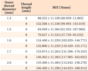
Descriptive statistics for MIT with length and outer dia meter of the screw thread evaluated at covariate cor- tical bone thickness (1.5190 mm) are shown in Table 2.

In bivariate correlations, MIT was significantly posi- tively correlated with the depth of the thread (r = 0.413;

p < 0.001), the pitch of the thread (r = 0.402; p < 0.001), TSF (r = 0.403; p < 0.001), the outer diameter of the implant thread (r = 0.398; p < 0.001), and cortical bone thickness (r = 0.409; p < 0.001), whereas it was not significantly correlated with the length of the implant thread or the lead angle of the thread.

The multiple linear regression model showed that while controlling for other variables, significant predictors for higher MIT were a larger implant diameter (p < 0.001), a higher lead angle (p < 0.001), and thicker cortical bone (p < 0.001; Table 3). The unique contribution of these factors in accounting for the MIT variability was 12.3, 10.7, and 24.7%, respectively. The whole model accounted for 44.2% of the variability in MIT.

DISCUSSION

In-office decision making regarding selection of a specific implant type is related to host characteristics, namely, bone density and cortical thickness,

11

and choo- sing the most appropriate geometrical screw design Table 2. Descriptive statistics for maximum insertion

torque with length and outer diameter of the screw thread evaluated at covariate cortical bone thickness*

Outer thread diameter

(mm)

Thread length

(mm) MIT (Nmm)

1.4 6 88.552 ± 11.330 (66.039−11.065) 8 122.506 ± 11.330 (99.993−145.019) 1.5 6 84.949 ± 11.584 (61.932−107.966) 8 70.637 ± 11.524 (47.739−93.535) 1.6 6 124.400 ± 11.259 (102.029−146.771)

8 132.800 ± 11.259 (110.429−155.171) 1.7 6 153.974 ± 11.263 (131.596−176.353) 8 140.363 ± 11.303 (117.905−162.822) 2.0 6 135.460 ± 11.484 (112.642−158.278) 8 166.469 ± 11.296 (144.023−188.914) Values are presented as mean ± standard error (95% confi- dence interval).

*1.5190 mm.

Table 3. Multiple linear regression model for prediction of maximum insertion torque

Unstandardized

coefficients (B) SE Standardized coefficients

(Beta) Significance Correlations

Zero-order Partial Part

Constant −757.934 146.055 < 0.001

Thread length 0.957 3.765 0.020 0.800 0.113 0.026 0.019

Outer thread diameter 233.475 50.075 0.985 < 0.001 0.398 0.433 0.350

Lead angle 59.091 13.557 0.497 < 0.001 −0.050 0.410 0.327

TSF −242.796 148.766 −0.285 0.106 0.403 −0.166 −0.123

Cortical bone thickness 58.310 8.805 0.527 < 0.001 0.409 0.564 0.497

R = 0.685; R2 = 0.470; adjusted R2 = 0.442; F = 16.662; p < 0.001.

By multiple linear regression analysis.

SE, Standard error; TSF, thread shape factor.

(5)

combination will enhance the primary stability of the TAD and thus, the success of orthodontic therapy.

Our study showed high vari ability in the MIT in the manual procedure of miniscrew insertion but revealed that significant unique contributors to higher MIT values were thicker cortical bone, a larger outer implant thread diameter, and a higher thread lead angle.

Similar conclusions regarding cortical bone thickness and implant diameter as important predictors for MIT are found in other studies.

8-10,12

It has been noted that overall implant shape (conical vs. cylindrical) influences MIT,

12

but the unique predictive value of specific geo- metrical design characteristics has not been analyzed in detail previously.

In bivariate correlations, statistically significant corre- lations were found between MIT and the depth of the thread, the pitch of the thread and TSF, but not between MIT and lead angle of the thread.

These results indicated an association between two variables without controlling for the influence of other possible cofactors. Therefore, a multiple regression model was included to control for the influence of other possible cofactors. This advanced model established no significant predictive value for TSF but strengthened the predictive value of cortical bone thickness and confirmed a positive correlation with lead angle, whose unique contribution in the prediction of MIT was 10.7%. This multiple regression model explains only 44.2% of the MIT prediction, leaving a large portion of the variability of MIT unaccounted for. We used the forced-entry regression method, where all predictors are forced into the model simultaneously. This method is appropriate for theory testing

13

because the experimenter does not determine the entrance order of variables. Predictors are chosen based on previous studies and analysis of inter- correlations and multicollinearity.

9,10,12,14,15

Further studies might include bone density, angle of implantation, and changes in implant design when trying to combine the best individual features to maximize implant efficiency.

During the statistical analysis, we encountered the pro blem of multicollinearity because the lead angle cor- related with the outer diameter and pitch of the thread, TSF correlated to depth and pitch of the thread, and the depth of the thread correlated with the outer dia- meter. Therefore, depth and pitch of the thread were not accounted for in the multiple regression analysis;

instead, TSF was taken into account as a combined value of both variables. The depth/pitch ratio (TSF) is not linearly related to changes in lead angle. It seems that higher lead angle improves cutting efficiency during implantation because the lead defines the axial travel for a single revolution,

15

whereas a larger thread surface (represented through TSF) is more resistant to pullout forces.

8,9

Values for the length and outer diameter of the screw thread are provided by every manufacturer, but the manu facturer’s data regarding depth, pitch, and lead angle of the thread are rarely provided. Results of all mea sured geometrical design characteristics show very small variations within the same screw type, making their use highly recommended. It should be noted that the actual size of the outer thread diameter of the tested Aarhus Anchorage System mini-implants (1.5 × 6 mm; 1.5 × 8 mm) is close to the diameter of the tested Dual Top

TM

mini-implants (1.6 × 6 mm; 1.6 × 8 mm);

thus, using mini-implants of different sizes may not be so different in actual in-office work. The greater value of the larger implant should be taken into account when implanting near tooth roots.

All implantations were done manually with no pilot hole because all of the manufacturers recommend direct implantation for self-drilling mini-implants in most bony areas in the oral cavity, except for mandibular and palatal areas, where use of a pilot drill or cortical bone punch is recommended because of the presence of dense cortical bone. Many researchers recommend insertion without a guide hole for self-drilling implants with diameters up to 1.6 mm.

7

Others state that a diameter smaller than 1.5 mm significantly increases the risk for implant fracture.

16

Several studies showed that self-drilling mini-implants have better primary stability when inserted without a pilot hole.

2,14

The majority of routine in-office orthodontic mini-implants are placed manually. Additionally, the measurement of MIT is not possible in every situation, and orthodontic implantation often proceeds until no thread is visible and the transmucosal neck is seated in the surrounding soft tissue. Therefore, practitioners have only one “mea- sure” for primary stability available, which is feeling the resistance, especially the final resistance, of the bone

Figure 5. Representative curve for manual insertion of

orthodontic mini-implants.

(6)

during insertion, which corresponds to the MIT. Results for the MIT in this study are somewhat higher compared to previously recommended insertion torques

7

but are far below the fracture torque values of the respective mini-implant types shown in another study.

17

These values are in agreement with the recommendations for using a pilot hole for larger diameters in order to achieve better long-term stability (secondary stability) because excessive insertion forces could cause later bone necrosis.

9

The representative curve for manual insertion of orthodontic mini-implants shows oscillations in insertion torque as a result of the repositioning of the screwdriver in the surgeon’s hand (Figure 5). Inter- mittent force,

18

lower speed,

19

and smaller cortical thick- ness

20

reduce the chance for heat production at the border between the screw and bone, which is a known factor for the failure of implantation. On the other hand, during manual implantation, excessive insertion tor que and mechanical injury can be controlled only by the ope rator’s subjective judgment. Although the motor- driven method of insertion showed better results, it is not recommended for beginners and is more complicated and expensive overall.

21

Therefore, one should control MIT by combining data from the implantation site (av ailable space, cortical bone thickness) and implant design characteristics.

It is widely accepted that MIT is an important and measurable factor of primary stability of orthodontic miniscrews

6,14

because other measurements described in the literature−radiological examination, Periotest®

(Medizintechnik Gulden, Modautal, Germany), and removal torque

8

−deliver insufficiently precise measurements or have limited areas of application,

22

while the resonance frequency analysis (RFA) by Osstell

TM

(Integration Diag- nostics Ltd., Goteborgsvagen, Sweden) is currently not applicable for orthodontic mini-implants.

23

This study and others

23-26

used swine ribs because their ratio between cortical and spongious bone is similar to those in the alveolar processes of the maxilla and mandible. The cortical bone thickness was on average 1.5 ± 0.4 mm, which represents variations in tested biological material similar to the range in human jaw bones and enables analysis of the influence of variability of cortical bone thickness on overall MIT values.

11,25

The primary stability of a miniscrew is important for im mediate loading, and because it is intended for tem- porary use, it is desirable to achieve good primary stability.

6,7,9

Findings from this study indicate possible choices regarding implant design and host characteristics in order to achieve proper primary stability without compromising long-term stability because all measure- ments were conducted in simulated in-office conditions.

Our analysis of various implant types showed that lead angle may be an important factor of MIT values,

so manufacturers should consider increasing the lead angle of the implant to improve the implant design and achieve better primary stability in cases where the operator cannot use a larger implant diameter.

CONCLUSION

The MIT values can be controlled by choosing an implant diameter and lead angle of the thread according to the assessed thickness of cortical bone and the available space.

ACKNOWLEDGMENTS

A part of tested mini-implants was donated by the manufacturers FORESTADENT, MEDICON eG, and Jeil Medical Corp., to whom authors hereby thank for the donation.

This research is part of the University of Rijeka research project 13.06.2.1.53, principal investigator Stjepan Špalj.

REFERENCES

1. Kanomi R. Mini-implant for orthodontic anchorage.

J Clin Orthod 1997;31:763-7.

2. Motoyoshi M. Clinical indices for orthodontic mini- implants. J Oral Sci 2011;53:407-12.

3. Tseng YC, Hsieh CH, Chen CH, Shen YS, Huang IY, Chen CM. The application of mini-implants for orthodontic anchorage. Int J Oral Maxillofac Surg 2006;35:704-7.

4. Poggio PM, Incorvati C, Velo S, Carano A. “Safe zones": a guide for miniscrew positioning in the maxil lary and mandibular arch. Angle Orthod 2006;

76:191-7.

5. Cheng SJ, Tseng IY, Lee JJ, Kok SH. A prospective study of the risk factors associated with failure of mini-implants used for orthodontic anchorage. Int J Oral Maxillofac Implants 2004;19:100-6.

6. Meredith N. Assessment of implant stability as a prognostic determinant. Int J Prosthodont 1998;11:

491-501.

7. Motoyoshi M, Hirabayashi M, Uemura M, Shimizu N.

Recommended placement torque when tightening an orthodontic mini-implant. Clin Oral Implants Res 2006;17:109-14.

8. Pithon MM, Nojima MG, Nojima LI. In vitro evalua- tion of insertion and removal torques of ortho dontic mini-implants. Int J Oral Maxillofac Surg 2011;

40:80-5.

9. Wilmes B, Drescher D. Impact of bone quality,

implant type, and implantation site preparation on

insertion torques of mini-implants used for ortho-

dontic anchorage. Int J Oral Maxillofac Surg 2011;

(7)

40:697-703.

10. Lim SA, Cha JY, Hwang CJ. Insertion torque of orthodontic miniscrews according to changes in shape, diameter and length. Angle Orthod 2008;78:

234-40.

11. Xu Z, Zhao L, Wu Y, Wei X, Wang J, Tang N, et al.

Histomorphometric and biomechanical analyses of the osseointegration of loaded orthodontic micro- screws inserted at different cortical bone thickness sites. Oral Surg Oral Med Oral Pathol Oral Radiol 2012. doi: 10.1016/j.oooo.2012.06.007. [Epub ahead of print]

12. Pithon MM, Nojima MG, Nojima LI. Primary stability of orthodontic mini-implants inserted into maxilla and mandible of swine. Oral Surg Oral Med Oral Pathol Oral Radiol 2012;113:748-54.

13. Studenmund AH, Cassidy HJ. Instructor's manual to accompany. Using econometrics, a practical guide.

Boston: Little Brown; 1987.

14. Miyawaki S, Koyama I, Inoue M, Mishima K, Suga- hara T, Takano-Yamamoto T. Factors associated with the stability of titanium screws placed in the posterior region for orthodontic anchorage. Am J Orthod Dentofacial Orthop 2003;124:373-8.

15. American Gear Manufacturers Association, American National Standards Institute. Gear nomenclature, definitions of terms with symbols: AGMA standard.

Alexandria: American Gear Manufacturers Associa- tion; 2005.

16. Dalstra M, Cattaneo PM, Melsen B. Load transfer of miniscrews for orthodontic anchorage. Orthodontics 2004;1:53-62.

17. Wilmes B, Panayotidis A, Drescher D. Fracture resistance of orthodontic mini-implants: a biome- chanical in vitro study. Eur J Orthod 2011;33:396- 401.

18. Kondo S, Okada Y, Iseki H, Hori T, Takakura K, Koba yashi A, et al. Thermological study of drilling bone tissue with a high-speed drill. Neurosurgery 2000;46:1162-8.

19. Brisman DL. The effect of speed, pressure, and time on bone temperature during the drilling of implant sites. Int J Oral Maxillofac Implants 1996;11:35-7.

20. Toews AR, Bailey JV, Townsend HG, Barber SM. Ef- fect of feed rate and drill speed on temperatures in equine cortical bone. Am J Vet Res 1999;60:942-4.

21. Kim JS, Choi SH, Cha SK, Kim JH, Lee HJ, Yeom SS, et al. Comparison of success rates of orthodontic mini-screws by the insertion method. Korean J Orthod 2012;42:242-8.

22. Cha JY, Yu HS, Hwang CJ. The validation of Periotest values for the evaluation of orthodontic mini- implants' stability. Korean J Orthod 2010;40:167- 75.

23. Aparicio C, Lang NP, Rangert B. Validity and clinical significance of biomechanical testing of implant/

bone interface. Clin Oral Implants Res 2006;17 (Suppl 2):2-7.

24. Kim SJ, Yoo J, Kim YS, Shin SW. Temperature change in pig rib bone during implant site prepa- ration by low-speed drilling. J Appl Oral Sci 2010;

18:522-7.

25. Marković A, Mišić T, Miličić B, Calvo-Guirado JL, Aleksić Z, Ðinić A. Heat generation during implant placement in low-density bone: effect of surgical technique, insertion torque and implant macro de- sign. Clin Oral Implants Res 2013;24:798-805.

26. Friberg B, Sennerby L, Roos J, Johansson P, Strid

CG, Lekholm U. Evaluation of bone density using

cutting resistance measurements and microradio-

graphy: an in vitro study in pig ribs. Clin Oral Im-

plants Res 1995;6:164-71.

수치

Figure 2. Tested mini-implants (from left to right): Dual  Top™ (Jeil Medical Corp., Seoul, Korea) 1.4 × 6 mm, and  1.4 × 8 mm; Aarhus (MEDICON eG, Tuttlingen, Germany)  1.5 × 6 mm, and 1.5 × 8 mm; Dual Top™ 1.6 × 6 mm, and  1.6 × 8 mm; Ortho Easy® (FOREST
Table 1. Geometrical design characteristics of orthodontic mini-implants assessed with optical stereomicroscope  Implant type
Table 3. Multiple linear regression model for prediction of maximum insertion torque Unstandardized
Figure 5. Representative curve for manual insertion of  orthodontic mini-implants.

참조

관련 문서

Torque &amp; power delivered by hydraulic motors Torque &amp; power delivered by hydraulic motors Performance of hydraulic motors.. Comparison of variable performance

For an exacting investigation of the distance from root apex to outer surface of buccal cortical bone of mandibular first molar with 2R2C (2 separate

3 Mean percentages of bone implant contact ratio in the control group and experimental groups at 6 and 12 weeks after placement of the

In particular, the dependences of maximum temperature increase and decay time constant on the laser pulse width, beam diameter, and absorption coefficient

By analysing the dependences of the maximum increase in temperature and the maximum acceleration on the medium properties and the information of the damage

Evaluating parameters of osseointegrated dental implants using finite element analysis-a two- dimentional comparative study examining the effects of implant

Histologic evaluation of early human bone response to different implant surfaces2. Histologic evaluation of human bone integration on machined and

Greco-Roman wrestlers of high school, college, and the professionals players showed significant differences depending on their performance when measuring the maximum muscle