aFormer Resident, bProfessor, cAssistant Professor, dFull-time Lecturer, Department of Orthodontics, Dental Hospital, East-West Neo Medical Center.
Corresponding author: Jong-Hyun Nahm.
Department of Orthodontics, Dental Hospital, East-West Neo Medical Center, 149, Sangil-dong, Gangdong-gu, Seoul 134- 727, Korea.
+82 2 440 6205; e-mail, [email protected].
Received January 30, 2008; Last Revision June 8, 2009;
Accepted June 13, 2009.
DOI:10.4041/kjod.2009.39.4.248
Effects of compressive stress on the expression of M-CSF, IL-1β, RANKL and OPG mRNA in periodontal ligament cells
Ji-Woong Kim, DMD, MSD,a Ki-Soo Lee, DMD, MSD, PhD,b Jong-Hyun Nahm, DMD, MSD, PhD,c Yoon-Goo Kang, DMD, MSD, PhDd
Objective: The aim of this study was to determine if human PDL cells can produce osteoclastogenic mRNA and examine how compressive stress affects the expression of osteoclastogenic mRNA in human PDL cells. Methods: Human PDL cells were obtained from biscupids extracted for orthodontic treatment. The compressive force was adjusted by increasing the number of cover glasses. PDL cells were subjected to a compressive force of 0.5, 1.0, 2.0, 3.0 or 4.0 g/cm2 for 0.5, 1.5, 6, 24 or 48 hours. Reverse transcription polymerase chain reaction (RT-PCR) analysis was performed to examine levels of M-CSF, IL-1β, RANKL, OPG mRNA expression. Results: Human PDL cells could produce M-CSF mRNA. Human PDL cells under compressive stress showed increased M-CSF, IL-1β and RANKL mRNAs expression in a force (up to 2 g/cm2) and time-dependent manner. However, OPG mRNA expression was constant regardless of the level and duration of stress. Conclusions: Continuous compressive stress induced the mRNA expression of os- teoclastogenic cytokines including M-CSF, RANKL, IL-1β in PDL cells. Together with an unchanged OPG mRNA level, these results suggest that compressive stress-induced osteoclastogenesis in vivo is partly controlled by M-CSF, RANKL and IL-1β expression in PDL cells. (Korean J Orthod 2009;39(4):248-256) Key words: Human PDL cell, Mechanical stress, Osteoclastogenesis
INTRODUCTION
Orthodontic tooth movement occurs during the se- quential periodontal tissue remodeling, especially al- veolar bone, induced by therapeutic mechanical stress.1 It is a generally accepted that the periodontal ligament tissue plays a key role in tooth movement as a re- sponse to an applied mechanical stress, due to parad-
ental tissue remodeling including bone resorption and formation. “Ankylosed teeth”, in which the cementum of the tooth root is connected directly to the alveolar bone, cannot be moved by therapeutic mechanical stress due to the lack of a periodontal ligament.2 Osteoclastogenesis has been an important subject in the field of bone cell biology for a long time. Recent- ly, the molecular determinants of osteoclastogenesis were identified. Membrane-bound proteins, receptor ac- tivator of nuclear factor κB ligand (RANKL), and soluble macrophage colony-stimulating factor (M-CSF) are considered essential factors for osteoclastogenesis produced by osteoblasts and bone marrow stromal cells.3-6 In contrast, osteoprotegerin (OPG), a soluble tumor necrosis factor (TNF) receptor homolog, was found to inhibit osteclastogenesis by competing with the binding of RANKL to the RANK (receptor of RANKL).7
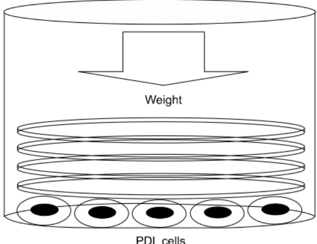
Fig 1. Method used to apply a compressive stress.
Pre-cultured PDL cells were compressed continuously using a different number of round-shaped cover glas- ses. Round-shaped cover glasses were placed over a confluent cell layer in each well of a 6-well plate. The amount of compressive force was adjusted by increas- ing or decreasing the number of cover glasses placed.
Cultured cells derived from the PDL, mostly fibro- blasts, can express and produce the cytokines asso- ciated with osteoclastogenesis. Hasegawa et al8 re- ported that PDL cells derived from deciduous and per- manent teeth synthesized both RANKL and OPG, and could regulate the differentiation of osteoclasts. It was also reported that PDL cells secrete M-CSF in re- sponse to TNF-α stimulation.9 Wada et al10 showed that human PDL fibroblastic cells have the capacity to produce and secrete OPG. Furthermore, it was reported that inflammatory cytokines, such as PGE2, IL-1α, IL-1β, IL-6 and TNF-α, are produced by mechan- ically stimulated PDL cells.11-13
Previous reports showed that PDL cells can partic- ipate in osteoclastogenesis but the production of the re- lated cytokines in PDL cells has not been fully charac- terized. In particular, for M-CSF and IL-1β, their pro- duction in mechanically stimulated PDL cells has not been documented. This study examined the mode of osteoclastogenetic cytokine production including MCSF, RANKL, IL-1β and OPG in PDL cells in response to compressive mechanical stimulation.
MATERIAL AND METHODS Primary human PDL cells
Biscupids extracted from 5 patients for orthodontic reasons were used in this study. Informed consent was obtained from all volunteers. Immediately after ex- traction, the teeth were placed in α-MEM containing 15% FBS (Sigma-aldrich, St. Louis, MO, USA) and 3-fold-reinforced antibiotics (Antibiotics and Antimy- cotics, Gibco BRL, Grand Island, NY, USA) in a 50 ml conical tube (Corning, NY, USA). Using a No. 15 surgical blade, a piece of PDL was obtained exclu- sively from the middle of the tooth roots in order to exclude the intermixture of gingivae and dental pulp.9,10 The PDL tissue obtained was treated with 1.10 unit/ml dispase (Gibco BRL, Grand Island, NY, USA) and 264 unit/ml collagenase (Collagenase Type II;
Gibco BRL, Grand Island, NY, USA) for 1 hour at 37oC. After washing with α-MEM, ligament samples were cultured on a 100 mm primary culture dish (Corning, NY, USA) in α-MEM containing 15% FBS
and antibiotics. The cells proliferating from the extracts were passaged. For all experiments, PDL cells from the 4th to 8th passages were used.
Cell culture
All PDL cells were cultured in α-MEM containing 15% FBS and antibiotics at 37oC in a 5% CO2
incubator. The culture medium was changed twice a week throughout the experiment.
Application of compressive stress
After sufficient cultivation, PDL cells from each pa- tient were transferred to 6-well plates. Each well of a 6-well plate contained 1 × 10⁶ PDL cells. Two days later, the PDL cells were allowed to adhere to the well plate base, and PDL cells were compressed con- tinuously using the uniform compression method illus- trated in Fig 1.
Briefly, round-shaped cover glasses (30 mm diame- ter, Marienfeld, Louda-Könlgshofen, Germany) were placed over a confluent cell layer in each well of a 6-well plate. Each cover glass weighed 0.245 gm. The compressive force was adjusted by increasing or de-
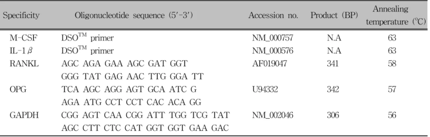
Table 1. Primers used for RT-PCR
Annealing Specificity Oligonucleotide sequence (5'-3') Accession no. Product (BP)
temperature (oC)
M-CSF DSOTM primer NM_000757 N.A 63
IL-1β DSOTM primer NM_000576 N.A 63
RANKL AGC AGA GAA AGC GAT GGT AF019047 341 58
GGG TAT GAG AAC TTG GGA TT
OPG TCA AGC AGG AGT GCA ATC G U94332 342 57
AGA ATG CCT CCT CAC ACA GG
GAPDH CGG AGT CAA CGG ATT TGG TCG TAT NM_002046 306 56
AGC CTT CTC CAT GGT GGT GAA GAC
Sequence of DSOTM primer is not shown according to patent protection law.
creasing the number of cover glasses. Because the di- ameter of a well in a 6-well culture plate is approx- imately 35 mm, the periphery of each well was not fully covered by the cover glass. However, the un- covered area was minor and did not appear to affect the results of the study. The PDL cells were subjected to a compressive force of 0.5, 1, 2, 3 or 4 g/cm2 (total given force was 3.43, 7.015, 14.21, 21.315, 28.175 gm, which was 0.49, 1.01, 2.01, 3.01, 3.99 g/cm2) for 0.5, 1.5, 6, 24 or 48 hours. The PDL cells without com- pressive stimulation served as the control group.
RNA extraction and first-strand comple- mentary DNA synthesis
After each culture period, the total RNA was ex- tracted from each culture. After removing the culture medium, the PDL cells from each well were homogen- ized using Trizol reagent (Invitrogen Co., Carlsbad, CA, USA). After homogenization, 0.2 ml of chloro- form (Sigma-aldrich, St. Louis, MO, USA) per 1 ml of Trizol reagent was added. The samples were centri- fuged at 4oC, 12,000 rpm for 15 minutes. An aqueous phase was transferred to a fresh tube, and 0.5 ml of isopropyl alcohol (Sigma-aldrich, St. Louis, MO, USA) per 1 ml of Trizol reagent was added. The samples were centrifuged again under the same conditions.
Removing the supernatant, the RNA pellets were wash- ed with 75% alcohol (Sigma-aldrich, St. Louis, MO, USA), and dried for 5 to 10 minutes. The RNA was
dissolved in 0.1% diethyl pyrocarbonate (DEPC) water (Fermentas, Glen Burnie, MD, USA).
For complementary DNA synthesis, a mixture of 500 ng mRNA, 2μl of 10μM Oligo dT (Fermentas, Glen Burnie, MD, USA) and 3μl DEPC water was in- cubated at 80oC for 3 minutes, and chilled on ice for 2 minutes. Subsequently, 4μl of 5X RT buffer (Fermentas, Glen Burnie, MD, USA), 20 units of RNase inhibitor (Fermentas, Glen Burnie, MD, USA), 200 units of RevertAidTM M-MuLV RT (Fermentas Inc., Glen Burnie, MD, USA) and 4μl of 2.5 mM dNTP Mix (Fermentas, Glen Burnie, MD, USA) were added to the mixture and incubated at 42oC for 90 minutes.
Reverse transcription polymerase chain re- action assays
First-stranded complementary DNA was subjected to polymerase chain reaction (PCR) amplification using gene specific PCR primers. PCR for M-CSF and IL-1β was carried out using a GeneXPTM kit (Seegene, Seoul, Korea). Each 10μl reaction mixture contained 2μl of 5X Human CYTO-X DSOTM primer, 5μl of 2X mas- ter mix and 20 ng of cDNA. Each cycle consisted of the following: heat denaturation at 94oC for 30s, an- nealing at 63oC for 90s and extension at 72oC for 90s.
PCR amplification can only start if all two parts of the DSOTM (Dual Specificity Oligonucleotide; Seegene, Seoul, Korea) primer bind to cDNA, which result in
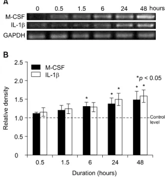
Fig 2. Compressive stress up-regulated M-CSF and IL-1β mRNAs expression in PDL cells. A, RT-PCR analysis of PDL cells. The PDL cells were loaded at different compressive stress (0, 0.5, 1, 2, 3 or 4 g/cm2) for 48 hours; B, Densitometry analysis. The results are expressed as the mean ratio to GAPDH expression of five independent experiments. M-CSF and IL-1β mRNAs expression at 2 g/cm2 was significantly differ- ent compared to those of the control.
Fig 3. Compressive stress up-regulated M-CSF and IL-1β mRNAs expression in a time dependent manner. A, RT-PCR analysis of PDL cells. The PDL cells were loaded at a constant compressive stress (2 g/cm2) for 0, 0.5, 1.5, 6, 24 or 48 hours; B, Densi- tometry analysis. The results are expressed as the mean ratio to GAPDH expression of five independent experiments. M-CSF and IL-1β mRNAs expression was significantly different from the control after 6 hours and 24 hours, respectively.
higher specificity and sensitivity than when using the usual primer.
In PCR for RANKL and OPG, 2X PCR Master Mix (Fermentas, Glen Burnie, MD, USA) was used for the reaction. Each 50μl reaction mixture contained 20 pmol of the sense and antisense PCR primers, 200 ng of cDNA and 25μl of 2X PCR Master Mix. Each cy- cle consisted of the following: denaturation at 94oC for 30s, annealing at a temperature optimized for each pri- mer pair (Table 1) for 90s and extension at 74oC for 90s.
The PCR products were electrophoresed and vi- sualized on a 2% agarose gel containing ethidium bro- mide with UV light illumination. The relative intensity of the gel bands was measured using Scion Image (Scion, Frederick, MD, USA) for Windows XP.
Statistical treatment
Statistical significance was evaluated by analysis of variance (ANOVA) and a multiple-comparison test (Scheffé's test) using SPSS V.13 (SPSS inc., Chicago, IL, USA). A p value < 0.05 was considered signi- ficant. The values are expressed as the mean ± SD.
RESULTS
Effects of various compressive stress and duration on the expression of M-CSF and IL-1β mRNAs
The expression of M-CSF and IL-1β mRNAs in hu- man PDL cells was assessed after applying various mechanical compressive stresses for various durations.
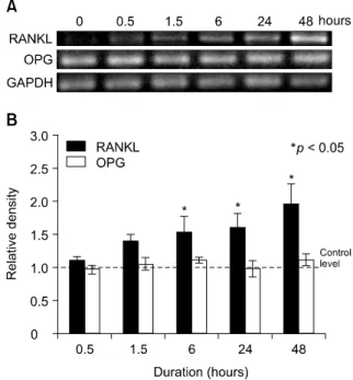
Fig 4. Compressive stress up-regulated RANKL mRNA expression in PDL cells. In contrast, OPG mRNA ex- pression did not change. A, RT-PCR analysis of PDL cells. The PDL cells were loaded at different com- pressive stresses (0, 0.5, 1, 2, 3 or 4 g/cm2) for 48 hours; B, Densitometry analysis. The results are ex- pressed as the mean ratio to GAPDH expression of five independent experiments. RANKL mRNA ex- pression was significantly different from the control at 2 g/cm2. In contrast, OPG mRNA expression was con- stant throughout the experiment.
Fig 5. Compressive stress up-regulated RANKL mRNA expression in a time dependent manner. In contrast, OPG mRNA expression did not change. A, RT-PCR analysis of PDL cells. The PDL cells were loaded at a constant compressive stress (2 g/cm2) for 0, 0.5, 1.5, 6, 24 or 48 hours; B, Densitometry analysis. The re- sults are expressed as the mean ratio to GAPDH ex- pression of five independent experiments. RANKL mRNA expression was significantly different from the control after 6 hours. In contrast, OPG mRNA ex- pression was constant throughout the experiment.
PDL cells under compression showed an increase in M-CSF and IL-1β mRNAs expression in a force-de- pendent manner up to 2 g/cm2, but the expression de- creased after applying 3 g/cm2. The expression of M- CSF and IL-1β mRNAs in the experimental groups reached a maximum of 1.7- and 1.5-fold, respectively, at a compressive load of 2 g/cm2, and were signifi- cantly different from those of the control group which was without compressive stress (0 g/cm2) (Fig 2).
M-CSF and IL-1β mRNAs expression was similar to that of the control group at 0.5 hour of 2 g/cm2 com- pression but a time-dependent increase was evident af- ter 6 and 24 hours, respectively, for up to 48 hours (Fig 3).
Effects of various compressive forces and duration on the expression of RANKL and OPG mRNAs
Expression of RANKL and OPG mRNA in human PDL cells was assessed after applying various com- pressive mechanical stresses for various durations. PDL cells under compression showed increased RANKL mRNA expression in a force-dependent manner up to 2 g/cm2, but expression was decreased after 3 g/cm2. The expression of RANKL mRNA in the experimental groups reached a maximum of 2.1-fold at 2 g/cm2, and was significantly different from those of the control group (Fig 4). RANKL mRNA expression was similar to that of the control group after 0.5 hours of 2 g/cm2 compression. However, an increase was evident after 6 hours, and it increased in a time-dependent manner for
up to 48 hours (Fig 5). In contrast, OPG mRNA ex- pression did not change, regardless of the amount of compressive force applied and the duration of com- pression (Figs 4 and 5).
DISCUSSION
In this study, human PDL cells were cultured under various levels of compressive stress for different peri- ods to determine the effects of mechanical stress on the expression of M-CSF mRNA in PDL cells. A RT- PCR assay was used to analyze target mRNA expression.
Generally, PDL includes multipopulation cells con- sisting mainly of fibroblasts with high alkaline phos- phatase activity,14 and fibroblasts from PDL share many physiological characteristics with osteoblasts.15,16 Some studies already described methods to isolate PDL cells from PDL, and an almost identical method was used in this study. It is possible that osteoblastic cells in the PDL subpopulation transduce mechanical stress.
However, the effects of other types of PDL cells were negligible in this experiment because most cells in the population in this culture system were spindle-shaped fibroblastic cells.
Many researchers have already examined the effects of mechanical stress on PDL cells. Various methods have been used to impart mechanical stress to cultured cells in the manner of tension, compression and fluid shear stress, etc.17-26 Among the manner of mechanical stress, compressive stress was used to confirm the role of PDL cell in the tissue remodeling process of the compressive side during orthodontic tooth movement with particular focus on osteoclastogenesis. Hydrostatic compression,22,23 reverse-tension compression,24 and di- rect contact compression methods19,25,26 were used to provide compressive stress on cultured cells. The direct contact compression method appeared to mimic most in vivo systems in orthodontic treatment because a stat- ic compressive force is applied to the tooth in daily or- thodontic practice and that PDL tissue is compressed directly between two hard structures, bone and cemen- tum. Therefore, this study adopted the direct contact compression method, which provides a compression force by squeezing the cells between two hard surfa-
ces, the culture dish surface and cover glass.
Kanzaki et al.19 used glass cylinder and lead gran- ules to provide compression stimulation. They placed a glass cylinder over a confluent cultured PDL cell layer and adjusted the compressive force by adding lead granules to the cylinder. Similarly, Yamaguchi et al.25 applied compressive stimulation by placing a 30 mm cell disk over a confluent cell layer followed by a glass cylinder on top of it. They also used lead gran- ules to control the amount of compressive force by placing them into the glass cylinder. The present meth- od used only cover glasses in order to simplify the equipment. In addition, by stacking same diameter glasses, a more uniform force could be delivered to the underlying cells than the lead granules, which can roll around in the glass cylinder. In addition, the cover glass weight was small enough (0.245 g) to control the force in a more accurate manner.
The M-CSF and RANKL evaluated in this study are essential factors for osteoclastogenesis produced by os- teoblasts and bone marrow stromal cells.3-6 M-CSF is essential for not only the proliferation of osteoclast progenitors, but also for differentiation into mature os- teoclasts and survival in vitro.27 In contrast, OPG was reported to inhibit osteclastogenesis competing with the binding of RANKL to the RANK. IL-1β is a proin- flammatory cytokine that stimulates M-CSF and PGE2
production,28 and up-regulated PGE2 mediates RANKL expression in PDL cells under a static compressive stress.19
However, there is some controversy regarding the effects of M-CSF in osteoclastogenesis. Although M- CSF is an essential factor for the formation of osteo- clasts, it also has been reported that the addition of M-CSF to a culture inhibits osteoclast formation.
Udagawa et al.3 reported that M-CSF, while stimulat- ing murine osteoclast survival, did not induce pit-form- ing activity. Perkins et al.29 showed that the addition of exogenous M-CSF to a co-culture of mouse spleen cells and ST2 cells caused a dose-dependent decrease in the number of osteoclasts formed, and was accom- panied by an increase in the number of macrophages.
Recently, Yasuda et al.30 reported that a high concen- tration of M-CSF (> 40 ng/ml) suppressed osteoclast formation in murine spleen cell cultures stimulated
with soluble RANKL. Considering that these studies examined osteoclast formation in animal models, spe- cies differences and different requirements for M-CSF between the different culture systems can account for these divergent findings.
Through RT-PCR assays, PDL cells under a com- pressive stress showed increased M-CSF, IL-1β and RANKL mRNAs expression in a force- (up to 2 g/
cm2) and time-dependent manner, but OPG mRNA ex- pression was constant regardless of the amount of compressive stress or duration of compression. This can be explained by the fact that PDL cells under compressive stress regulate bone remodeling through an increase in the production of osteoclastogenetic cy- tokines but not OPG. In addition, compressive stress- induced intracellular changes are not involved in OPG mRNA expression. However, it was reported that ten- sion type stimulation up-regulates OPG mRNA ex- pression.31,32 Therefore compression and tension stim- ulation appears to have separate signal transduction pathways.
These results partly coincide with previous reports.
Kanzaki et al.19 reported that compressive stress up to 2 g/cm2 up-regulated RANKL mRNA in a time-de- pendent manner. They also measured the level of OPG and reported no change. Nishijima et al.26 examined levels of RANKL and OPG in gingival crevicular fluid during orthodontic tooth movement and found that the RANKL level was elevated while the OPG level was decreased. They also performed an in vitro study and reported that compression stressed PDL cells increased the secretion of RANKL in a time-dependent manner but decreased the secretion of OPG in a time-depend- ent manner. Nakajima et al.33 carried out a similar compressive stimulation study on PDL cells. They also reported that RANKL production was elevated in a time and force-dependent manner up to 4 g/cm2, the maximal force that they used. On the other hand, they reported an increase in OPG production in com- pression-stressed PDL cells. Overall, it appears that compressive stress up-regulates RANKL mRNA and increases the production of RANKL in PDL cells.
However, for OPG, there are discrepancies between studies, which will need more investigation to establish the mode of OPG production in PDL cells.
With the exception of Nakajima et al.'s33 study, oth- er studies examining compressive stress effects on PDL cells reported that a 2 g/cm2 force is the optimal level of force that can elicit a maximal PDL cell response.19,23,26 These results also support the optimal force level of 2 g/cm2. At an applied force magnitude of 3 g/cm2, the level of M-CSF, IL-1β and RANKL mRNA expression decreased. This is probably due to the 3 g/cm2 applied force being too heavy making cell survival difficult. Indeed, a microscopic observation af- ter applying 4 g/cm2 showed that the compressive stress was so heavy that PDL cells were partially dam- aged and?the number of cells decreased (data not shown). Nakao et al.23 suggested that 2 g/cm2 was suit- able but 7.0 g/cm2 was too heavy for cell survival.
This in vitro result can be applied clinically. Although the applied orthodontic force cannot be distributed uni- formly on the desired tooth root surface, the area of root surface that faces a compressive orthodontic force according to the type of tooth movement can be esti- mated and the desirable amount of force can be calcu- lated by multiplying 2 g/cm2. The validity of this as- sumption may be proven by another systemized clin- ical trial investigation.
After 6 hours of 2 g/cm2 compressive mechanical stress, there was a significant increase in the level of M-CSF and RANKL mRNA expression compared to the control. However, the level of IL-1β mRNA ex- pression increased significantly after 24 hours. This means that IL-1β mRNA up-regulation is delayed compared to M-CSF and RANKL. In addition, other cytokines might mediate signal transduction during a compressive mechanical stress. To the best of our knowledge, there are no reports on the effect of com- pressive stress on the expression of M-CSF and IL-1β mRNA. These results suggest that the expression of M-CSF and IL-1β mRNA increases in a time- and force-dependent manner (up to 2 g/cm2). In addition, it appears that a compressive force directly regulates M-CSF mRNA expression but indirectly regulates IL-1β mRNA expression. More signal transduction studies will be needed to confirm the mechanism of com- pressive force induced M-CSF and IL-1β mRNA expression.
CONCLUSION
Continuous compressive stress up-regulates M-CSF, IL-1β and RANKL mRNA expression in cultured hPDL cells in a force- and time-dependent manner.
However, a compressive force does not regulate OPG mRNA expression in PDL cells. These results suggest that a compressive stimulated PDL cells regulate osteo- clastogenesis by up-regulating the stimulatory cytokines but not down-regulating the OPG production. There- fore, in orthodontic tooth movement, PDL cells partic- ipate in bone resorption by up-regulating osteoclastoge- netic cytokines, including M-CSF, IL-1β and RANKL.
- 국문초록 -
압박력이 치주인대 세포의 M-CSF, IL-1β, RANKL 및 OPG mRNA 발현에 미치는 영향
김지웅aㆍ이기수bㆍ남종현cㆍ강윤구d
이 연구의 목적은 배양된 사람 치주인대 세포에서 파골세 포의 형성에 관련된 물질을 합성, 분비할 수 있는지를 알아 보고 압박력이 M-CSF, IL-1β, RANKL 및 OPG mRNA의 발현에 미치는 영향을 알아보고자 하였다. 교정치료를 목적 으로 발치된 소구치에서 얻은 치주인대세포를 배양한 후 다 양한 크기(0.5, 1.0, 2.0, 3.0, 4.0 g/cm2)의 기계적 자극을 다 양한 기간(0.5, 1.5, 6, 24, 48 hours) 동안 적용하고, M-CSF, IL-1β, RANKL, OPG mRNA 발현양의 변화를 검사하였다.
각각의 실험군에서 얻어진 mRNA에 대해 역전사 중합효소 연쇄반응검사를 시행하였다. 검사 결과 압박력은 사람 치주 인대 세포에서 M-CSF mRNA를 발현시켰으며 M-CSF, IL-1β, RANKL mRNA의 발현양은 자극의 크기와 기간에 따라 증가 하였다. 그러나 압박력은 사람 치주인대 세포에서 OPG mRNA의 발현양에 영향을 미치지 않는 것으로 나타났다. 이 상의 결과는 기계적 자극이 치주인대 세포에서 M-CSF, IL-1β, RANKL mRNA의 발현양을 조절함으로 파골세포의 분화에 영향을 미칠 수 있음을 시사한다.
주요 단어: 치주인대세포, 기계적 자극, 파골세포형성
REFERENCES
1. Roberts WE, Goodwin WC, Heiner SR. Cellular response to
orthodontic force. Dent Clin North Am 1981;25:3-17.
2. Mitchell DL, West JD. Attempted orthodontic movement in the presence of suspected ankylosis. Am J Orthod 1975;68:
404-11.
3. Udagawa N, Takahashi N, Jimi E, Matsuzaki K, Tsurukai T, Itoh K, et al. Osteoblasts/stromal cells stimulate osteoclast acti- vation through expression of osteoclast differentiation fac- tor/RANKL but not macrophage colony-stimulating factor.
Bone 1999;25:517-23.
4. Suda T, Takahashi N, Martin TJ. Modulation of osteoclast differentiation. Endocrine Rev 1992;13:66-80.
5. Matsuzaki K, Udagawa N, Takahashi N, Yamaguchi K, Yasuda H, Shima N, et al. Osteoclast differentiation factor (ODF) induces osteoclast-like cell formation in human periph- eral blood mononuclear cell cultures. Biochem Biophys Res Commun 1998;246:199-204.
6. Jimi E, Nakamura I, Amano H, Taguchi Y, Tsurukai T, Tamura M, et al. Osteoclast function is activated by osteo- blastic cells through a mechanism involving cell-to-cell contact. Endocrinology 1996;137:2187-90.
7. Simonet WS, Lacey DL, Dunstan CR, Kelley M, Chang MS, Luthy R, et al. Osteoprotegerin: a novel secreted protein in- volved in the regulation of bone density. Cell 1997;89:309-19.
8. Hasegawa T, Kikuiri T, Takeyama S, Yoshimura Y, Mitome M, Oguchi H, et al. Human periodontal ligament cells derived from deciduous teeth induce osteoclastogenesis in vitro. Tissue Cell 2002;34:44-51.
9. Yongchaitrakul T, Lertsirirangson K, Pavasant P. Human pe- riodontal ligament cells secrete macrophage colony-stimulating factor in response to tumor necrosis factor-alpha in vitro. J Periodontol 2006;77:955-62.
10. Wada N, Maeda H, Tanabe K, Tsuda E, Yano K, Nakamuta H, et al. Periodontal ligament cells secrete the factor that in- hibits osteoclastic differentiation and function: the factor is os- teoprotegerin/osteoclastogenesis inhibitory factor. J Periodont Res 2001;36:56-63.
11. Lowney JJ, Norton LA, Shafer DM, Rossomando EF.
Orthodontic forces increase tumor necrosis factor alpha in the human gingival sulcus. Am J Orthod Dentofacial Orthop 1995;108:519-24.
12. Bumann A, Carvalho RS, Schwarzer CL, Yen EH. Collagen synthesis from human PDL cells following orthodontic tooth movement. Eur J Orthod 1997;19:29-37.
13. Yamaguchi M, Shimizu N. Identification of factors mediating the decrease of alkaline phosphatase activity caused by ten- sion-force in periodontal ligament cells. Gen Pharmacol 1994;
25:1229-35.
14. Lekic P, McCulloch CA. Periodontal ligament cell populations:
the central role of fibroblasts in creating a unique tissue. Anat Rec 1996;245:327-41.
15. McCulloch CA, Melcher AH. Cell density and cell generation in the periodontal ligament of mice. Am J Anat 1983;
167:43-58.
16. Kawase T, Sato S, Miake K, Saito S. Alkaline phosphatase of human periodontal ligament fibroblastic cells. Adv Dent Res 1988;2:234-9.
17. Kanai K, Nohara H, Hanada K. Initial effects of continuously applied compressive stress to human periodontal ligament
fibroblasts. J Jpn Orthod Soc 1992;51:153-63.
18. Watanabe K, Saito I, Hanada K. Effects of conditioned me- dium of continuously compressed human periodontal ligament fibroblasts on MC3T3-E1 cells. J Jpn Orthod Soc 1998;57:
173-9.
19. Kanzaki H, Chiba M, Shimizu Y, Mitani H. Periodontal liga- ment cells under mechanical stress induce osteoclastogenesis by receptor activator of nuclear factor κB ligand up-regu- lation via prostaglandin E2 synthesis. J Bone Miner Res 2002;17:210-20.
20. Brown TD. Techniques for mechanical stimulation of cells in vitro: a review. J Biochem 2000;33:3-14.
21. Basso N, Heersche JNM. Characteristics of in vitro osteob- lastic cell loading models. Bone 2002;30:347-51.
22. Nakao K, Goto T, Gunjigake KK, Konoo T, Kobayashi S, Yamaguchi K. Intermittent force induces high RANKL ex- pression in human periodontal ligament cells. J Dent Res 2007;86:623-8.
23. Nakao K, Goto T, Gunjigake K, Konoo T, Kobayashi S, Yamaguchi K. Neuropeptides modulate RANKL and OPG ex- pression in human periodontal ligament cells. Orthodontic Waves 2007;66:33-40.
24. Choi HS. Effects of tension and compression force on PGE2 of human periodontal ligament cells in vitro. [PhD thesis]
Seoul; Ewha Womans University: 2000.
25. Yamaguchi M, Ozawa Y, Nogimura A, Aihara N, Kojima T, Hirayama Y, et al. Cathepsins B and L increased during re- sponse of periodontal ligament cells mechanical stress in vitro.
Connect Tissue Res 2004;45:181-9.
26. Nishijima Y, Yamaguchi M, Kojima T, Aihara N, Nakajima R, Kasai K. Levels of RANKL and OPG in gingival crevicular fluid during orthodontic tooth movement and effect of com-
pression force on releases from periodontal ligament cells in vitro. Orthod Craniofac Res 2006;9:63-70.
27. Suda T, Udagawa N, Nakamura I, Miyaura C, Takahashi N.
Modulation of osteoclast differentiation by local factors. Bone 1995;17(Suppl):87-91.
28. Sato K, Fujii Y, Asano S, Ohtsuki T, Kawakami M, Kasono K, et al. Recombinant human interleukin 1 alpha and beta stimulate mouse osteoblast-like cells (MC3T3-E1) to produce macrophage-colony stimulating activity and prostaglandin E2. Biochem Biophys Res Commun 1986;141:285-91.
29. Perkins SL, Kling SJ. Local concentrations of macrophage col- ony-stimulating factor mediate osteoclastic differentiation. Am J Physiol 1995;269:E1024-30.
30. Yasuda H, Shima N, Nakagawa N, Yamaguchi K, Kinosaki M, Mochizukt S, et al. Osteoclast differentiation factor is a ligand for osteoprotegerin/osteoclastogenesis inhibitory factor and is identical to TRANCE/RANKL. Proc Natl Acad Sci USA 1998;95:3597-602.
31. Tsuji K, Uno K, Zhang GX, Tamura M. Periodontal ligament cells under intermittent tensile stress regulate mRNA ex- pression of osteoprotegerin and tissue inhibitor of matrix met- alloprotease-1 and -2. J Bone Miner Metab 2004;22:94-103.
32. Lee KJ, Lee SI, Hwang CJ, Ohk SH, Tian YS. The effect of progressive tensional force on mRNA expression of osteoprote- gerin and receptor activator of nuclear factor κB ligand in the human periodontal ligament cell. Korean J Orthod 2005;35:
262-74.
33. Nakajima R, Yamaguchi M, Kojima T, Takano M, Kasai K.
Effects of compression force on fibroblast growth factor-2 and receptor activator of nuclear factor kappa B ligand production by periodontal ligament cells in vitro. J Periodontal Res 2008;43:168-73.