Ⅰ. Introduction
The pathways of periodontal tissue de- struction during the course of destructive periodontal diseases have been fairly well described at the cellular level1). According to the current paradigm of infectious etiology, the events associated with tissue destruction could be most briefly summarized as follows:
an external stimulus, usually toxins and an- tigens from bacteria of the dental biofilm, can exert direct effects on the periodontal tissues, but acts mainly by triggering a com- plex array of inflammatory and immune re- actions that ultimately leads to the loss of supporting apparatus of affected teeth2).1)
There is evidence supporting a direct par- ticipation of PDL cells on pathologic break- down of extracellular matrix from both min-
eralized and non-mineralized periodontal tissues. Chang et al.3) showed synthesis and secretion of matrix metalloproteinase 2 (MMP-2, type IV collagenase, gelatinase A) as well as indication of some synthesis of MMP-9 (also a type IV collagenase, gelati- nase B) by PDL cells. Domeij et al.4) also found similar results in gingival fibroblasts, demonstrating increase in secretion of both MMP-1 and MMP-3 by these cells after stimulation with IL-1βâ, TNF-αá and epi- dermal growth factor (EGF). By using spe- cific pharmacological inhibitors, they also report indirect evidence of the involvement of both tyrosine kinase and p38 MAP kinase on the signaling pathways for the proin- flammatory stimuli in these cells.
Collagenase 3 (or MMP-13) is a recently described protein in the MMP family and al- though type II collagen is the main sub-
* corresponding author; Young Joon Kim, Dept. of Periodontology, School of Dentistry, Chonnam National University, 5, Hak 1 dong, Dong-gu, Gwangju,501-746, Korea, E-mail:[email protected])
대한치주과학회지 : Vol. 36, No. 1, 2006
Role of MAP kinase on MMP-13 expression in rat periodontal ligament cells
Chan Gil Chung1, De-Zhe Cui2, Hyun-Ju Chung1,2, Young Joon Kim1,2 Department of Periodontology School of Dentistry1, Dental Science Research Institute2,
Chonnam National University
strate target for this enzyme, it also de- grades collagen types I, III and V, which are all major components of cartilage and bone5). In periodontal inflammation, several resi- dent cell types have been shown to im- munoreactive to MMP-13, including fibro- blasts and macrophages. MMP-13 has been shown to exist at higher levels in perio- dontal infected tissues than in healthy con- trol tissues6). In peri-implantitis models of bone loss, implants having >3 mm of bone loss had higher levels of MMP-13 from the periimplant fluid at these sites7). Thus, MMP-13 expression may be related not only to collagen degradation, but also directly or indirectly to bone resorption.
It was shown recently that proin- flammatory TNF-α increases MMP-13 mRNA expression in human PDL cells, with the maximal effect observed with a concen- tration of 10 ng/㎖8).
The study of the signal transduction path- way leading to expression of MMP-13 mRNA may give further insight into the possible cross-talk by proteins involved in signal transduction, which may also activate other genes related to bone resorption.
Proinflammatory cytokines stimulate mul- tiple intracellular signaling pathways, in- cluding the p38 MAP kinase pathways, NF-kB pathway, extracellular regulated kin- ase (ERK), and stress-activated protein kin- ase/c-Jun N-terminal kinase (SAPK/JNK)9,10). Although much attention has focused on the NF-kB pathway of cytokine gene activation, relatively less attention has been spent on p38 MAP kinase which is also involved in
inflammation and environmental stress in- duced signaling11, 12). The p38 kinases con- stitute a distinct MAP Kinase subfamily that plays a role in adaptation, homeostasis, and stress responses. In recent years, the MAP kinase system has been shown to mediate, in part, the actions of proinflammatory cyto- kines as well as acting a principal mediator of the inflammatory response to LPS in os- teoblastic cells13). The role of p38 MAP Kinase in MMP-13 gene regulation is becom- ing more evident14), although the mecha- nisms of gene regulation are not entirely clear.
PDL cells play a potential central role in tissue destruction associated with perio- dontal disease15). The relative lack of data related to signal transduction pathways of both inflammatory and infectious external signals in these cells, and the relevance of metaloproteinases, specifically of MMP-13.
This study was accomplished as an initial step towards a better understanding of the role of p38 MAP Kinase on the expression of MMP-13 secreted by PDL cells. Thus, the purpose of this study is to investigate the role of p38 MAP Kinase on MMP-13 mRNA expression through RT-PCR and western blot assays.
Ⅱ. Material and Method
1. Cell culture
Rat periodontal ligament fibroblasts (PDL) obtained as described by Matsuda et al. 16) were cultured in DMEM (Invitrogen, U.S.A) medium, supplemented with 10%
heat-inactivated fetal bovine serum (FBS;
Invitrogen, U.S.A), 100 U/㎖ penicillin and 100 U/㎖ streptomycin (Penicillin-Streptomycin, GibcoBRL, U.S.A), at 37℃ in a humidified atmosphere of 5% CO2 in air. Cells were routinely deinduced in culture media with- out FBS for 6-8 hours prior to stimulation with proinflammatory cytokines.
2. Reverse transcription-polymerase chain reaction (RT-PCR) analysis Cells were grown to near confluency in 6-well dishes, deinduced and then treated with the following proinflammatory and oth- er substances: recombinant human inter- leukin 1β (IL-1β; 1 nM), recombinant human tumour necrosis factor-α (TNF-α 10 nM), re- combinant human bone morphogenic pro- tein-7 (BMP-7, 100 ng/ml), parathyroid hormon (PTH, 10 nM). A negative control represented by the vehicle used to dilute these substances also were included.
After the treatment periods, total RNA were collected with TrizolⓇ reagent (Invitrogen, U.S.A), according to the instructions pro- vided by the manufacturer. Briefly, approx- imately 1 ㎖ of TrizolⓇ was added to each well to cell lysis and this cell lysate was ho- mogenized by passing several times through a pipette. This solution was transferred to eppendorf tubes, incubated at room temper- ature (RT) for 15-30 min, and then 0.2 ㎖ of chloroform per ㎖ of TrizolⓇ was added.
After vigorously shaking the tubes and cen- trifugation at 10,000 xg for 15 min at 2-8℃, the aqueous phase was collected and trans- ferred to a fresh tube. Total RNA was pre-
cipitated by adding isopropyl alcohol, wash- ed in 75% ethanol and redissolved in RNAse-free water for storage at -70℃ until use.
Using spectrophotometry, 5 ㎍ of RNA was quantitated (read at an absorbance of 260 nm) and used for cDNA synthesis with oligo (dT) 12-18 primer by reverse transcriptase(RT) reactions (RT-PCR primers: MMP-13 328bp S5-GACTTCACGATGGCATTGCTG-3, A5-GCATCAACCTGCTGAGGATGC-3 GAPDH 418bp S5-ATTCCATGGCACCGTCAAGGCT-3, A5-TCAGGTCCACCACTGACACGTT-3 ).
Approximately 2 ㎕ of RT product was used as a template for the polymerase chain re- action (PCR).
The PCR products were checked by 2%
agarose gel electrophoresis, stained by ethi- dium bromide. Semi-quantitative PCR was performed. Semi-quantitative differences of MMP-13 expression were normalized by GAPDH.
3. mRNA stability Experiments
To determine the relative role of p38 MAP Kinase on IL-1β-induced MMP-13 mRNA stability, the rate of MMP-13 mRNA decay was determined in the presence and absence of the p38 inhibitor, SB203580 (10 μìM).
PDL cells were pre-treated with SB203580 (10 μM) for 60 minutes, then treated with IL-1β(1 nM) for 16-18 hours. Subsequently, actinomycin D (2 ㎍/㎖), DNA polymerase inhibitor, was added to prevent further transcription. Total RNA was harvested and analyzed by RT-PCR as described above af- ter 0, 60, 120, 240 minutes treated with ac- tinomycin D.
4. Western blot analysis
To analyze the cell signaling pathways as- sociated with the different stimuli, the cells were treated with the same substances (IL-1 β, TNF-α, BMP-7 and PTH) for shorter time periods. For this experiment, PDL cells were grown to near confluency in 6-well dishes and de-induced as described previously.
After treatment, whole cell lysates were collected for Western blotting analysis of both active (phosphorylated) and inactive (non-phosphorylated) MAP kinase. Western blotting was conducted as described in the literature. Briefly, after removal of media by aspiration, cells were washed with ice-cold PBS (pH 7.4), and 100 ㎕ of SDS-sample buffer was added to each well with removal of cells by scraping the wells with a disposable cell scraper. The cell ly- sate was transferred to eppendorf tubes, boiled at 100℃ for 5 min, placed on ice, so- nicated for 10-15 seconds (three times) and stored at -20℃ until use.
Following sample preparation (by boiling at 100℃ for 5 min and placing on ice), SDS-PAGE was performed using pre-stained molecular weight markers to verify electro- transfer and biotinylated protein markers to determine molecular weights. After running the gel, proteins were blotted to a nitro- cellulose membrane.
The immunoblotting protocol was per- formed as described by the supplier of the commercial antibody kits. The nitrocellulose membrane was blocked for non-specific bind- ing, washed and incubated with the primary mouse anti-human antibodies (for MAP kin-
ases; Cell Signaling, U.S.A) using gentle agitation overnight at 4℃. After three washes, the membrane was incubated with the secondary anti-mouse antibodies con- jugated to Horseradish peroxidase (Cell Signaling, U.S.A) and anti-biotin HRP-con- jugated antibody (to detect biotinylated pro- tein markers) with gentle agitation for 1 hour at room temperature. After three con- secutive washes, the membrane was in- cubated with the enzyme substrate for 1 mi- nute, wrapped in plastic film and exposed to X-ray film (2 mins).
Ⅲ. Results
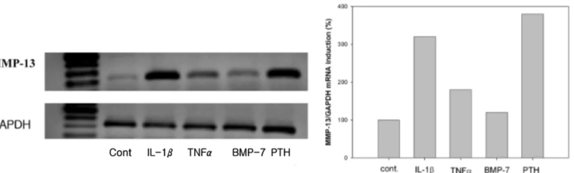
1. MMP-13 mRNA was expressed by proinflammatory cytokines and PTH In semi-quantitive RT-PCR analysis, MMP-13 was induced to different extents by IL-1β, TNFα, rh BMP-7, and PTH. When levels of GAPDH were used to normalize the data, IL-1β, TNFα, and PTH-induced MMP-13 expression was increase to 320%, 180%, 380%, respectively. But BMP-7 had little effect on MMP-13 expression (120%) (Figure 1).
2. p38 was reqiuired for IL-β-induced MMP-13 mRNA induction
MMP-13 mRNA gene expression was de- termined by RT-PCR. The result was shown that p38 is necessary dor IL-1β-induced MMP-13 mRNA synthesis. When PDL cells were pre-treated with SB203580, the p38 MAPK inhibitor, and then stimulated with
IL-1β, the result was shown to approx- imately 40% reduction in IL-1β-induced MMP-13 mRNA levels compared to IL-1β treated cells only (from 3.5 fold to 2.0 fold).
There was no effect on PTH pre-treated cells (Figure 2).
3. p38 was needed to mediate IL-1β -induced MMP-13 mRNA stability.
To determine if p38 MAP Kinase regula- teed IL-1β induced MMP-13 stability, we used Actinomycin D. The results indicate
that IL-1β increased the half-life of MMP-13, with 120 minutes, but in the pres- ence of SB203580, the half life was de- creased to 60 minutes following normal- ization to GAPDH (Figure 3).
4. p38 MAP Kinase and JNK were acti vated by IL-1β
Western blot analysis was utilized to ex- amine whether IL-1β, TNFα, BMP-7 and PTH activated MAP Kinase. Western blot analysis of antibodies indicated that IL-1β Figure 3. MMP-13 mRNA stability experi-
ment using Actinomycin D in PDL cells. P38 MAP kinase mediates IL-1 β-induced MMP-13 stability. * in- dicates half lifes of MMP-13 mRNA in the presence of SB203580.
T im e (h o u r)
0 1 2 3 4 5
% MMP-13 mRNA Remaining
0 2 0 4 0 6 0 8 0 1 0 0 1 2 0
IL -1 b S B + IL -1 b
*
Figure 2. Semiquantitative RT-PCR of rat periodontal ligament cells showing steady state MMP-13 mRNA levels. (lane 1;DNA ladder, 2; control, 3; IL-1β(1 nM), 4;
PTH, 5; SB203580(10mM) + IL-1, 6:
SB203580(10mM) + PTH)
Time(hr) 0 1 2 4 0 1 2 4
IL-1β + + + + + + + +
SB - - - - + + + +
Figure 1. Semiquantitative RT-PCR of rat periodontal ligament cells showing steady state MMP-13 mRNA levels. (lane 1;DNA ladder, 2; control, 3; IL-1β(1 nM), 4; TNFα(10 nM), 5; BMP-7(50 ng/㎖), 6: PTH(10 nM))
Cont IL-1β TNFα BMP-7 PTH
activates both phosphorylated form of p38 MAP Kinase and JNK MAP Kinase, but not ERK MAP Kinase. Neither BMP-7 nor PTH activates p38, JNK, and ERK MAP Kinase (Figure 4~6).
5. Activation of p38 MAP Kinase was in- hibited with treatment with SB203580
Western blot analysis of antibodies in- dicated that anti-phospho p38 and non- phospho p38 MAP Kinase are activated by IL-1β in PDL cells. Importantly, the p38 in-
hibitor, SB203580 was able to block IL-1β -induced p38 MAP Kinase (Figure 7).
Ⅳ. Discussion
Proinflammatory cytokine production in periodontal disease, as well as other chronic inflammatory bone diseases, such as rheu- matoid arthritis, results in a net bone loss that ultimately negatively affects host func- tion17). Complex cytokine networking and cellular cross-talk dictate cellular responses involved in bone remodeling. Several media- Figure 4. Western blot analysis of phos-
phorylated and unphosphorylated p38 MAP Kinase. (lane 1;MW, 2; control, 3; IL-1β (1 nM), 4; TNF-α (10 nM), 5; BMP-7 (50 ng/㎖), 6: PTH (10 nM))
Figure 5. Western blot analysis of phosphory- lated and unphosphorylated JNK. JNK is activated by IL-1β. (lane 1;MW, 2; control, 3; IL-1β (1 nM), 4; TNF-α (10 nM), 5;
BMP-7 (50ng/㎖), 6: PTH (10 nM))
Figure 6. Western blotanalysis of phosphory- lated and unphosphorylated ERK. (lane 1;MW, 2; control, 3; IL-1β(1 nM), 4;
TNF-α(10 nM), 5; BMP-7 (50 ng/㎖), 6:
PTH (10 nM))
Figure 7. Western blot analysis of phos- phorylated and unphosphorylated p38 MAP Kinase. p38 MAP Kinase is activated by IL-1b and subsequent p38 inhibition by treatment with SB203580. (lane 1;MW, 2;
control, 3; IL-1β(1 nM), 4; SB203580 (10 mM) only, 5; SB203580 (10 mM)+ IL-1)
Cont IL-1βTNF-αBMP-7 PTH
Cont IL-1β TNF-αBMP-7 PTH
Cont IL-1β TNF-αBMP-7 PTH
Cont IL-1β SB SB IL-1β
tors of bone resorption and remodeling have been identified in response to proin- flammatory cytokines, such as IL-1, lip- opolysaccharide, as well as other bone-re- sorptive agents18).
In fact, there is evidence supporting a di- rect participation of PDL cells on pathologic breakdown of extracellular matrix from both mineralized and non-mineralized periodontal tissues15, 19). In most systems, collagenase expression is not constitutive but is induced by variety of agents, including cytokines, tumour promoters, and bone-resorbing agents. Bone resorptive agents, IL-1, IL-6, PTH, and TNF-α enhance the expression of collagenase 3 in mouse calvaria, primary rat osteoblastic cells, and other osteoblastic model systems14, 20, 21).
Several studies showed that the synthesis of MMP-13 by rat osteoblasts is regulated by systemic hormones and by cytokines. But there are few reports that the expression of MMP-13 by various proinflammatory cyto- kines in PDL cells. Thus we examined the effects of various stimuli, including proin- flammatory cytokines such as IL-1β, TNF-α, and BMP-7, and PTH on MMP-13 in PDL cells. In this study, MMP-13 steady state gene expression in PDL cells was de- termined by semi-quantitative RT-PCR.
MMP-13 was induced to different extents by IL-1β, TNF-α, BMP-7 and PTH. IL-1β and PTH stimulated MMP-13 in PDL cells.
However, in contrast to IL-1β and PTH, BMP-7-induced MMP-13 expression were weak. This result is consistent with results obtained other investigators20, 22, 23) where PTH enhanced expression of MMP-13 but
bone morphogenic proteins suppressed ex- pression of MMP-13 in rat osteoblasts. In the present study, TNF-α also induced ex- pression of MMP-13. This result is con- sistent with Kirkwood's report20) that TNF- α-induced MMP-13 expression in rat osteo- blasts was more enhanced than untreated control.
The signaling cascade from the external membrane to the nucleus is extremely com- plex and can involve a great number of dif- ferent proteins, depending on the external factor being considered. Nevertheless, some components of the signaling cascade have been extensively studied, such as the cyto- plasmic protein kinases collectively known as the mitogen-activated protein (MAP) kin- ases, which up to now includes the following kinases: SAPK/JNK, ERK1/2 and p3813).
In recent years, the MAP Kinase system has been shown to mediate, in part, the ac- tions of pro-inflammatory cytokines, al- though the mechanisms of gene regulation are not entirely clear. Moreover, there are many questions unanswered regarding the role for specific MAP Kinase involvement in bone metabolism. No studies to date have directly addressed the role of p38 MAP Kinase in periodontal disease models.
Understanding p38 MAP kinase-mediated signaling pathways is essential to aid in generating therapeutics capable of treating diseases mediated by MMP-13 and other mediators. In this study, through semi-quantitative RT-PCR in PDL cells, IL-1β-induced MMP-13 expression was re- duced in the presence of specific p38 MAP Kinase inhibitor, SB203580 by 50%.
However in contrast IL-1β, PTH-induced MMP-13 repression was not observed in the presence of SB203580. This result showed that p38 MAP Kinase was involved in IL-1β -induced MMP-13 steady state mRNA levels.
From this result , although a number of cy- tokines and hormones can induce MMP-13 expression, they do not always activate the same signaling events that mediate these effect. For example, PTH-induced MMP-13 expression requires activation of protein kinase C and protein kinase A pathway. In contrast, MMP-13 induction by IL-1 re- quired nuclear factor (NF) -κB trans- location, p38 MAP kinase activity in chon- drocytes24). Thus different signaling path- ways may be activated in MMP-13 expression. This result also showed that specific inhibition of p38 MAP Kinase pre- vents MMP-13 induction by IL-1β, indicating the importance of this pathway in MMP-13 gene expression in PDL cells.
In figure 3 to 5, it was shown that IL-1β only induced phosphorylation of p38 MAP Kinase and c-Jun N-terminal Kinase (JNK) in PDL cells by Western blot analysis. It im- plicates that IL-1β-induced MMP-13 requires p38 MAP Kinase activity and JNK activity.
This result is consistent with Mengshol's study24) that IL-1-induced-MMP-13 ex- pression requied NF-κB, p38 and JNK acti- vation in chondrocytes. In this study, TNF- α was not activate p38 MAP kinase nor ERK. According to Johansson et al25), TNF- α stimulation of MMP-13 expression in kera- tinocytes led to activation of ERK, JNK, p38 activation, but there is also a report26) that ERK activation did not mediate MMP-13 ex-
pression in fibroblast-like synoviocytes. A possible explanation of different results may be different intracellular mechanism which affect MMP-13 expression. Thus, although a number of cytokines and hormones can in- duce MMP-13 expression, they do not al- ways activate the same signaling events in different cell lines.
Through further Western blot analysis, it was shown that SB203580 inhibited the abil- ity of IL-1β to induce p38 phosphorylation in PDL cells. Specific inhibition of p38 MAP kinase prevents MMP-13 induction by IL-1β, indicating the importance of this pathway in MMP-13 gene expression.
The mechanism of p38 regulation of MMP-13 mRNA expression could occur at the transcriptional or post-transcriptional level. To determine whether p38 stabilized MMP-13 mRNA by post-transcriptional reg- ulation, mRNA degradation studies were performed. SB203580 altered IL-1β-induced MMP-13 mRNA decay rates in PDL cells. It means that in PDL cells, p38 MAP kinase appears to mediate its effects on enhancing MMP-13 gene expression mainly through the stabilization of MMP-13 mRNA. As shown in Figure 3, p38 MAP kinase mediates IL-1β -induced MMP-13 mRNA stability. But SB203580 reduces the half life of the MMP-13 from approximately 120 minutes to less than 60 minutes in IL-1β-stimulated cells in the presence of Actinomycin D, an inhibitor of DNA-primed RNA polymerase, to arrest transcription.
This study demonstrates that activation of MMP-13 gene expression is induced by IL-1 β, TNF-α and PTH in PDL cells, but only
IL-1β-induced MMP-13 activation through p38 MAP kinase. It implicates that p38 MAP kinase pathway is a central signal pathway which controls IL-1β-induced MMP-13 expression.
Further studies will be needed to eluci- date that MAP Kinases pathway would be a central signalling pathway controlling IL-1β -induced MMP-13 expression at the gene promotor level. Understanding signalling mechanism which contributes towards MMP-13 expression may provide therapeutic targets in the management of inflammatory bone diseases such as periodontitis and rheumatoid arthritis
Ⅴ. Conclusion
Matrix metalloproteinases (MMPs) play an important role both in the maintenance and degradation of extracellular matrix within the periodontium. MMP-13 has been shown to positively correlate with perio- dontal disease. Mitogen activated protein (MAP) Kinases play an important role in the induction of pro-inflammatory cytokines such as IL-1β and TNF-α. This study is ac- complished as an initial step towards a bet- ter understanding of the role of MMP-13 in the pathogenesis of periodontal disease and possible regulation of MMP-13 through MAP Kinase signaling by different stimuli.
MMP-13 mRNA expression in PDL cells was determined by semi-quantitative RT-PCR and MAP Kinases expression were de- termined by Western blot.
The results are as follows;
1. In semi-quantitive RT-PCR analysis, IL-1 β, TNF-α, and PTH-induced MMP-13 expression was increase to 320%, 180%, 380%, respectively. But rh BMP-7 had little effect on MMP-13 expression (120%).
2. When PDL cells were pre-treated with SB203580 (10 μM), the p38 inhibitor, IL-1β-induced MMP-13 mRNA level was reduced by 40% in compared to IL-1β-treated cells only. There was no effect on PTH pre-treated cells.
3. IL-1β increased the half-life of MMP-13 mRNA with 120 minutes, but in the presence of SB203580, the half life was decreased to 60 minutes.
4. In western blot analysis, IL-1β acti- vates both phosphorylated form of p38 MAP Kinase and JNK, but not of ERK.
Neither BMP-7 nor PTH activates p38, JNK, and ERK MAP Kinase. IL-1β -induced p38 MAP Kinase phosphor- ylation is inhibited by SB203580.
This results suggest that p38 MAP Kinase pathway would be a central signal pathway which controls IL-1β-induced MMP-13 expression.
Ⅵ. References
1. Page RC. and Schroeder HE. Pathogenesis of inflammatory periodontal disease. A summary of current work. Lab. Invest 1976;34:235-249.
2. Dennison DK. and Van Dyke TE. The acute inflammatory response and the role of phagocytic cells in periodontal
health and disease. Periodontol. 2000;
14:54-78.
3. Chang Y-C, Yang S-F, Lai C-C, Liu J-Y, Hsieh YS. Regulation of matrix metalloproteinase production by cyto- kines, pharmacological agents and perio- dontal pathogens in human periodontal ligament fibroblast cultures. J. Periodont.
Res. 2002 ;37:96-203.
4. Domeij H, Yucel-Lindberg T, Moder T.
Signal pathways involved in the pro- duction of MMP-1 and MMP-3 in human gingival fibroblasts. Eur J Oral Sci.
2002;110: 302-306.
5. Freije J M P, Diez-Itza I, Balbin M et al. Molecular cloning and expression of collagenase-3, a novel human matrix metalloproteinase produced by breast carcinomas. J Biol Chem. 1994;269:
16766-16773.
6. Tervahartiala T, Pirila E, Ceponis A et al. The in vivo expression of the collage- nolytic matrix metalloproteinases (MMP-2, -8, -13, and -14) and matrilysin (MMP-7) in adult and localized juvenile periodontitis. J Dent Res. 2000;79:1969 -1977.
7. Ma J, Kitti U, Teronen O, Sorsa T et al.
Collagenases in different categories of peri-implant vertical bone loss. J Dent Res. 2000;79:1870-1873.
8. Nishikawa M, Yamaguchi Y, Yoshitake K, Saeki Y. Effects of TNFalpha and prostaglandin E2 on the expression of MMPs in human periodontal ligament fibroblasts. J Periodontal Res. 2002;37:
167-76.
9. Gonzalez G.A. and Montminy M.R.
Cyclic AMP stimulates somatostatin
gene transcription by phosphorylation of CREB at serine 133. Cell 1989;59:675-680.
10. Haskill S et al. Characterization of an immediate-early gene induced in adher- ent monocytes that encodes I kappa B-like activity. Cell 1991;65:1281-1289.
11. Han Z, Boyle DL, Chang L et al. c-Jun N-terminal kinase is required for metal- loproteinase expression and joint de- struction in inflammatory arthritis. J Clin Invest. 2001;108:73-81.
12. Patil C, Zhu X, Rossa C Jr, Kim YJ, Kirkwood KL. p38 MAPK regulates IL-1beta induced IL-6 expression through mRNA stability in osteoblasts.
Immunol Invest. 2004;33:213-233.
13. Seger R. and Krebs EG. The MAPK sig- naling cascade. FASEB 1995;9:726-735.
14. Lee HS, Miau LH, Chen CH et al.
Differential role of p38 in IL-1alpha in- duction of MMP-9 and MMP-13 in an established liver myofibroblast cell line.
J Biomed Sci. 2003;10:757-765.
15. Ejeil AL, Gaultier F, Igondjo-Tchen S et al. Are cytokines linked to collagen breakdown during periodontal disease progression? J Periodontol 2003;74:196-201.
16. Matsuda N, Kumar M, Ramakrishnan PR et al. Evidence for up-regulation of epidermal growth factors on rat perio- dontal ligament fibroblastic cells asso- ciated with stabilization of phenotype in vitro. Arch Oral Biol. 1993;38: 559-569.
17. Dayer J.M. and H. Fenner. The role of cytokines and their inhibitors in arthritis.
Baillieres Clin Rheumatol. 1992;6:485-516.
18. Horwood N.J et al. Osteotropic agents regulate the expression of osteoclast dif- ferentiation factor and osteoprotegerin
in osteoblastic stromal cells. Endocrinology 1998;139:4743-6.
19. Lekic P. and McCulloch CA. Periodontal ligament cell population: the central role of fibroblasts in creating a unique tissue. Anat Rec. 1996;245:327-341.
20. Kirkwood K, Martin T, Agnello K, Kim YJ. Differential regulation of MMP-13 by chemical modified tetracyclines in osteoblasts. J Int Acad Periodontol.
2004;6:39-46.
21. Shlopov BV, Stuart JM, Gumanovskaya ML, Hasty KA. Regulation of cartilage collagenase by doxycycline. J Rheumatol.
2001;28:835-42.
22. Canalis E, Rydziel S, Delany A, Varghese S, Jeffrey J. Insulin-like growth factors inhibit interstitial colla- genase synthesis in bone cell cultures.
Endocrinology 1995;136: 1348-1354.
23. Gazzerro E, Rydziel S, Canalis E.
Skeletal bone morphogenetic proteins suppress the expression of collagenase-3
by rat osteoblasts. Endocrinology. 1999;140:
562-567.
24. Mengshol JA, Vincenti MP, Brinckerhoff CE.
IL-1 induces collagenase-3 (MMP-13) promoter activity in stably transfected chondrocytic cells: requirement for Runx-2 and activation by p38 MAPK and JNK pathways. Nucleic Acids Res. 2001;29:
4361-4372.
25. Johansson N, Ala-aho R, Uitto V et al.
Expression of collagenase-3 (MMP-13) and collagenase-1 (MMP-1) by trans- formed keratinocytes is dependent on the activity of p38 mitogen-activated protein kinase. J Cell Sci. 2000;113:227 -235.
26. Pillinger MH, Rosenthal PB, Tolani SN et al. Cyclooxygenase-2-derived E pros- taglandins down-regulate matrix metal- loproteinase-1 expression in fibroblast- like synoviocytes via inhibition of ex- tracellular signal-regulated kinase activation. J Immunol. 2003;171:6080-6089
-Abstract-
백서 치주인대세포에서의 MMP-13 mRNA에 대한 MAP kinase의 역할
정찬길1, 최득철2, 정현주1,2, 김영준1,2
전남대학교 치의학전문대학원 치주과학교실1 및 치의학연구소2
Matrix metalloproteinases (MMPs)는 치주조직내에 존재하는 세포외기질의 유지와 분해에 중요한 역할 을 담당하고 있으며 이중 MMP-13은 치주질환의 진행과 깊은 관계가 있다고 알려져 있다.
이번 연구는 치주질환의 진행에 있어서 MMP-13의 활성에 대한 mitogen activated protein(MAP) Kinase의 역할을 구명하기 위해 시행되었다. 백서 치주인대세포에서의 MMP-13 mRNA의 발현은 RT-PCR 에 의하여, 그리고 MAP Kinase의 발현은 Western blot에 의하여 측정하였다.
Interleukin-1β(IL -1β), Tumor necrosis factorα(TNF-α)와 parathyroid hormon(PTH)는 MMP-13 mRNA 발현을 각각 320%, 180%, 380% 증가시켰으나 bone morphogenetic pro- tein-7(BMP-7)은 MMP-13 mRNA의 발현을 증가시키지 않았다. p38 MAP Kinase 억제제인 SB203580 은 IL -1β 유도 MMP-13의 발현을 약 40%정도 억제시켰으나, PTH-유도 MMP-13 mRNA의 발현은 억제 하지 못했다. IL -1β는 MMP-13 mRNA의 반감기를 약 2시간 정도로 증가시켰으나, p38 MAP Kinase 억 제제로 전처치한 경우에는 반감기가 60분으로 줄어들었다. IL-1β는 p38 MAP kinase와 JNK의 인산화 활성 을 증가시켰으나 PTH, TNF-α와 BMP-7은 p38, JNK, ERK의 활성을 증가시키지 못했다.
이상의 연구결과는 p38 MAP Kinase가 백서 치주인대세포에서의 MMP-13 mRNA 발현을 조절하는데 중 요한 역할을 담당함을 시사하였다.2)
주요어:rat periodontal ligament cell, MMP-13, MAP Kinase