Received on January 28, 2009. Revised on February 23, 2009. Accepted on March 4, 2009.
*Corresponding Author. Tel: 82-53-620-4363; Fax: 82-53-653-6628; E-mail: [email protected] Keywords: 15d-PGJ2, IL-8/CXCL8, vascular smooth muscle cells
15-Deoxy-Δ 12,14 -Prostaglandin J 2 Upregulates the Expression of LPS-Induced IL-8/CXCL8 mRNA in Vascular Smooth Muscle Cells from Spontaneously Hypertensive Rats
Jung Hae Kim and Hee Sun Kim*
Department of Microbiology, College of Medicine, and Aging-associated Vascular Disease Research Center, Yeungnam University, Daegu, Korea
Background: 15d-PGJ2 has been known to act as an anti-in- flammatory agent and has anti-hypertensive effects. As a re- sult of these properties, we examined the effect of 15d-PGJ2
on the LPS-induced IL-8/CXCL8 mRNA expression in VSMCs from SHR. Methods: Effect and action mechanism of 15d-PGJ2 on the expression of LPS-induced IL-8/CXCL8 mRNA in VSMCs from SHR and WKY were examined by us- ing real-time polymerase chain reaction, electrophoretic mo- bility shift assay for NF-κB avtivity, Western blotting analy- sis for ERK and p38 phosphorylation and flow cytometry for NAD(P)H oxidase activity. Results: 15d-PGJ2 decreased the expression of LPS-induced IL-8/CXCL8 mRNA in WKY VSMCs, but increased the expression of LPS-induced IL-8/CXCL8 mRNA in SHR VSMCs. The upregulatory effect of 15d-PGJ2 in SHR VSMCs was mediated through PPARγ, and dependent on NF-κB activation and ERK pho- sphorylation. However, inhibition of the p38 signaling path- way augmented the upregulatory effect of 15d-PGJ2 on LPS-induced IL-8/CXCL8 mRNA. A NAD(P)H oxidase in- hibitor inhibited the upregulatory effect of 15d-PGJ2 on LPS- induced IL-8/CXCL8 mRNA expression in SHR VSMCs, and an increase in NAD(P)H oxidase activity was detected in SHR VSMCs treated with 15d-PGJ2/LPS. Conclusion: Our results indicate that the upregulatory effect of 15d-PGJ2 on LPS-induced IL-8/CXCL8 expression in SHR VSMCs is mediated through the PPARγ and ERK pathway, and may be related to NAD(P)H oxidase activity. However, p38 in- activation may also play an important role in 15d-PGJ2/ LPS-induced IL-8/CXCL8 expression in SHR VSMCs.
[Immune Network 2009;9(2):64-73]
INTRODUCTION
Infiltration of inflammatory cells and oxidative stresses in vas- cular walls have been shown to contribute to the patho- genesis of hypertension (1-4), and monocytes/macrophages infiltration and the proliferation of VSMCs and endothelial cells in arterial walls are mediated by chemokines (5,6).
Chemokine IL-8/CXCL8 is known to play an important role in the migration of monocytes into the subendothelial space in the early phase of atherosclerosis, and along with MCP-1/CCL2, plays an important role in the pathogenesis of atherosclerosis (7). In addition, elevated levels of IL-8/CXCL8 are associated with an increased risk of future coronary artery disease (8). IL-8/CXCL8 may directly enhance membrane per- meability to Ca2+; thus, inducing vasoconstriction in the smooth muscle cells (9). Moreover, we have previously shown that expression of IL-8/CXCL8 in SHR VSMCs is stron- ger than in WKY VSMCs (10). Therefore, it has been sug- gested that IL-8/CXCL8 is also involved in the pathogenesis and maintenance of hypertensive vascular wall formation in hypertension.
PPAR ligands have been known to reduce systemic blood pressure (11-13), increase production of a potent endogenous vasodilator, NO and act as an anti-inflammatory agent (14,15).
15d-PGJ2, a metabolite of prostaglandin PGD2, is a natural and high affinity ligand for PPARγ (16). It is known to pro- duce anti-hypertensive effects, such as inhibiting cell migra- tion and proliferation in rat and human VSMCs, and stimulat- ing HO-1 expression in rat VSMCs (17-19). 15d-PGJ2 also in- hibits the production of inflammatory mediators (TNF-α,
65
IL-6, IL-1β, IL-2, IP-10, MCP-1, gelatinase B, and cyclo- oxygenase-2) and reduces the expression of iNOS (15,20-22).
As a result of these effects, 15d-PGJ2 has been suggested to be a potential therapeutic compound for use as an anti-in- flammatory agent. However, there are also evidences that 15d-PGJ2 can promote inflammation (23-26). Thus, the role and effects of 15d-PGJ2 on inflammation is complex and re- mains controversial. Moreover, the precise role of 15d-PGJ2
in hypertensive VSMCs is not yet fully understood.
Therefore, the aim of this study is to investigate the action mechanism of 15d-PGJ2 on LPS-induced IL-8/CXCL8 expre- ssion in VSMCs from SHR.
MATERIALS AND METHODS Reagents
Trizol reagent, lipofectamine 2000 and rat p38 siRNA oligom- ers were purchased from Invitrogen (Carlsbad, CA). Dulbec- co's phosphate-buffered saline (PBS), Dulbecco's modified Eagle's medium (DMEM), penicillin-streptomycin and fetal bovine serum (FBS) were purchased from Gibco/BRL (Life Technologies, Gaithersburg, MD). 15d-PGJ2 and GW9662 were purchased from Biomol (Plymouth Meeting, PA).
Escherichia coli LPS (O111:B4), diphenyleneiodonium chlor- ide (DPI), dithiothreitol (DTT), phenylmethylsulfonyl fluoride (PMSF), pepstatin, leupeptin, autipain, and aprotinin were ob- tained from Sigma Chemical Co. (St. Louis, MO). MAPK in- hibitors, 2'-amino-3'methoxyflavone (PD98059), 4-(4-fluorophenyl)- 2-(4-nitrophenyl)-5-(4-pyridyl)-1H-imidazole (PD169316), and NF-κB inhibitor, (E)3-[(4-Methylphenyl)sulfonyl]-2-propene- nitrile (Bay 11-7082) were purchased from Calbiochem (San Diego, CA). Dichlorofluorescein diacetate (DCF-DA) was ob- tained from Molecular probes (Eugene, OR). Nitrocellulose transfer membranes were obtained from Schleicher & Schuell Bioscience (Dassel, Germany). [α-32P]dCTP was purchased from Dupont-New England Nuclear (Boston, MA). Oligonu- cleotide primers for polymerase chain reaction (PCR) of IL-8/CXCL8, PPAR and β-actin were synthesized by Bionics (Seoul, Korea). The LightCycler FastStart DNA SYBR Green I Mix was obtained from Roche (Mannheim, Germany).
Anti-NF-κB, Phospho-ERK and phospho-p38 antibodies were obtained from Cell Signaling Technology (Danvers, MA). The γ-tubulin antibody was obtained from Sigma Chemical Co.
(St Louis, MO). All other reagents were from pure-grade com- mercial preparations.
Experimental animal
Specific pathogen-free male inbred WKY and SHR, 20 to 30 weeks of age, were purchased from Japan SLC Inc.
(Shizuoka, Japan). All experimental animals received auto- claved food and bedding to minimize exposure to viral or microbial pathogens. The rats were cared for in accordance with the Guide for the Care and Use of Experimental Animals of Yeungnam Medical Center.
VSMCs preparation
VSMCs were obtained from the thoracic aortas of 20- to 30-week-old male WKY and SHR as described previously (25). VSMCs were cultured in Dulbecco's modified Eagle's medium (DMEM) that was supplemented with 10% FBS and 1% penicillin-streptomycin. Cells were detached using 0.25%
trypsin/EDTA and seeded into 75-cm2 tissue culture flasks at a density of 105 cells per ml. All experiments were conducted at cell passage 3 to 7. Prior to stimulation, 95% confluent VSMCs were serum-starved overnight by incubating in DMEM with 0.1% FBS. Cell cultures were incubated in a humidified incubator at 37oC and 5% CO2 in the presence or absence of stimuli for the indicated times.
Preparation of total RNA and real-time polymerase chain reaction (real-time PCR)
Total RNA was extracted using a Trizol reagent in accordance with the manufacturer's instructions. The quantity of total RNA obtained was determined by measuring optical density (OD) at 260 and 280 nm. Real-time PCR for the amplification of IL-8/CXCL8 and PPARγ in VSMCs was performed using a LightCycler (Roche). RNA was reverse transcribed to cDNA from 1 μg of total RNA, and then subjected to real-time PCR.
PCR was performed in triplicate. The total PCR volume was 20 μl and contained the LightCycler FastStart DNA SYBR Green I mix (Roche), appropriate primer and 2 μl of cDNA.
Prior to PCR amplification, the mixture was incubated at 95oC for 10 min, and the amplification step consisted of 45 cycles of denaturation (10 s at 95oC), annealing (5 s at the pri- mer-appropriate temperature), and extension (10 s at 72oC) with fluorescence detection at 72oC after each cycle. After the final cycle, melting point analyses of all samples were per- formed over the range of 65 to 95oC with continuous fluo- rescence detection. β-actin expression levels were used for sample normalization. Results for each gene were expressed as the relative expression level compared with β-actin. The primers used for PCR were as follows: for IL-8/CXCL8 (365
bp) sense, 5'-gaagatagattgcaccga-3'; antisense, 5'-catagcctctca- cacatttc-3', for PPARγ (359 bp): sense, 5'-tgaggagaagtca- cactctg-3'; antisense, 5'-tgggtcagctcttgtgaatg-3' and for β-actin (101 bp): sense, 5'-tactgccctggctcctagca-3'; antisense, 5'-tgga- cagtgaggccaggatag-3'. The level of IL-8/CXCL8 mRNA was de- termined by comparing experimental levels to the standard curves and was expressed as the fold of relative expression.
Electrophoretic mobility shift assay (EMSA)
Nuclear extracts were prepared as described previously (25).
Cells were washed three times with cold PBS, then scraped and harvested by centrifugation. Cell pellets were re- suspended and incubated on ice for 15 min in 400 μl of hypotonic buffer A (10 mmol/l HEPES, 10 mmol/l KCl, 1.5 mmol/l MgCl2, 0.5 mmol/l DTT, 0.1 mmol/l PMSF, 10 μg/ml pepstatin, 10 μg/ml leupeptin, 10 μg/ml autipain, and 10 μg/ml aprotinin). Nonidet P-40 was then added to a final concentration of 2.5%, and the cells were vortexed for 10 s.
Nuclei were separated from the cytosol by centrifugation at 12,000 g for 15 s. Pellets were resuspended in 40 μl of hypo- tonic buffer C (20 mmol/l HEPES, 25% glycerol, 0.4 mol/l NaCl, 1 mmol/l EDTA, 1 mmol/l EGTA, 0.5 mmol/l DTT, 0.1 mmol/l PMSF, 10 μg/ml pepstatin, 10 μg/ml leupeptin, 10 μg/ml autipain, and 10 μg/ml aprotinin). Samples were so- nicated at level 3-4 for 2-3 s, followed by centrifugation for 10 min at 4oC. The nuclear protein concentration was de- termined using the Bradford assay (Bio-Rad, Richmond, CA).
A consensus sequence for the NF-κB DNA binding site (5'- AGTTGAGGGGACTTTAGGC-3') (sc-2505; Santa Cruz Bio- technology, Santa Cruz, CA) was labeled with [α-32P]dCTP using a random-primed DNA labeling kit (Roche). The mutant NF-κB binding sequence was identical to sc-2505 except for a "G" → "C" substitution in the NF-κB DNA binding motif (sc-2511; Santa Cruz Biotechnology). DNA The labeled DNA was purified over an S-200 HR column (Pharmacia, Piscataway, NJ) to remove unbound nucleotides. Nuclear protein extracts were incubated at room temperature for 20 min with approx- imately 50,000 cpm of labeled oligonucleotides that were sus- pended in the binding buffer [200 mmol/l HEPES, 500 mmol/l KCl, 10 mmol/l EDTA, 50% glycerol, 10 mmol/l DTT, 1 mg/ml BSA, 1 μg/μl poly (dI-dC)]. Following this in- cubation, samples were resolved on 4% polyacrylamide gels at 140 V and exposed to film.
Western blotting
Total lysates were prepared in a PRO-PREP buffer (iNtRON,
Seoul, Korea). Protein concentrations were determined by a Bradford assay using bovine serum albumin as a standard.
Thirty-micrograms of the protein samples were separated on 10% SDS-polyacrylamide gels, and then transferred to nitro- cellulose membranes. The membranes were soaked in 5%
nonfat dried milk in TBST (10 mmol/l Tris-HCl pH 7.5, 150 mmol/l NaCl and 0.05% Tween-20) for 1 h and then in- cubated for 16∼18 h with primary antibodies against phos- pho-ERK, phospho-p38 and γ-tubulin at 4oC. Membranes were washed three times with TBST for 10 min and then in- cubated with horseradish peroxidase-conjugated secondary antibody for 1 h at 4oC. After incubation with the secondary antibody, the membranes were rinsed three times with TBST for 10 min and antigen-antibody complex was detected using an enhanced chemiluminescence detection system (LAS-3000, Fujifilm, Tokyo, Japan).
Small interfering RNA
To confirm whether the p38 pathway contributes to the in- hibitory effect of 15d-PGJ2 on LPS-induced IL-8/CXCL8 ex- pression, p38 expression was silenced using small interfering RNA (siRNA). VSMCs were plated on 24-well plates and grown to 90% confluence. VSMCs were then transfected with p38 siRNA oligomers (20 nmol/l) using lipofectamine 2000 in accordance with the manufacturer's instructions. After 24 h of incubation, VSMCs were placed in growth medium for 24 h before the experiments. Cells were then cultured in the presence or absence of stimuli for 4 h. Sense and antisense oligonucleotides corresponding to the rat p38 siRNA se- quence: sense, 5'-uacauuugcgaaguucaucuucggc-3'; antisense, 5'-gccgaagaugaacuucgcaaaugua-3' was purchased from Invi- trogen (Carlsbad, CA).
Flow cytometry for ROS generation
ROS production was measured using flow cytometric analysis of DCF-DA-stained cells. In brief, VSMCs were grown to 70%
confluence in serum-enriched DMEM at 37oC in 5% CO2. The medium was then replaced with serum-free DMEM and the cells were cultured in the presence or absence of stimuli for the indicated times. Cells were incubated in the dark with DCF-DA (50 μmol/l) for 1 h at 37oC, scraped, and re- suspended in PBS. Fluorescence was monitored using flow cytometry (FACScan, Becton Dickison).
Statistical analysis
Results are expressed as means±SD from at least three or
67 Figure 1. Effect of 15d-PGJ2 on the expression of LPS-induced IL-8/CXCL8 mRNA in VSMCs from SHR and WKY, and the time course of 15d-PGJ2/LPS-induced IL-8/CXCL8 mRNA expression in SHR VSMCs. (A) VSMCs were untreated (NT) or treated with LPS (1 μg/ml) or/and 15d- PGJ2 (10 μM) for 4 h, and the total RNA was analyzed by real-time PCR. Bars represent means±SD from three independent experiments. *p<0.05 vs. VSMCs treated with LPS alone. (B) SHR VSMCs were untreated (NT) or treated with LPS (1 μg/ml) or LPS plus 15d-PGJ2 (10 μM) simul- taneously (15d-PGJ2/LPS) for the indicated times and the total RNA was analyzed by real-time PCR. Bars represent means±SD from three independent experiments.
four independent experiments. For comparison between mul- tiple groups, statistical significance was determined by the Mann-Whitney test using the SPSS version 12.0.
RESULTS
Effect of 15d-PGJ2 on the LPS-induced IL-8/CXCL8 expression in SHR VSMCs
We examined the differential effect of LPS on IL-8/CXCL8 mRNA expression in SHR VSMCs in comparison to WKY VSMCs. From this experiment, we found that the expression of LPS-induced IL-8/CXCL8 mRNA was greater in SHR VSMCs than WKY. Real-time PCR was performed on VSMCs after they were untreated (NT) or treated with LPS (1 μg/ml), 15d- PGJ2 (10 μM) or LPS plus 15d-PGJ2 simultaneously (15d-PGJ2/ LPS) for 4 h. 15d-PGJ2 treatment had different effects on SHR VSMCs relative to WKY VSMCs, where 15d-PGJ2 had upregu- latory effect on LPS-induced IL-8/CXCL8 mRNA expression in SHR VSMCs and suppressive effect on LPS-induced IL-8/CXCL8 mRNA expression in WKY VSMCs. 15d-PGJ2 alone did not induce IL-8/CXCL8 mRNA expression in SHR VSMCs sig- nificantly (Fig. 1A). The time course of 15d-PGJ2/LPS-induced IL-8/CXCL8 mRNA expression was determined in SHR VSMCs over a 0 to 8 h time period. In this experiment, we found that the expression of IL-8/CXCL8 mRNA induced by 15d-PGJ2/ LPS was almost the same as that for cells treated with LPS alone until 2 h after treatment. However, the expression lev- els of IL-8/CXCL8 mRNA induced by 15d-PGJ2/LPS were sig-
nificantly greater than those in the cells treated with LPS alone from the 4 h period (Fig. 1B).
Action mechanisms of 15d-PGJ2 on LPS-induced IL-8/CXCL8 expression in SHR VSMCs
It is widely accepted that 15d-PGJ2 exerts its effects on pro-in- flammatory genes in cells through PPARγ dependent or PPARγ independent mechanisms (27-29). Before evaluating whether the mechanism of the upregulatory action of 15d-PGJ2 in SHR VSMCs was PPARγ-dependent, we de- termined the expression pattern of PPARγ mRNA in SHR VSMCs treated with 15d-PGJ2/LPS. There was no meaningful difference between the level of PPARγ expression induced by 15d-PGJ2/LPS and that induced by 15d-PGJ2 alone (Fig.
2A). To evaluate whether the upregulatory effect of 15d-PGJ2
in SHR VSMCs is mediated by PPARγ, the effect of GW9662, a PPARγ antagonist, was tested in SHR VSMCs. GW9662 blocked LPS-induced IL-8/CXCL8 mRNA expression at the dose of 10 μM. And, although 10 μM of GW9662 did not block the upregulatory effect of 15d-PGJ2 on LPS-induced IL-8/CXCL8 mRNA expression, the high doses (40 and 100 μM) of GW9662 inhibited the upregulatory effect of 15d-PGJ2 on LPS-induced IL-8/CXCL8 mRNA expression (Fig. 2B).
To further understand the nature of the upregulatory effect of 15d-PGJ2 on LPS-induced IL-8/CXCL8 expressions in SHR VSMCs, the role of NF-κB activation was investigated.
Bay11-7082 is known to selectively block the phosphorylation of IκB-α; thus, preventing the activation and nuclear trans-
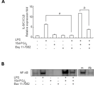
Figure 3. Upregulatory effect of 15d-PGJ2 on LPS-induced IL-8/CXCL8 mRNA expression is dependent on NF-κB activation in SHR VSMCs.
(A) VSMCs were untreated or treated with LPS (1 μg/ml) and/or 15d-PGJ2 (10 μM) in the absence or presence of Bay11-7082 (10 μ M) for 4 h. Bars represent means±SD from three independent real-time PCR experiments. a: p<0.05 vs. VSMCs treated with LPS alone. b: p<0.05 vs. VSMCs treated with 15d-PGJ2/LPS. (B) Specific binding activity of NF-κB from nuclear extracts was assessed by EMSA. Aliquots of the nuclear extract were incubated with a 100-fold excess of the mutant probe (m) or with 2 μg of the anti NF-κB antibody before EMSA. Data shown are representative of four independent experiments.
Figure 2. Upregulatory effect of 15d-PGJ2 on LPS-induced IL-8/CXCL8 mRNA expression is dependent on the PPARγ pathway in SHR VSMCs. (A) VSMCs were untreated (NT) or treated with LPS (1 μg/ml) and/or 15d-PGJ2 (10 μM) for 4 h, and the total RNA was analyzed by real-time PCR. Bars represent means±SD from three independent experiments. (B) VSMCs were untreated or treated with LPS (1 μg/ml) and/or 15d-PGJ2 (10 μM) in the absence or presence of GW9662 (10, 40, and 100 μM) for 4 h. Bars represent means±SD from three independent experiments. *p<0.05 vs. VSMCs treated with 15d-PGJ2/ LPS.
location of NF-κB. Real-time PCR and EMSA were performed on VSMCs after they were untreated or treated with LPS (1 μg/ml) and/or 15d-PGJ2 (10 μM) in the absence or pres- ence of Bay11-7082 (10 μM) for 4 h. Bay11-7082 remarkably blocked the upregulatory effect of 15d-PGJ2 on LPS-induced IL-8/CXCL8 mRNA expression. And it also remarkably blocked LPS-induced IL-8/CXCL8 expression (Fig. 3A). However, in spite of negative NF-κB activity in cells treated 15d-PGJ2
alone, NF-κB activity in SHR VSMCs treated with15d-PGJ2/ LPS was remarkably increased compared to the activity in cells treated with LPS alone (Fig. 3B).
Next we investigated whether the MAPK signaling path- ways are involved in the upregulatory effect of 15d-PGJ2 in SHR VSMCs. After VSMCs were untreated (NT) or pretreated with PD98059 (ERK inhibitor, 10 μM, 4A), or PD169316 (p38 inhibitor, 10 μM, 5A) for 30 min, cells were untreated or treated with LPS (1 μg/ml) and/or 15d-PGJ2 (10 μM) for 4 h. Real-time PCR was then performed on these treated cells.
In addition, these results were further confirmed by inves- tigating the phosphorylation of MAP kinases in VSMCs that had been treated with 15d-PGJ2/LPS. The expression of 15d-
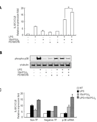
PGJ2/LPS-induced IL-8/CXCL8 mRNA was decreased by the ERK inhibitor PD98059. And the expression of LPS alone-in- duced IL-8/CXCL8 mRNA was also decreased by PD98059 (Fig. 4A). However, although ERK phosphorylation in cells treated 15d-PGJ2 alone was not detected, a remarkable in- crease in ERK phosphorylation in VSMCs that were treated with 15d-PGJ2/LPS relative to VSMCs that were treated with LPS alone was also detected (Fig. 4B). PD169316 increased the IL-8/CXCL8 mRNA expression in VSMCs stimulated with 15d-PGJ2/LPS, rather than inhibiting IL-8/CXCL8 expression (Fig. 5A). Moreover, 15d-PGJ2 decreased LPS-induced p38 phosphorylation (Fig. 5B). More specifically, blocking p38 phosphorylation caused an increase in 15d-PGJ2/LPS-induced IL-8/CXCL8 expression. To confirm this result, real-time PCR using p38-directed small interfering RNA (siRNA) was performed. In this experiment we found that while LPS in- hibited the expression of IL-8/CXCL8 mRNA, 15d-PGJ2 in- duced IL-8/CXCL8 expression and 15d-PGJ2/LPS increased IL-8/CXCL8 mRNA expression in p38 siRNA transfected SHR VSMCs (Fig. 5C).
69 Figure 4. Upregulatory effect of 15d-PGJ2 on LPS-induced IL-8/CXCL8
expression is mediated through the ERK pathway in SHR VSMCs. (A) VSMCs were untreated (NT) or pretreated with PD98059 (ERK inhibitor, 10 μM) for 30 min, and then untreated or treated with LPS (1 μg/ml) and/or 15d-PGJ2 (10 μM) for 4 h. Real-time PCR was performed after total mRNAs were isolated. Bars represent means±SD from three independent experiments. a: p<0.05 vs. VSMCs treated with LPS alone. b: p<0.05 vs. VSMCs treated with 15d-PGJ2/LPS. (B) Cell lysates were separated on 10% SDS-polyacrylamide gels and then immunoblotted with the phospho-ERK antibody. The data shown are representative of three independent experiments.
Figure 5. Blocking of p38 phosphorylation increased the expression of 15d-PGJ2/LPS-induced IL-8/CXCL8 mRNA expression in SHR VSMCs.
(A) VSMCs were untreated (NT) or pretreated with PD169316 (p38 inhibitor, 10 μM) for 30 min, and then untreated or treated with LPS (1 μg/ml) and/or 15d-PGJ2 (10 μM) for 4 h. Real-time PCR was per- formed after total mRNAs were isolated. Bars represent means±SD from three independent experiments. *p<0.05 vs. VSMCs treated with 15d-PGJ2/LPS. (B) Cell lysates were separated on 10% SDS-poly- acrylamide gels and then immunoblotted with the phospho-p38 anti- body. Data shown are representative of four independent experiments.
(C) VSMCs were plated on 24-well plates, grown to 90% confluence and then transfected with p38 siRNA oligomers (20 nmol/l). VSMCs were then untreated or treated with LPS (1 μg/ml) and/or 15d-PGJ2
(10 μM) for 4 h. Bars represent means±SEM from three independent experiments.
Effect of NAD(P)H oxidase activity on 15d-PGJ2/LPS- induced IL-8/CXCL8 expression in SHR VSMCs VSMCs generate reactive oxygen species (ROS), which play an important role in the pathogenesis of hypertensive vas- cular injury. A major source of ROS is NAD(P)H oxidase.
Therefore, we investigated whether NAD(P)H oxidase activity is related to the upregulatory effect of 15d-PGJ2 on LPS-in- duced IL-8/CXCL8 expressions in SHR VSMCs.
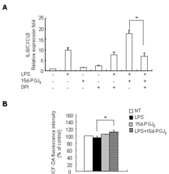
Real-time PCR was performed on VSMCs SHR after they were untreated or treated with LPS (1 μg/ml) and/or 15d- PGJ2 (10 μM) in the absence or presence of flavin containing oxidase inhibitor, DPI (10 μM) for 4 h. DPI remarkably de- creased the expression of 15d-PGJ2/LPS-induced IL-8/CXCL8 mRNA; thus, blocking the upregulatory effects of 15d-PGJ2 on LPS-induced IL-8/CXCL8 expression (Fig. 6A). To support these results, the ability of 15d-PGJ2/LPS to induce NAD(P)H oxidase activity was examined in SHR VSMCs. ROS gen- eration in SHR VSMCs was measured by flow cytometric anal- ysis of DCF-DA-stained VSMCs. DCF-DA fluorescence can be also used as a measurement of NAD(P)H oxidase activity.
From this analysis, SHR VSMCs treated with 15d-PGJ2/LPS was shown to increase DCF-DA fluorescence slightly compared to those treated with LPS alone (Fig. 6B).
DISCUSSION
In relation to IL-8/CXCL8 expression, 15d-PGJ2 was shown to have pro-inflammatory effects in SHR VSMCs and anti-in- flammatory effects in WKY VSMCs. Wakino et al. (30) also observed similar differential effects of PPARγ ligands on vas- cular tissues from SHR and WKY. They demonstrated that pioglitazone reduces the stimulated Rho-kinase activity in the vascular tissue from SHR, but not WKY. Troglitazone mark-
Figure 6. Activity of NAD(P)H oxidase mediates the upregulatory effect of 15d-PGJ2 on the expression of LPS-induced IL-8/CXCL8 mRNA in SHR VSMCs. (A) VSMCs were untreated or treated with LPS (1 μg/
ml) and/or 15d-PGJ2 (10 μM) in the absence or presence of DPI (10 μM) for 4 h. Bars represent means±SD from three independent experiments. *p<0.05 vs. VSMCs treated with 15d-PGJ2/LPS. (B) VSMCs were untreated or treated with LPS (1 μg/ml) and/or 15d-PGJ2
(10 μM) for 4 h, stained with DCF-DA (50 μM) for ROS detection, and subjected to flow cytometry. Bars represent means±SD from four independent experiments. *p<0.05 vs. VSMCs treated with LPS alone.
edly decreased the expression of TGFβ-1, PDGF, or bFGF mRNAs in SHR VSMCs, but not in WKY VSMCs (12). Therefore, these different effects of PPAR ligands on SHR and WKY VSMCs indicate that diverse, complex pathways mediate the action of PPARg ligands on hypertension.
The upregulatory effect of 15d-PGJ2 on LPS-induced IL-8/CXCL8 expression was mediated through PPARg in SHR VSMCs. 15d-PGJ2 and other cyclopentenone prostaglandin, such as prostaglandin A1 (PGA1) are known to exert effects on cytokine genes through PPARγ-dependent and PPARγ- independent mechanisms (25,26,28,29,31,32). In our previous studies (25,26), the upregulatory effect of 15d-PGJ2 on LPS-in- duced MIP-2/CXCL2 and KC/CXCL1 gene expression was found to not mediated by the PPARγ pathway, but the effect of PGA1 on LPS-induced IL-10 expression was dependent on PPARγ in mouse peritoneal macrophages. In a separate VSMCs study, 15d-PGJ2-induced HO-1 expression was shown to be independent of PPARγ (19).
In the EMSA, an increase in NF-κB activity in SHR VSMCs treated with 15d-PGJ2/LPS relative to VSMCs treated with LPS alone was detected, and the expression of 15d-PGJ2/LPS-in- duced IL-8/CXCL8 mRNA was decreased by the presence of Bay11-7082. These results indicate that the upregulatory effect of 15d-PGJ2 in SHR VSMCs is dependent on NF-κB acti- vation. It is widely accepted that 15d-PGJ2 exerts its effects on inflammatory mediated-genes in cells by either inhibiting or activating NF-κB signaling (24,26,33-35). The anti-in- flammatory activity of 15d-PGJ2 has been shown to be medi- ated mainly through the inhibition of NF-κB activation (33, 34), but 15d-PGJ2 has also been shown to upregulate IL-8/
CXCL8 and MIP-2/CXCL2 expression through NF-κB activa- tion (24,26).
Among the MAPK signaling pathways, the ERK pathway is known to be associated with the stimulatory activity of 15d- PGJ2 on the expression of some cytokine genes (23,24,36).
15d-PGJ2 induces the rapid activation of the ERK pathway in VSMCs (37). We also detected an increase in ERK phosphor- ylation in SHR VSMCs treated with 15d-PGJ2/LPS. Up-regu- lation of LPS-induced IL-8/CXCL8 expression by 15d-PGJ2 was mediated through the ERK signaling pathway in SHR VSMCs.
Inhibition or activation by 15d-PGJ2 on p38 MAP kinase ap- pears to be target gene-, cell type- and stimulation con- dition-dependent (19,38,39). Activation of the p38 MAP kin- ase has been shown to be involved in 15d-PGJ2-induced HO-1 expression (19) and in IL-1β-induced IL-8/CXCL8 gene ex- pression in human VSMCs (38). However, 15d-PGJ2 is known to inhibit IL-1-induced p38 MAP kinase expression in human astrocytes (39). In this study, blocking p38 phosphorylation increased 15d-PGJ2/LPS-induced IL-8/CXCL8 expression in SHR VSMCs. 15d-PGJ2 inhibited p38 phosphorylation in SHR VSMCs treated with LPS. Furthermore, while the expression of LPS-induced IL-8/CXCL8 mRNA was abolished in SHR VSMCs that were transfected with p38 siRNA, 15d-PGJ2/LPS- induced IL-8/CXCL8 expression increased. Therefore, LPS-in- duced IL-8/CXCL8 mRNA expression appears to be related to p38 activation, but 15d-PGJ2 itself induces IL-8/CXCL8 mRNA expression without p38 activation. These combined results in- dicate that the upregulatory effect of 15d-PGJ2 on LPS-induced IL-8/CXCL8 expression is related to p38 inactivation. Although the upregulatory effect of 15d-PGJ2 on LPS-induced IL-8/
CXCL8 expression is mediated by the ERK pathway, an un- known mechanism via p38 inactivation may play an im- portant role in 15d-PGJ2/LPS-induced IL-8/CXCL8 expression in SHR VSMCs.
71
NAD(P)H oxidase is a known major source of reactive oxy- gen species (ROS). ROS are not only harmful cellular metabo- lites but are also essential molecules in cell signaling and reg- ulation (40). Excessive ROS generation by NAD(P)H oxidase is known to play an important role in the pathogenesis of hypertensive vascular injury and has also been implicated in the pathogenesis of hypertension (1,41-44). Cruzado et al.
(41) demonstrated that ROS generation was enhanced in SHR VSMCs during the development of hypertension. Therefore, we examined the effect of NAD(P)H oxidase activity on IL-8/
CXCL8 expression in SHR VSMCs treated with 15d-PGJ2/LPS.
An inhibitor of the flavin-containing oxidases, DPI, remark- ably decreased the expression of 15d-PGJ2/LPS-induced IL-8/
CXCL8 mRNA. Although a significant production of ROS by 15d-PGJ2 has been reported in Sprague-Dawley rat VSMCs (19) and ROS generation by LPS alone was detected in WKY VSMCs (data not shown), ROS generation did not increase in SHR VSMCs after treatment with 15d-PGJ2 or LPS alone.
However, 15d-PGJ2/LPS did increase ROS generation in SHR VSMCs. These results suggest that the upregulatory effect of 15d-PGJ2 on LPS-induced IL-8/CXCL8 expression in SHR VSMCs may be related to NAD(P)H oxidase activity.
In conclusion, this is the first study to report on the upregu- latory effect of 15d-PGJ2 on LPS-induced IL-8/CXCL8 gene ex- pression in SHR VSMCs and the inhibitory effect in WKY VSMCs. In addition, we showed that the upregulatory effect of 15d-PGJ2 in SHR VSMCs is mediated through PPARγ path- way, NF-κB and ERK activation, and p38 inactivation may play an important role in 15d-PGJ2/LPS-induced IL-8/CXCL8 expression. These results provide new insight into the poten- tial diverse effects of 15d-PGJ2 on hypertensive vascular smooth muscle cells.
ACKNOWLEDGEMENT
This work was supported by the Korean Science and Engineering Foundation (KOSEF) grant funded by the Korean government (MEST) (No. R13-2005-005-02002-0(2008)).
CONFLICTS OF INTEREST
The authors have no financial conflict of interest.
REFERENCES
1. Alexander RW: Hypertension and the pathogenesis of
atherosclerosis. Oxidative stress and the mediation of arte- rial inflammatory response: a new perspective. Hyperten- sion 25;155-161, 1995
2. Capers Q 4th, Alexander RW, Lou P, De Leon H, Wilcox JN, Ishizaka N, Howard AB, Taylor WR: Monocyte chemo- attractant protein-1 expression in aortic tissues of hyper- tensive rats. Hypertension 30;1397-1402, 1997
3. Rodríguez-Iturbe B, Vaziri ND, Herrera-Acosta J, Johnson RJ: Oxidative stress, renal infiltration of immune cells and salt-sensitive hypertension: all for one and one for all. Am J Physiol 286;F606-F616, 2004
4. Zhang Y, Griendling KK, Dikalova A, Owens GK, Talyor WR: Vascular hypertrophy in angiotensin II-induced hyper- tension is mediated by vascular smooth muscle cell-derived H2O2. Hypertension 46;732-737, 2005
5. Gerszten RE: Pleiotropic effects of chemokines in vascular lesion development. Artherioscler Thromb Vasc Biol 22;
528-529, 2002
6. Luster AD: Chemokines - chemotactic cytokines that medi- ate inflammation. N Engl J Med 338;436-445, 1998 7. Gerszten RE, Garcia-Zepeda EA, Lim YC, Yoshida M, Ding
HA, Gimbrone MA, Luster AD, Luscinskas FW, Rosenzweig A: MCP-1 and IL-8 trigger firm adhesion of monocytes to vascular endothelium under flow conditions. Nature 398;
718-723, 1999
8. Boekholdt S, Peters R, Hack CE, Day NE, Luben R, Bingham SA, Wareham NJ, Reitsma PH, Khaw K: IL-8 plas- ma concentrations and the risk of future coronary artery disease in apparently healthy men and women: the EPIC-Norfolk prospective population study. Arterioscler Thromb Vasc Biol 24;1503-1508, 2004
9. Buemi M, Marino D, Floccari F, Ruello A, Nosto L, Aloisi C, Marino MT, Di Pasquale G, Corica F, Frisina M: Effect of interleukin 8 and ICAM-1 on calcium-dependent outflow of K+ in erythrocytes from subjects with essential hyper- tension. Curr Med Res Opin 20;19-24, 2004
10. Kim HY, Kang YJ, Song IH, Choi HC, Kim HS: Upregu- lation of Interleukin-8/CXCL8 in vascular smooth muscle cells from spontaneously hypertensive rats. Hypertens Res 31;515-523, 2008
11. Dobrian AD, Schriver SD, Khraibi AA, Prewitt RL: Pioglita- zone prevents hypertension and reduces oxidative stress in diet-induced obesity. Hypertension 43;48-56, 2004 12. Fukuda N, Hu WY, Teng J, Chikara S, Nakayama K,
Kanmatsuse K: Troglitazone inhibits growth and improves insulin signaling by suppression of angiotensin II action in vascular smooth muscle cells from spontaneously hyper- tensive rats. Atherosclerosis 163;229-239, 2002
13. Wakino S, Hayashi K, Tatematsu S, Hasegawa K, Takamatsu I, Kanda T, Homma K, Yoshioka K, Sugano N, Saruta T:
Pioglitazone lowers systemic asymmetric dimethylarginine by inducing dimethylarginine dimethylaminohydrolase in rats. Hypertens Res 28;255-262, 2005
14. Calnek DS, Mazzella L, Roser S, Roman J, Hart CM:
Peroxisome proliferatorsactivated receptor gamma ligands increase release of nitric oxide from endothelial cells.
Arterioscler Thromb Vasc Biol 23;52-57, 2003
15. Jiang C, Ting AT, Seed B: PPAR-gamma agonists inhibit
production of monocyte inflammatory cytokines. Nature 391; 82-86, 1998
16. Chastine Bell-Parikh L, Ide T, Lawson JA, Mcncmara P, Reilly M, Fitzgerald GA: Biosynthesis of 15-deoxy-delta 12,14- PGJ2 and the ligation of PPARgamma. J Clin Invest 112;945-955, 2003
17. Benkirane K, Amiri F, Diep QN, Mabrouk ME, Schiffrin EL:
PPAR-gamma inhibits ANG II-induced cell growth via SHIP2 and 4E-BP1. Am J Physiol Heart Circ Physiol 290;H390- H397, 2006
18. Law RE, Goetze S, Xi, XP, Jackson S, Kawano Y, Demer L, Fishbein MC, Meehan WP, Hsueh WA: Expression and function of PPARgamma in rat and human vascular smooth muscle cells. Circulation 101;1311-1318, 2000 19. Lim HJ, Lee KS, Lee S, Park JH, Choi HE, Go SH, Kwak
HJ, Park HY: 15d-PGJ2 stimulates HO-1 expression through p38 MAP kinase and Nrf-2 pathway in rat vascular smooth muscle cells. Toxicol and Appl Pharmacol 223;20-27, 2007 20. Cuzzocrea S, Wayman NS, Mazzon E, Dugo L, Paola R, Serraino I, Britti D, Chatterjee PK, Caputi AP, Thiemermann C: The cyclopentenone prostaglandin 15-deoxy-Delta(12,14)- prostaglandin J(2) attenuates the development of acute and chronic inflammation. Mol Pharmacol 61;997-1007, 2002 21. Reilly CM, Oates JC, Suidan J, Crosby MB, Halushka PV,
Gilkeson GS: Prostaglandin J2 inhibition of mesangial cell iNOS expression. Clin Immunol 3;337-345, 2001
22. Sawano H, Haneda M, Sugimoto T, Inoki K, Koya D, Kikkawa R: 15-Deoxy-Delta12,14-prostaglandin J2 inhibits IL-1beta-induced cyclooxygenase-2 expression in mesangial cells. Kideny Int 61;1957-1967, 2002
23. Fu Y, Luo N, Lopes-Virella MF: Upregulation of inter- leukin-8 expression by prostaglandin J2 (15d-PGJ2) in hu- man THP-1 macrophages. Atherosclerosis 160;11-20, 2002 24. Harris SG, Smith RS, Phipps RP: 15-deoxy-Delta 12,14-PGJ2
induces IL-8 production in human T cells by a mi- togen-activated protein kinase pathway. J Immunol 168;
1372-1379, 2002
25. Kim HY, Kim HK, Kim JR, Kim HS: Upregulation of LPS-in- duced chemokine KC expression by 15-deoxy-delta12,14- prostaglandin J2 in mouse peritoneal macrophages. Immu- nol Cell Biol 83;286-293, 2005.
26. Kim HY, Kim HS: Upregulation of MIP-2 (CXCL2) ex- pression by 15-deoxy-Delta(12,14)-prostaglandin J(2) in mouse peritoneal macrophages. Immunol Cell Biol 85;60- 67, 2007
27. Kelly G, Robert B, Chris R, Gary G, Perry H, James C:
Differential effects of 15-deoxy-Delta(12,14)-PGJ2 and a peroxisome proliferators-activated receptor gamma agonist on macrophage activation. J Leukoc Biol 69;631-638, 2001 28. Ricote M, Huang JT, Welch JS, Glass CK: The peroxisome
proliferator-activated receptor gamma (PPARgamma) as a regulator of monocyte/macrophage function. J Leukoc Biol 66;733- 739, 1999
29. Zhang X, Wang JM, Gong WH, Mukaida N, Young HA:
Differential regulation of chemokine gene expression by 15-deoxy-delta 12,14 prostaglandin J2. J Immunol 166;7104- 7111, 2001
30. Wakino S, Hayashi H, Kanda T, Tatematsu S, Homma K,
Yoshioka K, Taksmatsu I, Saruta T: Peroxisome pro- liferatior-activated receptor gamma ligands inhibit Rho/Rho kinase pathway by inducing protein tyrosine phosphatase SHP-2. Circulation 95;e45-e55, 2004
31. Guyton K, Bond R, Reilly C, Gileson G, Halushka P, Cook J: Differential effects of 15-deoxy-delta(12,14)-prostaglandin J2 and a peroxisome proliferator-activatedreceptor gamma agonist on macrophage activation. J Leukoc Biol 69;631- 638, 2001
32. Kim HY, Kim JR, Kim HS: Upregulation of lipopoly- saccharide-induced interleukin-10 by prostaglandin A1 in mouse peritoneal macrophages. J Microbiol Biotech 18;1170-1178, 2008
33. Bureau F, Desmet C, Melotte D, Jaspar F, Volanti C, Vanderplasschen A, Pastoret PP, Piette J, Lekeux P: A pro- inflammatory role for the cyclopentenone prostaglandins at low micromolar concentrations: oxidative stress-induced extracellular signal-regulated kinase activation without NF-kappa B inhibition. J Immunol 168;5318-5325, 2002 34. Ricote M, Li AC, Willson TM, Kelly CJ, Glass CK: The per-
oxisome proliferator-activated receptor-gamma is a negative regulator of macrophage activation. Nature 391;79-82, 1998 35. Straus DS, Pascual G, Li M, Welch JS, Ricote M, Hsiang CH,
Sengchanthalangsy LL, Ghosh G, Galss CK: 15-deoxy-delta 12,14-prostaglandin J2 inhibits multiple steps in the NF-kap- pa B signaling pathway. Proc Natl Acad Sci U S A 97;4844- 4849, 2000
36. Wilmer WA, Dixon C, Lu L, Hilbelink T, Rovin BH: A cyclo- pentenone prostaglandin activates mesangial MAP kinase independently of PPARgamma. Biochem Biophys Res Comm 281;57-62, 2001
37. Takeda K, Ichiki T, Tokunou T, Iino N, Takeshita A:
15-Deoxy-delta 12,14-prostaglandin J2 and thiazolidine- diones activate the MEK/ERK pathway through phosphati- dylinositol 3-kinase in vascular smooth muscle cells. J Biol Chem 276;48950-48955, 2001
38. Jung YD, Fan F, McConkey DJ, Jean ME, Liu W, Reinmuth N, Stoeltzing O, Ahmad SA, Parikh AA, Mukaida N, Ellis LM: Role of p38 MAPK, AP-1 and NF-kappaB in inter- leukin-1beta-induced IL-8 expression in human vascular smooth muscle cells. Cytokine 18;206-213, 2002 39. Zhao ML, Brosnan CF, Lee SC: 15-deoxy-delta (12,14)-PGJ2
inhibits astrocyte IL-1 signaling: inhibition of NF-kappaB and MAP kinase pathways and suppression of cytokine and chemokine expression. J Neuroimmunol 153;132-142, 2004 40. Thannickal VJ, Fanburg BL: Reative oxygen species in cell
signaling. Am J Physiol Lung Cell Mol Physiol 279;L1005- L1028, 2000
41. Cruzado MC, Risler NR, Miatello RM, Yao G, Schiffrin EL, Touyz RM: Vascular smooth muscle cell NAD(P)H oxidase activity during the development of hypertension: effect of angiotensin II and role of insulinkike growth factor-1 re- ceptor transactivation. Am J Hyertension 18;81-87, 2005 42. Kunsch C, Medford RM: Oxidative stress as a regulator of gene expression in the vasculature. Circ Res 85;753-766, 1999
43. Seshiah PN, Weber DS, Rocic P, Valppu L, Taniyama Y, Griendling KK: Angiotensin II stimulation of NAD(P)H oxi-
73
dase activity upstream mediators. Circ Res 91;406-413, 2002 44. Touyz RM, Schiffrin EL: Ang II-stimulated generation of re- active oxygen species in Human vascular smooth muscle
cells in mediated via PLD-dependent pathways. Hyperten- sion 34;976-982, 1999