ABSTRACT
Purpose: The objective of this study was to investigate whether phelligridin D could reduce glucose-induced oxidative stress, attenuate the resulting inflammatory response, and restore the function of human periodontal ligament cells (HPDLCs).
Methods: Primary HPDLCs were isolated from healthy human teeth and cultured. To investigate the effect of phelligridin D on glucose-induced oxidative stress, HPDLCs were treated with phelligridin D, various concentrations of glucose, and glucose oxidase. Glucose-induced oxidative stress, inflammatory molecules, osteoblast differentiation, and mineralization of the HPDLCs were measured by hydrogen peroxide (H2O2) generation, cellular
viability, alkaline phosphatase (ALP) activity, alizarin red staining, and western blot analyses. Results: Glucose-induced oxidative stress led to increased production of H2O2, with negative
impacts on cellular viability, ALP activity, and calcium deposition in HPDLCs. Furthermore, HPDLCs under glucose-induced oxidative stress showed induction of inflammatory molecules (intercellular adhesion molecule-1, vascular cell adhesion protein-1, tumor necrosis factor-alpha, interleukin-1-beta) and disturbances of osteogenic differentiation (bone morphogenetic protein-2, and -7, runt-related transcription factor-2), cementogenesis (cementum protein-1), and autophagy-related molecules (autophagy related 5, light chain 3 I/II, beclin-1). Phelligridin D restored all these molecules and maintained the function of HPDLCs even under glucose-induced oxidative stress.
Conclusions: This study suggests that phelligridin D reduces the inflammation that results from glucose-induced oxidative stress and restores the function of HPDLCs (e.g., osteoblast differentiation) by upregulating autophagy.
Keywords: Cementum; Inflammation; Osteogenesis; Periodontitis
INTRODUCTION
The periodontal ligament (PDL) is a specific connective tissue that unites the alveolar bone and cementum in order to support the teeth within the jaw. In addition to its role in maintaining the teeth, the PDL also helps to supply nutrients for the repair of damaged tissue and to maintain homeostasis in periodontal tissue [1,2]. The PDL consists of mixed cell types, including fibroblasts and undifferentiated mesenchymal cells, which differentiate
Research Article
Received: Jul 7, 2019 Revised: May 19, 2020 Accepted: Jun 24, 2020 *Correspondence: Ho-Keun YiDepartment of Oral Biochemistry, Institute of Oral Bioscience, Jeonbuk National University School of Dentistry, 567 Baekje-daero, Deokjin-gu, Jeonju 54896, Korea. E-mail: [email protected] Tel: +82-63-270-4033 Fax: +82-63-270-4004
†Ji-Eun Kim and Tae-Gun Kim contributed equally to the work.
Copyright © 2020. Korean Academy of Periodontology
This is an Open Access article distributed under the terms of the Creative Commons Attribution Non-Commercial License (https:// creativecommons.org/licenses/by-nc/4.0/). ORCID iDs Ji-Eun Kim https://orcid.org/0000-0003-4683-4775 Tae-Gun Kim https://orcid.org/0000-0001-5608-0132 Young-Hee Lee https://orcid.org/0000-0002-8081-4367 Ho-Keun Yi https://orcid.org/0000-0003-4403-3827 Funding
This work was supported by the National Research Foundation of Korea (NRF) funded by the Korean government (MSIP) (2018R1A2B6004744). This research was also supported by the “Research Base Construction Fund Support Program” funded by Jeonbuk National University in 2019.
Ji-Eun Kim †, Tae-Gun Kim †, Young-Hee Lee , Ho-Keun Yi *
Department of Oral Biochemistry, Institute of Oral Bioscience, Jeonbuk National University School of Dentistry, Jeonju, Korea
Phelligridin D maintains the function
of periodontal ligament cells through
autophagy in glucose-induced
oxidative stress
Author Contributions
Conceptualization: Tae-Gun Kim, Young-Hee Lee, Ho-Keun Yi; Formal analysis: Ji-Eun Kim, Young-Hee Lee, Ho-Keun Yi; Investigation: Ji-Eun Kim, Ho-Keun Yi; Methodology: Ji-Eun Kim, Tae-Gun Kim, Young-Hee Lee; Project administration: Tae-Gun Kim, Young-Hee Lee, Ho-Keun Yi; Writing - original draft: Ji-Eun Kim, Ho-Keun Yi; Writing - review & editing: Ho-Keun Yi.
Conflict of Interest
No potential conflict of interest relevant to this article was reported.
into cementoblast and osteoblast phenotypes [3,4]. Inflammation of the PDL is defined as periodontitis, which affects the supportive function of teeth [5]. Several studies have established that periodontitis is associated with diabetes mellitus (DM) [6-8]. More than three times as many DM patients have periodontitis than individuals without DM. A previous study found destruction of the periodontal tissue around the mandible and increased levels of inflammatory molecules in systemic DM-induced rats, suggesting that DM is involved in local periodontal tissue inflammation [9]. Therefore, periodontitis may be considered as a complication of DM [10].
DM is a chronic systemic disease commonly associated with neuropathy, nephropathy, retinopathy, and micro- and macro-vasculopathy [11]. The progress of diabetic complications is related to abnormal regulation of glucose levels, and DM contributes to serious periodontal diseases [12]. However, the mechanism through which DM contributes to chronic
periodontitis remains unclear.
Autophagy is a natural cellular process that recycles damaged proteins and organelles in order to maintain cellular homeostasis [13]. Similarly, autophagy plays a critical role in the maintenance of cellular homeostasis by regulating cell survival and death. If the autophagy system fails to remove intracellular debris from cells, cellular apoptosis will occur, resulting in tissue damage [14]. Consequently, dysfunction in autophagy contributes to the course of various diseases, including the progression of [15]. A previous study suggested that autophagy can protect cellular homeostasis against inflammatory conditions in PDL tissue and cells [16]. However, the role of autophagy in the inflammatory action and regulation of PDL function is poorly understood in human periodontal ligament cells (HPDLCs) affected by glucose-induced oxidative stress.
Phelligridin D is a compound isolated from the Phellinus baumii mushroom, which belongs
to the family Hymenochaetaceae. P. baumii has various biological properties, including
anti-influenza, anti-oxidant, anti-inflammatory, anti-obesity, and anti-platelet effects [17-20]. It has been conventionally consumed as a food source in East Asian countries, such as Korea, Japan, and China. Previous studies have shown that phelligridin D minimizes PDL inflammation by inhibiting the mitogen-activated protein kinase pathway and restores the function of HPDLCs in lipopolysaccharide -induced periodontitis [21]. Moreover, the implantation of phelligridin D-coated titanium dental implants in rat mandible led to increased osteogenesis and osseointegration of dental implants with bone [22]. In addition, it has been reported that P. baumii exerts anti-diabetic activity by improving insulin sensitivity
[23]. However, the effects of phelligridin D regarding the maintenance of PDL function and its relationship with autophagy have not been demonstrated yet.
In this study, we assumed that the functional and molecular alterations of HPDLCs caused by reactive oxygen species (ROS) in high-glucose conditions is similar to the cellular changes caused by DM. Therefore, the purpose of this study was to investigate the mechanism by which glucose-induced oxidative stress in HPDLCs causes dysfunction that is restored by phelligridin D.
MATERIALS AND METHODS
Materials
Alizarin red S, β-glycerophosphate disodium salt hydrate, dexamethasone, D-glucose, glucose oxidase, rapamycin (RAPA), and L-ascorbic acid were obtained from Sigma-Aldrich (St. Louis, MO, USA). Antibodies against intracellular adhesion molecule-1 (ICAM-1), vascular cell adhesion molecule-1 (VCAM-1), and interleukin-1-beta (IL-1β) were purchased from Santa Cruz Biotechnology (Santa Cruz, CA, USA). Bone morphogenetic protein-2 (2), BMP-7, runt-related transcription factor-2 (RUNX-2), and β-actin were obtained from Bioworld Technology (Louis Park, MN, USA). Tumor necrosis factor-alpha (TNF-α) and light chain 3 (LC3I/II) were obtained from Cell Signaling Technologies (Beverly, MA, USA). Cementum protein 1 (CEMP-1) was purchased from LifeSpan BioSciences (Seattle, WA, USA). Autophagy related 5 (ATG5) was obtained from Novus Biologicals (Littleton, CO, USA). Beclin-1 was purchased from Bethyl Laboratories (Montgomery, TX, USA).
Isolation and identification of phelligridin D
The isolation and identification of phelligridin D followed a previously described method [24]. Briefly, ground fruiting bodies of P. baumii were twice extracted at room temperature
with methanol. Next, the methanol was removed with reduced pressure, after which the remaining solution was divided between H2O and n-hexane, and between H2O and ethyl
acetate. The ethyl acetate-soluble fraction was loaded into a column in Sephadex LH-20 (Amersham Bioscience, Upsala, Sweden) and eluted with methanol. Phelligridin D was purified by a Sep-pak ODS cartridge (Amersham Bioscience), and eluted with 50%–60% aqueous methanol. Phelligridin D was collected and purified by reversed-phase
high-performance liquid chromatography (HPLC) (Hitachi L-2000 series, Hitachi, Tokyo, Japan) as previously reported [24].
Culture and treatment of HPDLCs with glucose oxidase to induce oxidative
stress
A freshly extracted third molar was collected for culture of HPDLCs. The Human Ethics Committee of Jeonbuk National University Hospital (CUH2015-11-03) approved the protocols. HPDLCs were isolated and cultured as previously reported [21]. Briefly, PDL tissue from the third molar was isolated, minced into small pieces with a surgical scissor, and then digested with 0.5 mg/mL trypsin and 3 mg/mL collagenase type II at 37°C for 10 minutes. The pieces of tissue were cultured in a 25-mm2 cell culture flask in Dulbecco's modified Eagle's medium (DMEM; Gibco,
Life Technologies, Grand Island, NY, USA) supplemented with 300 μg/mL L-glutamine, 10% fetal bovine serum, 100 μg/mL streptomycin, and 100 U/mL penicillin at 37°C with 5% CO2 in a
humidified atmosphere. After the cells' growth, migrated HPDLCs from the tissue were collected and subcultured. All the experiments were completed using HPDLCs between passages 3 and 8. Mineralization of cells was performed in osteogenic media (OM) by treatment with 10 mM β-glycerophosphate, 100 nM dexamethasone, and 50 μg/mL ascorbic acid.
Glucose-induced oxidative stress was established following previously described methods [25]. Briefly, 80% confluent cells were supplemented with low (5 mM) and high (50 mM) D-glucose in glucose-free media and treated with 5 mU/mL glucose oxidase to induce oxidative stress for 10 days. The medium was changed with high- glucose medium and glucose oxidase every 3 days to maintain glucose-induced oxidative stress. For the detection of autophagy, the cells were treated with 1 µM of phelligridin D or 1 µM of RAPA (autophagy inducer) for the indicated time intervals.
Measurement of H
2O
2production
The quantity of H2O2 production by glucose oxidase in DMEM was analyzed using a Biovision
hydrogen peroxide assay kit (Biovision Research Products, Milpitas, CA, USA) according to the manufacturer's instructions at 24 hours. Absorbance was measured at a wavelength of 570 nm with an enzyme-linked immunosorbent assay (ELISA) reader (Bio-TeK, Winooski, VT, USA).
Measurement of cell viability
Cell viability was evaluated by quantifying the reduction of 3-(4,5-dimethylthiazol-2-yl)-2,5-diphenyltetrazolium bromide (MTT) to formazan. Following the indicated culture periods, MTT (100 μg/100 μL of phosphate-buffered saline [PBS]) solution was added to each well and incubated for 3 hours. Dimethyl sulfoxide was then added in order to dissolve the formazan crystals. Absorbance was measured at a wavelength of 570 nm with an ELISA reader (Synergy 2, Bio-TeK).
Alizarin red staining
HPDLCs were cultured in 6-well culture plates for 24 hours at 37°C. After incubation, the cells were washed with PBS and treated with 0.1 and 1 μM phelligridin D followed by glucose-induced oxidative stress. After 14 days of mineralization induction, the HPDLCs were washed with PBS, air-dried, and fixed in 95% ice-cold ethanol at −20°C for 30 minutes. These cells were stained with 40 mM alizarin red S (pH 4.2) at room temperature for 1 hour, washed 5 times extensively with deionized water, and then rinsed with PBS (without magnesium or calcium) for 15 minutes.
Alkaline phosphatase (ALP) activity
An ALP activity assay was performed in the HPDLCs after treatment with 0.1 and 1 µM of phelligridin D followed by glucose-induced oxidative stress in OM for 3, 7, and 14 days. The cells were scraped with ice-cold PBS, transferred to an ice-cold bath and sonicated with a cell disruptor in the indicated time. ALP activity was assessed in the supernatant using a SensoLyte p-NPP Alkaline Phosphatase Assay Kit (AnaSpec, Fremont, CA, USA) at 3, 7, and 10 days. The absorbance of the assay was measured using an ELISA reader (Synergy 2, Bio-TeK) at 405 nm.
Western blot analysis
A previously described method was used for western blot analysis [26]. In brief, protein samples were electroblotted onto nitrocellulose membranes after being separated by 8%–15% sodium dodecyl sulfate-polyacrylamide gel electrophoresis under denaturing conditions. These electroblotted membranes were incubated with a specified primary antibody and then a horseradish peroxidase-conjugated secondary antibody. Bands were visualized by chemiluminescent detection (Amersham Pharmacia Biotech, London, UK). Equal protein loading was confirmed in the membrane by reprobing with an antiactin antibody. Protein expression was quantified with ImageQuantT TL 1D gel program (GE Healthcare, BioScience, Sweden).
Statistical analysis
All the results were expressed as the mean±standard deviation of at least 3 independent experiments. Statistical analysis was performed using SPSS version 15.0 (SPSS, Chicago, IL, USA). Differences in the mean values for H2O2, cell viability, and ALP activity among cells
treated with different amounts of glucose oxidase, different concentrations of glucose with or without glucose oxidase, or glucose with or without different concentration of phelligridin D were assessed by 1-way analysis of variance followed by Duncan's multiple range test. P values
RESULTS
Effects of glucose oxidase on H
2O
2production, cell viability and osteogenic
differentiation
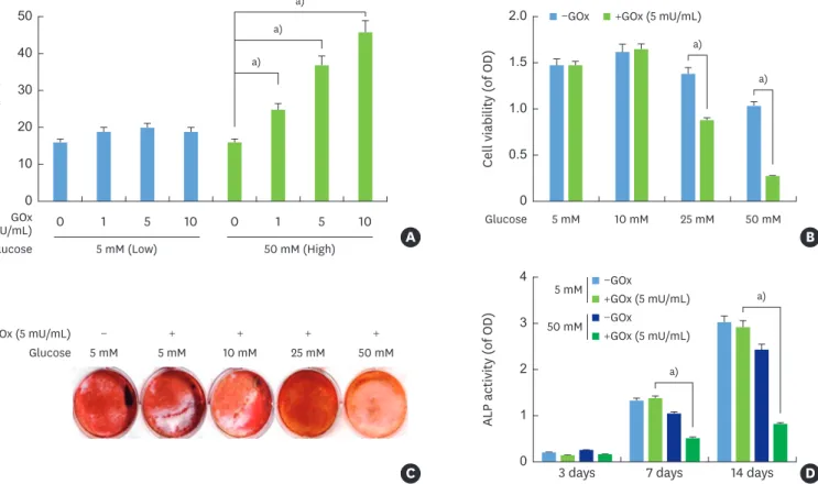
HPDLCs were treated with different concentrations of glucose and glucose oxidase, and high glucose levels showed significantly increased production of H2O2 in a
concentration-dependent manner (Figure 1A). Cell viability was detected by the MTT assay to rule out the cytotoxicity of the glucose concentration. Glucose concentrations of 25 mM and 50 mM with 5 mU/mL glucose oxidase demonstrated significantly lower cell viability than glucose levels of 5 mM and 10 mM with 5 mU/mL glucose oxidase (Figure 1B). Next, ALP activity and alizarin red staining were examined as markers of osteogenic differentiation in OM medium. ALP activity and alizarin red staining were inhibited under high-glucose conditions in the presence of glucose oxidase (Figure 1C and D).
Effects of glucose-induced oxidative stress on inflammation, osteogenic
differentiation, cementogenesis, and autophagy
The higher levels of H2O2 production induced by high glucose levels disturbed the cell
viability and osteogenic differentiation of HPDLCs. Similarly, the effects of high glucose levels on inflammation, osteogenic differentiation, and autophagy were also examined.
Cell viability (of OD)
0 0.5 1.0 2.0 1.5 5 mM Glucose 10 mM 25 mM 50 mM
−GOx +GOx (5 mU/mL)
B
a)
a) a) a)
ALP activity (of OD)
0 1 2 4 3
3 days 7 days 14 days D
C GOx (5 mU/mL) Glucose GOx (mU/mL) Glucose + 5 mM − + + 5 mM 10 mM 50 mM + 25 mM H2 O2 (µM ) 0 0 1 5 10 0 1 5 10 10 30 50 20 40 A −GOx +GOx (5 mU/mL) −GOx +GOx (5 mU/mL) 5 mM 50 mM 5 mM (Low) 50 mM (High) a) a) a)
Figure 1. H2O2 production by GOx at various glucose levels and the relationship of H2O2 production with cellular toxicity and mineralization in HPDLCs. (A) Cells were stimulated with GOx (1, 5, and 10 mU/mL) in the presence of low (5 mM) or high (50 mM) glucose levels. The amount of H2O2 production in the culture medium was then measured at 24 hours after treatment. (B) Cell viability was determined by an MTT assay at 24 hours. (C, D) Alizarin red staining and ALP activity were analyzed as mineralization markers at the indicated interval after stimulation with GOx according to the glucose levels in HPDLCs. Each value is reported as mean and standard error of the mean of 3 independent experiments.
H2O2: hydrogen peroxide, HPDLC: human periodontal ligament cell, MTT: 3-(4,5-dimethylthiazol-2-yl)-2,5-diphenyltetrazolium bromide, ALP: alkaline phosphatase, GOx: glucose oxidase, OD: optical density.
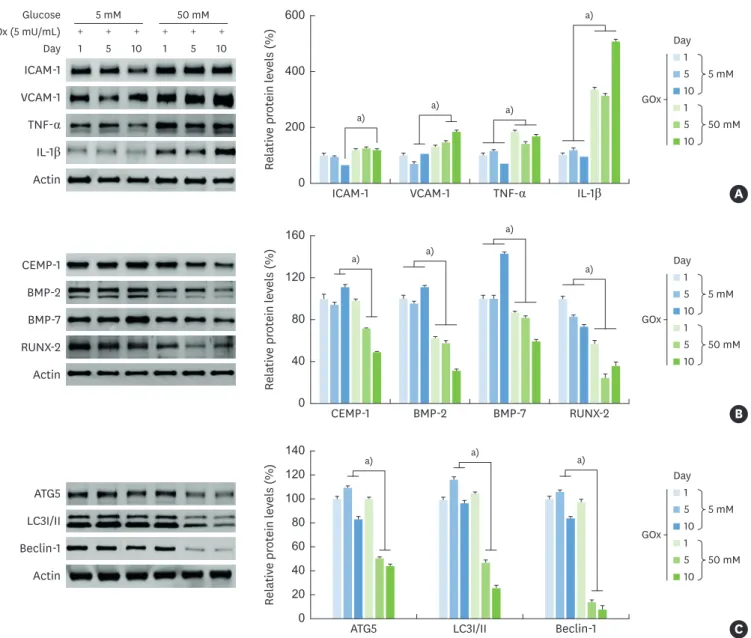
Inflammatory molecules such as ICAM-1, VCAM-1, TNF-α, and IL-1β were induced by high glucose levels with glucose oxidase in comparison with low-glucose conditions (Figure 2A). However, osteogenic differentiation-related molecules (BMP-2, BMP-7, and RUNX-2), CEMP-1, and autophagy-related molecules (ATG5, LC3I/II, and beclin-1) were gradually downregulated in a time-dependent manner under high- glucose conditions in the presence of glucose oxidase (Figure 2B and C).
Relativ e pr ot ein l ev els (%) 0 200 400 600 ICAM-1 a) a) a) a) 1 5 10 1 5 10 + + + + + + 5 mM GOx (5 mU/mL) Glucose Day Day ICAM-1 VCAM-1 TNF-α IL-1β Actin 50 mM VCAM-1 TNF-α IL-1β 1 5 10 1 5 10 5 mM GOx 50 mM A Day Relativ e pr ot ein l ev els (%) 0 40 80 160 120 CEMP-1 a) a) a) CEMP-1 BMP-2 BMP-7 RUNX-2 Actin BMP-2 BMP-7 RUNX-2 1 5 10 1 5 10 5 mM GOx 50 mM a) B Day Relativ e pr ot ein l ev els (%) 0 40 80 140 120 20 60 100 ATG5 a) ATG5 LC3I/II Beclin-1 Actin LC3I/II Beclin-1 1 5 10 1 5 10 5 mM GOx 50 mM a) a) C
Figure 2. Influence of glucose-induced oxidative stress on inflammation, osteogenic differentiation, and autophagy. Expression of inflammatory, osteogenic differentiation-related, and autophagy-related molecules in HPDLCs under glucose-induced oxidative stress. Cells were exposed to GOx (5 mU/mL) with a low (5 mM) or high (50 mM) glucose level at intervals of 5 days and 10 days. (A) Protein expression levels of inflammatory molecules were determined by western blot analyses at the indicated times. (B) Cementogenesis and osteogenic differentiation-related markers were determined by western blot analyses. (C) Western blot analyses of autophagy-related proteins after the indicated times. All data are representative of 3 separate experiments.
HPDLC: human periodontal ligament cell, GOx: glucose oxidase, ICAM: intracellular adhesion molecule, VCAM: vascular cell adhesion molecule, TNF: tumor necrosis factor, IL: interleukin, CEMP-1: cementum protein 1, BMP: bone morphogenetic protein, RUNX-2: runt-related transcription factor-2, ATG5: autophagy related 5, LC3I/II: light chain 3.
Effects of phelligridin D on cell viability and osteogenic differentiation of HPDLCs
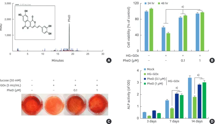
Phelligridin D extract from P. baumii exhibited potent antioxidant activity. The structure and
purification of phelligridin D by HPLC are demonstrated in Figure 3A. HPDLCs treated with high glucose levels and glucose oxidase showed significantly decreased cell viability in a time-dependent manner. However, treatment with phelligridin D at 0.1 or 1 μM led to the significant recovery of cell viability, even in HPDLCs that were treated with high glucose levels and glucose oxidase (Figure 3B). Alizarin red staining, as an index of bone mineralization, was also restored by treatment with phelligridin D in HPDLCs (Figure 3C). Phelligridin D also significantly restored ALP activity in HPDLCs that were treated with high glucose and glucose oxidase (Figure 3D).
Role of phelligridin D on inflammation, osteogenic differentiation, cementogenesis,
and autophagy in HPDLCs under glucose-induced oxidative stress
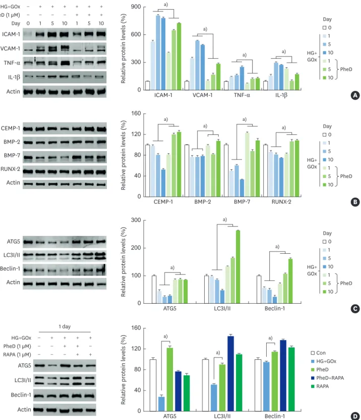
The levels of inflammatory molecules (ICAM-1, VCAM-1, TNF-α, and IL-1β) which were
increased by glucose-induced oxidative stress, were downregulated by phelligridin D in HPDLCs (Figure 4A). The anti-inflammatory properties of phelligridin D induced CEMP-1, 2, BMP-7, and RUNX-2 even under glucose-induced oxidative stress in HPDLCs (Figure 4B). In addition, phelligridin D led to the recovery of ATG5, LC3I/II, and beclin-1 expression levels, which were reduced by oxidative stress (Figure 4C). Specifically, phelligridin D restored the expression of autophagy- related proteins in the same way as RAPA in HPDLCs (Figure 4D).
a)
Cell viability (% of contr
ol) 0 40 80 120 − HG+GOx + + + − PheD (µM) − 0.1 1 24 hr 48 hr B a) a)
ALP activity (of OD)
0 1 2 4 3
3 days 7 days 14 days D
C GOx (5 mU/mL) Glucose (50 mM) PheD (µM) − + + + − − + + + − − + + + − 0.1 1 Mock HG+GOx HG+GOx PheD (0.1 µM) PheD (1 µM) m AU Minutes 0 5 10 15 20 25 30 1,000 2,000 3,000 A PheD
Figure 3. Effect of PheD on protection from cellular toxicity and mineralization in HPDLCs with glucose-induced oxidative stress. (A) Structure and HPLC analysis of PheD. (B) Cell viability was determined by an MTT assay at 24 and 48 hours. Mineralization assay was determined by (C) alizarin red S staining at 14 days, and (D) ALP activity at 3, 7, and 14 days. Each value is reported as the mean and standard deviation of 3 independent experiments.
HPDLC: human periodontal ligament cell, HPLC: high-performance liquid chromatography, ALP: alkaline phosphatase, HG: high glucose, GOx: glucose oxidase, PheD: phelligridin D, OD: optical density.
Relativ e pr ot ein l ev els (%) 0 300 600 900 ICAM-1 0 1 5 10 1 5 10 − − − − + + − + + + + + + + PheD (1 µM) HG+GOx Day ICAM-1 VCAM-1 TNF-α IL-1β
Actin VCAM-1 TNF-α IL-1β
a) a) a) a) A 1 5 10 1 5 10 HG+ GOx PheD Day 0 a) a) a) a) Relativ e pr ot ein l ev els (%) 0 40 80 160 120 CEMP-1 CEMP-1 BMP-2 BMP-7 RUNX-2 Actin BMP-2 BMP-7 RUNX-2 B 1 5 10 1 5 10 HG+ GOx PheD Day 0 a) a) a) Relativ e pr ot ein l ev els (%) 0 300 200 100 ATG5 ATG5 LC3I/II Beclin-1 Actin LC3I/II Beclin-1 C 1 5 10 1 5 10 HG+ GOx PheD Day 0 a) a) a) Relativ e pr ot ein l ev els (%) 0 40 80 160 120
ATG5 LC3I/II Beclin-1 D
HG+GOx PheD PheD+RAPA RAPA Con − − − + + − − + − − + + + + + PheD (1 µM) HG+GOx RAPA (1 µM) 1 day ATG5 LC3I/II Beclin-1 Actin
Figure 4. Effects of PheD on the expression of inflammatory, osteogenic, and differentiation and autophagy-related molecules in HPDLCs with glucose-induced oxidative stress. Protein levels were determined by western blot analyses. (A) Expression of inflammatory proteins at 1, 5, and 10 days. (B) Osteogenic differentiation-related proteins at 1, 5, and 10 days, (C) autophagy-differentiation-related proteins at 1, 5, and 10 days, and (D) expression of autophagy-differentiation-related proteins after treatment with PheD and RAPA (autophagy inducer) in HPDLCs with glucose-induced oxidative stress at 1 day. All data are representatives of 3 separate experiments.
HPDLC: human periodontal ligament cell, RAPA: rapamycin, ICAM: intracellular adhesion molecule, VCAM: vascular cell adhesion molecule, TNF: tumor necrosis factor, IL: interleukin, CEMP-1: cementum protein 1, BMP: bone morphogenetic protein, RUNX-2: runt-related transcription factor-2, ATG5: autophagy related 5, LC3I/II: light chain 3, HG: high glucose, GOx: glucose oxidase, PheD: phelligridin D.
DISCUSSION
This study was conducted to provide basic data on the ability of phelligridin D to restore the function of PDL cells affected by periodontitis as a complication of DM. The progression of DM leads to several oral diseases, including periodontal disease, dental caries, fungal infections, and soft-tissue lesions [27]. Oxidative stress is a physio-pathological factor that has been implicated in the development of DM [28]. Under normal conditions, glucose is metabolized into D-glucono-δ-lactone and H2O2 by glucose oxidase. During chronic
hyperglycemia, the excess production of free radicals by glucose oxidation exacerbates the complications of DM [29]. The results of this study demonstrated that exposure to glucose oxidase under high-glucose conditions resulted in high production of H2O2 in HPDLCs.
The functional and morphological alterations of HPDLCs due to the excessive production of H2O2 under high-glucose conditions are similar to those observed in diabetes. H2O2 is a
major index of oxidative stress that damages DNA, membrane lipids, and cellular proteins, and leads to cell death [30]. Therefore, treatment with glucose oxidase under high-glucose conditions induced oxidative stress similar to DM-mediated oxidative stress.
Previous studies have demonstrated that exposure to oxidative conditions increases
inflammation and disturbs cellular function [31,32]. The results of this study also verified that higher production of H2O2 by glucose oxidase under high-glucose conditions disturbed cell
viability and caused inflammation. Exposure of odontoblast or osteoblast cells to oxidative stress can disturb dental mineralization [33]. Consequently, excessive production of H2O2
caused HPDLCs to lose their differentiation potential to osteoblast and cementoblast phenotypes and reduced their levels of autophagy. These findings are similar to the results of previous studies [34] and suggest that high production of H2O2 initiates pathological
conditions and impairs the vitality of the PDL.
Many studies have suggested that anti-oxidant therapies reduce the complications of DM [25,35]. Phelligridin D possesses anti-oxidant, anti-inflammatory, and anti-diabetic effects [21,36]. In our previous study, phelligridin D showed typical antioxidant effects in terms of reducing ROS activity in HPDLCs [21]. The results of this study demonstrated that the decreased cellular viability and increased expression of inflammatory molecules (ICAM-1, VCAM-1, TNF-α, IL-1β) by glucose oxidase and high glucose were restored by phelligridin D in HPDLCs, implying that the anti-inflammatory properties of phelligridin D derive from its antioxidant characteristics. Furthermore, this study showed that phelligridin D upregulated cementogenesis (CEMP-1), osteogenesis (BMP-2, BMP-7, RUNX-2), and mineralization (alizarin red, ALP activity) even under glucose-induced oxidative stress. Antioxidants can protect or restore the osteogenic differentiation and mineralization of dental pulp cells in conditions of oxidative stress [37,38]. The result of this study indicate that phelligridin D enhances osteogenic differentiation and mineralization even under glucose-induced oxidative stress in HPDLCs.
Autophagy is also involved in cellular stress, inflammation, aging, development, and cancer [39]. Autophagy maintains cellular homeostasis in response to inflammation in PDL tissue and cells [40]. In this study, glucose-induced oxidative stress decreased autophagy-related markers such as ATG5, LC3I/II, and beclin-1, whereas phelligridin D restored the expression of all these molecules even under glucose- induced oxidative stress in HPDLCs. Moreover, the finding that phelligridin D led to increased expression of autophagy-related molecules in stress induced by glucose oxidase is similar to the findings that have been observed with
RAPA, an autophagy inducer, indicating that phelligridin D promoted autophagy under conditions of glucose-induced oxidative stress. Furthermore, phelligridin D enhanced cementogenesis and osteogenic differentiation in HPDLCs.
In conclusion, this study presents new insights related to the role of phelligridin D in glucose-induced oxidative stress in HPDLCs. In these cells, phelligridin D resulted in sustained functional improvement of PDL cells with the enhancement of autophagy, subsidization of inflammation, and promotion of cementogenesis and osteogenesis. Within the limitations of this study, these findings indicate that phelligridin D may have effects making it suitable for application as a supplement to improve diabetes-related periodontal disease. In the future, additional experiments related to the detailed mechanisms and an in vivo study are required to demonstrate the role of phelligridin D in the treatment of
DM-induced periodontal disease.
ACKNOWLEDGEMENTS
All of the authors would like to acknowledge Professor Bong-Sik Yun, Division of Biotechnology, Jeonbuk National University for isolation, purification, and provision of phelligridin D for this study.
REFERENCES
1. Bartold PM, McCulloch CA, Narayanan AS, Pitaru S. Tissue engineering: a new paradigm for periodontal regeneration based on molecular and cell biology. Periodontol 2000 2000;24:253-69.
PUBMED | CROSSREF
2. Hou J, Yamada S, Kajikawa T, Ozaki N, Awata T, Yamaba S, et al. Role of ferritin in the cytodifferentiation of periodontal ligament cells. Biochem Biophys Res Commun 2012;426:643-8.
PUBMED | CROSSREF
3. Hoz L, Romo E, Zeichner-David M, Sanz M, Nuñez J, Gaitán L, et al. Cementum protein 1 (CEMP1) induces differentiation by human periodontal ligament cells under three-dimensional culture conditions. Cell Biol Int 2012;36:129-36.
PUBMED | CROSSREF
4. Choi HD, Noh WC, Park JW, Lee JM, Suh JY. Analysis of gene expression during mineralization of cultured human periodontal ligament cells. J Periodontal Implant Sci 2011;41:30-43.
PUBMED | CROSSREF
5. Dahiya P, Kamal R, Gupta R, Bhardwaj R, Chaudhary K, Kaur S. Reactive oxygen species in periodontitis. J Indian Soc Periodontol 2013;17:411-6.
PUBMED | CROSSREF
6. Polak D, Shapira L. An update on the evidence for pathogenic mechanisms that may link periodontitis and diabetes. J Clin Periodontol 2018;45:150-66.
PUBMED | CROSSREF
7. Zheng J, Chen S, Albiero ML, Vieira GH, Wang J, Feng JQ, et al. Diabetes activates periodontal ligament fibroblasts via NF-κB in vivo. J Dent Res 2018;97:580-8.
PUBMED | CROSSREF
8. Preshaw PM, Bissett SM. Periodontitis and diabetes. Br Dent J 2019;227:577-84. PUBMED | CROSSREF
9. Lee YH, Kim JS, Kim JE, Lee MH, Jeon JG, Park IS, et al. Nanoparticle mediated PPARγ gene delivery on dental implants improves osseointegration via mitochondrial biogenesis in diabetes mellitus rat model. Nanomedicine (Lond) 2017;13:1821-32.
PUBMED | CROSSREF
10. Covani U, Marconcini S, Derchi G, Barone A, Giacomelli L. Relationship between human periodontitis and type 2 diabetes at a genomic level: a data-mining study. J Periodontol 2009;80:1265-73.
11. Jansson H, Lindholm E, Lindh C, Groop L, Bratthall G. Type 2 diabetes and risk for periodontal disease: a role for dental health awareness. J Clin Periodontol 2006;33:408-14.
PUBMED | CROSSREF
12. Mealey BL. Periodontal disease and diabetes. A two-way street. J Am Dent Assoc 2006;137 Suppl:26S-31S. PUBMED | CROSSREF
13. Hale AN, Ledbetter DJ, Gawriluk TR, Rucker EB 3rd. Autophagy: regulation and role in development. Autophagy 2013;9:951-72.
PUBMED | CROSSREF
14. White E, Karp C, Strohecker AM, Guo Y, Mathew R. Role of autophagy in suppression of inflammation and cancer. Curr Opin Cell Biol 2010;22:212-7.
PUBMED | CROSSREF
15. Mizushima N, Levine B, Cuervo AM, Klionsky DJ. Autophagy fights disease through cellular self-digestion. Nature 2008;451:1069-75.
PUBMED | CROSSREF
16. Wei W, An Y, An Y, Fei D, Wang Q. Activation of autophagy in periodontal ligament mesenchymal stem cells promotes angiogenesis in periodontitis. J Periodontol 2018;89:718-27.
PUBMED | CROSSREF
17. Kamruzzaman SM, Endale M, Oh WJ, Park SC, Kim TH, Lee IK, et al. Antiplatelet activity of Phellinus baummii methanol extract is mediated by cyclic AMP elevation and inhibition of collagen-activated
integrin-α(IIb) β3 and MAP kinase. Phytother Res 2011;25:1596-603.
PUBMED | CROSSREF
18. Hwang BS, Lee IK, Choi HJ, Yun BS. Anti-influenza activities of polyphenols from the medicinal mushroom Phellinus baumii. Bioorg Med Chem Lett 2015;25:3256-60.
PUBMED | CROSSREF
19. Jin QL, Zhang ZF, Lv GY, Cai WM, Cheng JW, Wang JG, et al. Antioxidant and DNA damage protecting potentials of polysaccharide extracted from Phellinus baumii using a delignification method. Carbohydr
Polym 2016;152:575-82. PUBMED | CROSSREF
20. Lee S, Lee D, Jang TS, Kang KS, Nam JW, Lee HJ, et al. Anti-inflammatory phenolic metabolites from the edible fungus Phellinus baumii in LPS-stimulated RAW264.7 cells. Molecules 2017;22:1583.
PUBMED | CROSSREF
21. Kim JE, Takanche JS, Yun BS, Yi HK. Anti-inflammatory character of phelligridin D modulates
periodontal regeneration in lipopolysaccharide-induced human periodontal ligament cells. J Periodontal Res 2018;53:816-24.
PUBMED | CROSSREF
22. Kim JE, Takanche JS, Kim JS, Lee MH, Jeon JG, Park IS, et al. Phelligridin D-loaded oral nanotube titanium implant enhances osseointegration and prevents osteolysis in rat mandible. Artif Cells Nanomed Biotechnol 2018;46:397-407.
PUBMED | CROSSREF
23. Cho EJ, Hwang HJ, Kim SW, Oh JY, Baek YM, Choi JW, et al. Hypoglycemic effects of exopolysaccharides produced by mycelial cultures of two different mushrooms Tremella fuciformis and Phellinus baumii in ob/ob
mice. Appl Microbiol Biotechnol 2007;75:1257-65. PUBMED | CROSSREF
24. Lee IK, Han MS, Lee MS, Kim YS, Yun BS. Styrylpyrones from the medicinal fungus Phellinus baumii and
their antioxidant properties. Bioorg Med Chem Lett 2010;20:5459-61. PUBMED | CROSSREF
25. Lee YH, Kim GE, Song YB, Paudel U, Lee NH, Yun BS, et al. Davallialactone reduces inflammation and repairs dentinogenesis on glucose oxidase-induced stress in dental pulp cells. J Endod 2013;39:1401-6. PUBMED | CROSSREF
26. Yu MK, Lee JC, Kim JH, Lee YH, Jeon JG, Jhee EC, et al. Anti-inflammatory effect of peroxisome proliferator activated receptor gamma on human dental pulp cells. J Endod 2009;35:524-8. PUBMED | CROSSREF
27. Manfredi M, McCullough MJ, Vescovi P, Al-Kaarawi ZM, Porter SR. Update on diabetes mellitus and related oral diseases. Oral Dis 2004;10:187-200.
PUBMED | CROSSREF
28. Giugliano D, Ceriello A, Paolisso G. Oxidative stress and diabetic vascular complications. Diabetes Care 1996;19:257-67.
PUBMED | CROSSREF
29. Baynes JW. Role of oxidative stress in development of complications in diabetes. Diabetes 1991;40:405-12. PUBMED | CROSSREF
30. Halliwell B, Clement MV, Long LH. Hydrogen peroxide in the human body. FEBS Lett 2000;486:10-3. PUBMED | CROSSREF
31. Nishimura F, Terranova V, Foo H, Kurihara M, Kurihara H, Murayama Y. Glucose-mediated alteration of cellular function in human periodontal ligament cells. J Dent Res 1996;75:1664-71.
PUBMED | CROSSREF
32. Kook SH, Lee D, Cho ES, Heo JS, Poudel SB, Ahn YH, et al. Activation of canonical Wnt/β-catenin signaling inhibits H2O2-induced decreases in proliferation and differentiation of human periodontal ligament fibroblasts. Mol Cell Biochem 2016;411:83-94.
PUBMED | CROSSREF
33. Lee DH, Lim BS, Lee YK, Yang HC. Effects of hydrogen peroxide (H2O2) on alkaline phosphatase activity and matrix mineralization of odontoblast and osteoblast cell lines. Cell Biol Toxicol 2006;22:39-46. PUBMED | CROSSREF
34. Kuang Y, Hu B, Feng G, Xiang M, Deng Y, Tan M, et al. Metformin prevents against oxidative stress-induced senescence in human periodontal ligament cells. Biogerontology 2020;21:13-27.
PUBMED | CROSSREF
35. Dene BA, Maritim AC, Sanders RA, Watkins JB 3rd. Effects of antioxidant treatment on normal and diabetic rat retinal enzyme activities. J Ocul Pharmacol Ther 2005;21:28-35.
PUBMED | CROSSREF
36. Wang WH, Wu FH, Yang Y, Wu N, Zhang JS, Feng N, et al. Hypoglycemic effect of ethanol and ethyl acetate extract of Phellinus baumii fruiting body in streptozotocin-induced diabetic mice. Evid Based
Complement Alternat Med 2015;2015:783460. PUBMED
37. Minamikawa H, Yamada M, Deyama Y, Suzuki K, Kaga M, Yawaka Y, et al. Effect of N-acetylcysteine on rat dental pulp cells cultured on mineral trioxide aggregate. J Endod 2011;37:637-41.
PUBMED | CROSSREF
38. Lee YH, Kang YM, Heo MJ, Kim GE, Bhattarai G, Lee NH, et al. The survival role of peroxisome proliferator-activated receptor gamma induces odontoblast differentiation against oxidative stress in human dental pulp cells. J Endod 2013;39:236-41.
PUBMED | CROSSREF
39. Boya P, Reggiori F, Codogno P. Emerging regulation and functions of autophagy. Nat Cell Biol 2013;15:713-20.
PUBMED | CROSSREF
40. An Y, Liu W, Xue P, Zhang Y, Wang Q, Jin Y. Increased autophagy is required to protect periodontal ligament stem cells from apoptosis in inflammatory microenvironment. J Clin Periodontol 2016;43:618-25. PUBMED | CROSSREF