PCDH 19 환자에서 효과적인 Corticosteroid 치료 사례
연세대학교 의과대학 세브란스병원 소아과학교실1, 국민건강보험 일산병원 소아청소년과2
이동민1,2・정희정2
Poster presented at the Spring Conference of the Korean Child Neurology Society in 2016.
Submitted: 28 February, 2018 Revised: 21 March, 2018 Accepted: 22 March, 2018
Correspondence to Hee Jung Chung, MD, PhD Department of Pediatrics, National Health Insurance Service, Ilsan Hospital, 100 Ilsanro, Ilsan Dong-gu, Goyang-si, Gyeonggi-do 10444, Korea
Tel: +82-31-900-0520, Fax: +82-31-900-0343 E-mail: agathac@nhimc.or.kr
A Case of Effective Treatment of a Patient with PCDH 19 - Related Epilepsy using Corticosteroid
An uncommon female-limited intractable epilepsy, protocadherin (PCDH)
19-related epilepsy, is characterized by mutations in the PCDH 19 gene, locatedon chromosome X. Clinical symptoms include early onset, fever sensitivity, focal seizures and psychomotor retardation. PCDH 19–related epilepsy is unresponsive to conventional antiepileptic drugs (AEDs), but corticosteroid is reported to be effective in a few cases. We report a case of a 25-month-old girl who was admitted to our hospital due to developmental regression, accompanied by aggravated seizures with fever. Although several conventional AEDs were administered, the frequency and severity of seizures increased with mild fever, and the symptoms did not improve.
Considering possible immune, and inflammatory involvement in seizure generation, the patient was administered corticosteroid treatment during the acute phase.
Corticosteroid dramatically improved seizures and her development gradually. The patient was finally diagnosed with
PCDH 19–related epilepsy in genomic evaluation.We observed the effect of corticosteroid on intractable epilepsy in patient with PCDH 19 mutation. If a female patient whose seizures are resistant to conventional AEDs or easily provoked by mild fever, has developmental delay or developmental regression, this may be an important clinical clue to the early diagnosis of
PCDH 19–related epilepsy
Key Words:
PCDH 19, Intractable epilepsy, X-linked mental retardation disorder, Female, Corticosteroids
Dong Min Lee, MD
1,2, Hee Jung Chung, MD, PhD
21
Department of Pediatrics, Yonsei University, College of Medicine, Seoul,
2Department of Pediatrics, National Health Insurance Service, Ilsan Hospital, Goyang, Korea
Copyright © 2018 by The Korean Child Neurology Society
http://www.cns.or.kr
Introduction
Mutations in the X-linked gene encoding protocadherin 19, protocadherin (PCDH) 19, cause female-limited epilepsy, with the males with PCDH 19 mutations being unaffected and phenotypically normal1,2). Females with heterozygous mutations are affected, but males with hemizygous mutations are unaffected1,2,3). This extraordinary mode of inheritance is explained by the pathogenic mechanism of "cellular interference". Only subjects with a combination of cells with mutant and wild-type PCDH 19 develop the symptoms4).
Although truncation mutations as well as deletions in other exons, have also been identified, most PCDH 19 mutations are found in exon 1, encoding the large
extracellular domain4,5,6). The clinical features of PCDH 19-related epilepsy include early onset (6-36 months of age), fever sensitivity, seizures occurring in clusters, and psychomotor retardation1,4,7).
Conventional AEDs fail to prevent these seizures. The exact pathogenesis and treatment of PCDH 19-related female-limited epilepsy remain unclear, but it has been suggested that corticosteroids are effective in controlling seizures8,9). We report a case of a 25-month-old girl with delayed development experi- encing intractable seizures who was eventually diagnosed with PCDH 19-related epilepsy, and effectively treated using
corticosteroid therapy.
Case report
A 25-month-old girl, undergoing physical therapy in the Department of Rehabilitation Medicine for gross motor retardation, was brought to our Pediatric Neurology clinic due to suspected seizures and developmental regression. Her ante- and perinatal histories were unremarkable, and there was no history of seizures. At the age of 11 months, she was taken to the Department of Rehabilitation Medicine of Severance Hospital due to delayed development detected on National Health Screening Program for Infants and Children. Rolling and creeping started at 7 and 9 months, respectively. She could not sit on her own and could not stand her own. After 1 year of physical therapy, she could walk with support, and the general development was improved gradually.
One month prior to visiting our Pediatric Neurology clinic, the parents had noticed that the child was gradually exhibiting a developmental regression in motor and verbal skills. The babbling, crawling, and walking she previously showed were
not observed any more. Moreover, her entire body often became flaccid and fell easily. During examination, she exhibited repetitive head drops, twitching of the eyes and facial muscles, and aimless movement of the upper and lower extremities, accompanied by staring and psychomotor arrest reaction.
The patient was admitted to our Pediatric Neurology department on the day of outpatient clinic visit for the differential diagnosis of seizures. The patient’s vital signs showed a pulse of 110 beats/min, respiratory rate of 24 breaths/
min, and body temperature of 37.0℃. The following measurements were recorded; height: 91 cm (75th–90th percentile), body weight: 12 kg (25th–50th percentile), and head circumference: 46 cm (5th–10th percentile). The neurological examination revealed that axial tone in the neck and trunk was decreased and distal tone in the lower extremities increased mildly. The results of routine laboratory tests for complete blood count and biochemistry were normal. The C-reactive protein concentration and erythrocyte sedimen tation rate were <0.3 (normal value, 0–8 mg/L) and 2 (normal value, 0–20 mm/hr) respectively. The ammonia (NH3) concentration was mildly elevated to 107 µg/dL (normal value, 12–66 µg/dL). In the initial metabolic study on hospital day 7, lactic acid in urine organic acid analysis was 397 mmol/mol (normal value, 7–150 mmol/
mol). However, follow-up metabolic study on hospital day 31 after protein-restricted diet, lactic acid in urine was normalized (119 mmol/mol).
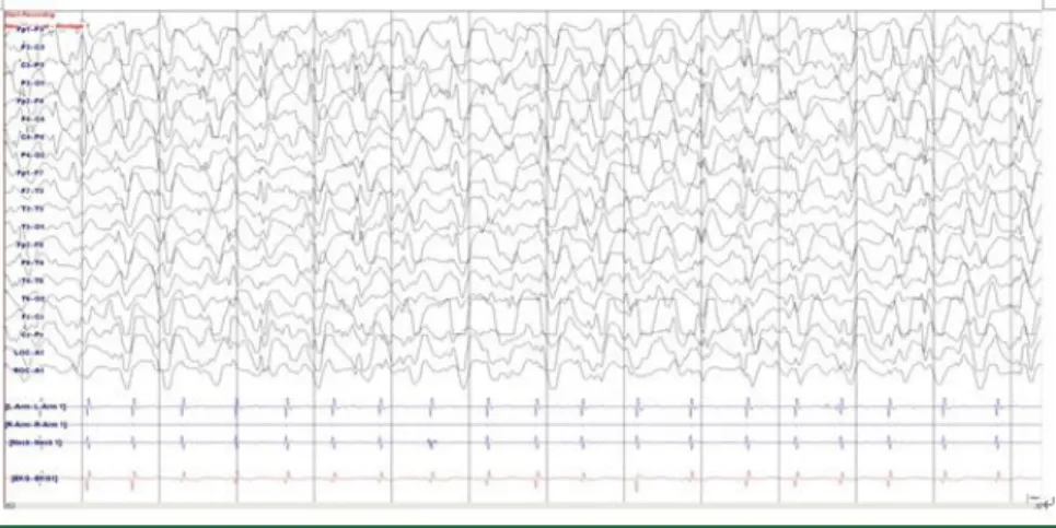
At the time of admission, her 8 hour-ambulatory electroence- phalography (EEG) results showed frequent generalized 1- to 1.5-Hz spike and slow wave bursts with predominance on both frontocentral areas (F3, F7, F4, and F8). Sometimes the frontal spike and slow wave bursts propagated to the centroparietal areas, which is associated with clinical or subclinical electro- graphic seizures (Fig. 1). Clinical seizures are presented with
Fig. 1. Electroencephalography showed a generalized 1- to 1.5-Hz spike and slow wave bursts,
with the predominance of both the frontocentral areas.
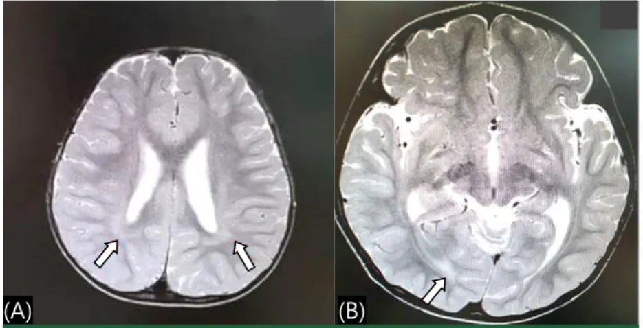
staring, head drops, generalized tonic posturing and atypical absence seizures. Brain magnetic resonance imaging (MRI) revealed mild bilateral peritrigonal white matter volume loss and suspicious subcortical white matter signal change in the right occipital lobe (Fig. 2).
On hospital day 1, systemic anticonvulsant treatment was started using phenobarbital and phenytoin loading. However, on hospital day 3, mild fever (37.8℃) started and the seizure frequency and severity were increased. Intravenous ceftriaxone (50 mg/kg/day) was initiated. Additional IV levetiracetam, valproic acid, and oral topiramate were tried because the seizures relapsed frequently. Nevertheless, the seizure clusters were resistant to those all antiepileptic drugs (AEDs) tried.
Oral prednisolone (2 mg/kg) was initiated on hospital day 6 and fever was subsided on the next day. In addition, clinical seizures were definitely diminished and finally stopped after 1 week of prednisolone administration. In the EEG performed 2
weeks after prednisolone administration, frequent generalized spikes and slow wave bursts and EEG seizures were no longer observed, but diffuse slowing of background rhythm was observed (Fig. 3). Oral prednisolone (2 mg/kg) was continued for 2 weeks and tapered over the following 2 weeks period (1 mg/kg for 1 week and 0.5 mg/kg for next 1 week). There were no remarkable adverse events during the cortico steroid therapy.
The patient was discharged after 31 days of hospitalization. In the EEG obtained 4 weeks after discharge, intermittent slowing of background rhythm was still noted without subclinical EEG seizures (Fig. 4). After discharge, the development improved, enabling walking with the support of both hands, as the seizures were controlled.
To ascertain the etiology, Next Generation Sequencing was performed, and a mutation in PCDH 19 (c.2250C>A, p.
Asp750Glu) was detected. Mutation screening of her parents revealed that the same mutation was founded in her father, who
(A) (B)
Fig. 2. Brain magnetic resonance imaging showed mild bilateral peritrigonal white matter
volume loss (A) and suspicious subcortical white matter signal change in the right occipital lobe (B).
Fig. 3. EEG performed 2 weeks after prednisolone administration did not show generalized spikes
and slow wave bursts, nor EEG seizures. However, diffuse slowing of background rhythm was
observed.
had been healthy. At the last follow-up visit on January 2018, the patient was 5 years and 1 month old, and she can walk with one hand holding and seizures have not recurred yet.
Discussion
The gene PCDH 19 is located on chromosome Xq22.110), and mutations in this gene are known to cause female-limited epilepsy and mental retardation (or Epilepsy, Female-restricted, with Mental Retardation; MIM 300088), and Dravet syndrome- like epileptic encephalopathies2,4). The clinical characteristics include early onset seizures (6–36 months of age), fever associated seizures, intractability of seizure clusters, delayed development, and behavioral problems10). Among these, two of the main clinical features are seizures clusters precipitated by fever and female-restricted epilepsy1,4,7.11). Although the patient's father also has this mutation, he has been healthy. The reason why symptoms appear only in our female patient is explained by the pathogenic mechanism of "cellular interference". A combination of cells with mutant and wild-type PCDH 19 develops the symptoms4). So females with hetero zygous mutations are affected, but males with hemizygous mutations are unaffected1-3).
In the present case, delayed development preceded the onset of PCDH 19-related epilepsy, and developmental regression was observed after onset of seizures in the midst of improvement of motor symptoms after physical therapy. Moreover, the frequency and intensity of seizures were easily increased by mild fever.
Based on the various neurological manifestations, seizures can be various, such as generalized tonic-clonic, tonic and focal seizures, with or without secondary generalization11). Focal seizures involve motion arrest, cyanosis, eye deviation,
respiratory alteration, systemic jerks, complex movement of the facial muscles and/or extremities, and tonic symptoms8). In a previous report, the electroclinical pattern of focal seizures, with affective symptoms, was considered a major feature of PCDH 19-related epilepsy12). In the present case, the patient exhibited twitching of the eyes and facial muscles with staring, head drops and involuntary movement of the upper/lower extremities. The baseline EEG showed 1–1.5Hz spike and slow wave bursts at both frontocentral areas (F3, F7, F4, and F8). Sometimes, it was propagated to the centroparietal areas, which was associated with clinical or subclinical EEG seizures. In the EEG performed after treatment with oral prednisolone, frequent generalized spikes and slow wave bursts and EEG seizures were no longer observed, but diffuse slowing of background rhythm was observed.
Clinicians should consider the possibility of PCDH 19-related epilepsy in female cases whose seizures are easily precipitated by fever, which may provide significant and characteristic clues for accurate diagnosis. Other representative cases of fever- sensitive epilepsy are observed in the SCN1A mutation- associated Dravet syndrome. However, in PCDH 19-related epilepsy, only a small number of patients exhibit a phenotype resembling Dravet syndrome4,12). Unlike Dravet syndrome, they rarely exhibit absences or myoclonic jerks or photosensitivity11). As a result, the ultimate approach for diagnosing PCDH 19-rela- ted epilepsy is the detection of a mutation in PCDH 19.
The exact pathogenesis of PCDH 19-related epilepsy is unclear. It has been suggested to be related to disruption of BBB integrity and its vulnerability9). Although no human evidence exists, in mouse, PCDH 19 gene is significantly expressed higher in the brain microvascular endothelial cells than in the liver or lung13). Higurashi et al.9) proposed the mechanism of seizure recurrence was that systemic inflammation might alter the Fig. 4. Electroencephalography (EEG) obtained 4 weeks after discharge still showed intermittent
slowing of background rhythm, without subclinical EEG seizures.
PCDH 19 expression in the BBB.
The antiepileptic treatments of patient with PCDH 19-related epilepsy are very challenging. In our patient, we have used AEDs such as IV phenobarbital, phenytoin, levetiracetam, valproic acid and oral topiramate, but have not had any effect until adding oral prednisolone. A previous study reported that the effect of IV phenytoin/fosphenytoin, phenobarbital and continuous administration of midazolam during the acute phase was often transient, however administration of methylprednisolone successfully stopped the seizures8). Higurashi et al.9) reported that corticosteroid dramatically improved seizure clusters: 4 out of 5 patients received intravenous infusion of methylpredni- solone and one patient received intravenous infusion of predni- solone. Seizures stopped dramatically in all of the 5 patients, but 3 out of 5 patients showed recurrence of seizures within a few weeks. However, our patient has not had a recurrence of seizure three years after the oral prednisolone has been withdrawn.
The possible mechanism of corticosteroid in PCDH 19-related epilepsy can be proposed as follows. First, corticosteroid may exert immunosuppression and feedback inhibition of cortico- tropin-releasing hormone secretion, which is well known mechanism of corticosteroid in West syndrome, Landau-Kleffner syndrome, or autoimmune epilepsy/encephalitis14). Second mechanism is an anti-inflammatory effect of corticosteroid. In chronic seizure condition, inflammatory mediators including cytokines and growth factors were released15). These inflamma- tory mediators could induce cerebrovascular endothelial injury and increased BBB permeability. Seizures are easily provoked by damages to BBB integrity, which can be diminished by cortico- steroid administration16), altering the expression of these inflammatory mediators15). Third mechanism is restoration of BBB integrity by corticosteroid17). In compromised BBB, seizures are easily provoked by mild disruptions of brain homeostasis16). As mentioned above, because PCDH 19 gene is significantly more expressed in the brain microvascular endothelial cells13), BBB integrity can be easily damaged if there is a PCDH 19 mutation. Higurashi et al.9) also reported that neuronal proteins produced by recurrent seizures enter the general circulation through compromised BBB, resulting in an elevated anti- neuronal autoantibody positivity in serum/cerebrospinal fluid. In this vulnerable situation of BBB integrity, corticosteroid induces expression of tight junction proteins, which increase transendo- thelial electrical resistance and finally it reduces the permeability of BBB.18) Thus, the seizures are controlled by improving integrity of the BBB.
In conclusion, we observed the effect of corticosteroid on intractable epilepsy in patient with PCDH 19 mutation. It is beneficial to try corticosteroids after a rapid diagnosis by genetic
analysis, if a female patient whose seizures are resistant to conventional AEDs or easily provoked by mild fever, has developmental delay or developmental regression.
요약
PCDH 19 돌연변이 관련 뇌전증은 드문 X 연관 질환으로서 생후 6-36개월 이내 조기발생, 열 민감성, 발달지연 및 국소경련, 항경련제 의 저항성 등을 특징으로 한다. 본 저자들은 운동발달 지연으로 재활 치료 중 발달퇴행을 보이고 열에 의해 악화되는 경련, 다양한 항경련 제 치료에 불응성을 가진 25개월 여아에서 경구 프레드니졸론의 효 과를 보고하게 되었다. 차세대 염기서열 분석을 통해 본 환아의 X 염 색체에서 c.2250C>A, p.Asp750Glu 를 발견하였으며 이는 환아의 아 버지로부터 기원했음을 추가적인 분석을 통하여 확인하였다.
PCDH 19 연관 뇌전증 환자에서는 다수의 항경련제 치료에도 불구 하고 경련발작의 조절이 잘 안되나, 스테로이드가 임상증상을 호전시 키는데 도움이 된다는 보고들이 있다. 여아에서 열에 의해 악화되는 경련 및 기존 항경련제에 저항성이 있으면서 발달지연이나 발달퇴보 를 보이는 경우 유전자 분석을 통한 빠른 진단 후 corticosteroid를 시도하는 것이 도움이 되겠다.
References
1) Scheffer IE, Turner SJ, Dibbens LM, Bayly MA, Friend K, Hodgson B, et al. Epilepsy and mental retardation limited to females: an under-recognized disorder. Brain 2008;131:918–27.
2) Dibbens LM, Tarpey PS, Hynes K, Bayly MA, Scheffer IE, Smith R, et al. X-linked protocadherin 19 mutations cause female-limited epilepsy and cognitive impairment. Nat Genet 2008;40: 776–81.
3) Juberg RC, Hellman CD. A new familial form of convulsive disorder and mental retardation limited to females. J Pediatr 1971;79:726–32.
4) Depienne C, Bouteiller D, Keren B, Cheuret E, Poirier K, Trouillard O, et al. Sporadic infantile epileptic encephalopathy caused by mutations in PCDH19 resembles Dravet syndrome but mainly affects females. PLoS Genet 2009;5:e1000381.
5) Marini C, Mei D, Parmeggiani L, Norci V, Calado E, Ferrari A, et al. Protocadherin 19 mutations in girls with infantile-onset epilepsy. Neurology 2010;75:646–53.
6) Jamal SM, Basran RK, Newton S, Wang Z, Milunsky JM. Novel de novo PCDH19 mutations in three unrelated females with epilepsy female restricted mental retardation syndrome. Am J Med Genet A 2010;152A:2475–81.
7) Christel D, Oriane T, Delphine B, Isabelle G, Karine P, Patrick B.
et al. Mutations and deletions in PCDH 19 account for various familial or isolated epilepsies in females. Hum. Mutat. 2011;32: E 1959-75
8) Higurashi N, Nakamura M, Sugai M, Ohfu M, Sakauchi M, Sugawara Y, et al. PCDH19-related female-limited epilepsy:
further details regarding early clinical features and therapeutic efficacy. Epilepsy Res 2013;106:191–9.
9) Higurashi N, Takahashi Y, Kashimada A, Sugawara Y, Sakuma H, Tomonoh Y, et al. Immediate suppression of seizure clusters by corticosteroids in PCDH19 female epilepsy. Seizure 2015;27: 1–5.
10) Vanhalst K, Kools P, Staes K, van Roy F, Redies C. delta-Proto- cadherins: a gene family expressed differentially in the mouse brain. Cell Mol Life Sci 2005;62:1247-59.
11) Higurashi N, Shi X, Yasumoto S, Oguni H, Sakauchi M, Itomi K, et al. PCDH19 mutation in Japanese females with epilepsy.
Epilepsy Res 2012;99:28–37.
12) Marini C, Darra F, Specchio N, Mei D, Terracciano A, Parmeg- giani L, et al. Focal seizures with affective symptoms are a major feature of PCDH19 gene-related epilepsy. Epilepsia 2012;53:
2111-9.
13) Daneman R, Zhou L, Agalliu D, Cahoy JD, Kaushal A, Barres BA.
The mouse blood-brain barrier transcriptome: a new resource for understanding the development and function of brain endothelial cells. PLoS One 2010;5:e13741.
14) Gupta R, Appleton R. Corticosteroids in the management of the paediatric epilepsies. Arch Dis Child 2005;90:379–84.
15) Gomes JA, Stevens RD, Lewin JJ 3rd, Mirski MA, Bhardwaj A.Glucocorticoid therapy in neurologic critical care. Crit Care Med 2005;33:1214-2.
16) Marchi N, Granata T, Freri E, Ciusani E, Ragona F, Puvenna V, et al. Efficacy of anti-inflammatory therapy in a model of acute seizures and in a population of pediatric drug resistant epileptics. PLoS One 2011;6:e18200.
17) Föster C, Waschke J, Burek M, Leers J, Drenckhahn D. Gluco- corticoid effects on mouse microvascular endothelial barrier permeability are brain specific. J Physiol 2006;573:413-25.
18) Salvador E, Shityakov S, Foster C. Glucocorticoids and endothelial cell barrier function. Cell Tissue Res 2014;355:597- 605.