KJTCVS
The Korean Journal of Thoracic and Cardiovascular SurgeryClinical Research Hemodynamic Performance of Pericardial Bioprostheses in the Aortic Position
Haeju Lee, M.D.
1, Ho Young Hwang, M.D., Ph.D.
1, Suk Ho Sohn, M.D.
1, Jae Woong Choi, M.D.
1, Jun-Bean Park, M.D., Ph.D.
2, Kyung Hwan Kim, M.D., Ph.D.
1, Ki-Bong Kim, M.D., Ph.D.
11
Department of Thoracic and Cardiovascular Surgery and
2Division of Cardiology, Department of Internal Medicine, Seoul National University Hospital, Seoul, Korea
ARTICLE INFO
Received December 30, 2019 Revised April 26, 2020 Accepted May 16, 2020 Corresponding author Ho Young Hwang Tel 82-2-2072-3020 Fax 82-2-2072-2340 E-mail scalpel@hanmail.net ORCID
https://orcid.org/0000-0002-8935-8118
Background: This study was conducted to evaluate the hemodynamic performance and the incidence of prosthesis-patient mismatch (PPM) after aortic valve replacement (AVR) using bovine pericardial valves (Carpentier-Edwards Perimount Magana and Magna Ease).
Methods: In total, 216 patients (mean age, 70.0±10.5 years) who underwent AVR using stented bovine pericardial valves and had follow-up echocardiography between 3 months and 2 years (mean, 12.0±6.6 months) after surgery were enrolled. The implanted valve sizes were 19, 21, 23, and 25 mm in 32, 56, 99, and 29 patients, respectively.
Results: On follow-up echocardiography, the mean transvalvular pressure gradients for the 19-mm, 21-mm, 23-mm, and 25-mm valves were 13.3±4.4, 12.6±4.2, 10.5±3.9, and 10.2±
3.7 mm Hg, respectively. The effective orifice area (EOA) was 1.25±0.26, 1.54±0.31, 1.81±0.41, and 1.87±0.33 cm
2, respectively. These values were smaller than those suggested by the manufacturer for the corresponding sizes. No patients had PPM, when based on the ref- erence EOA. However, moderate (EOA index ≤0.85 cm
2/m
2) and severe (EOA index ≤0.65 cm
2/m
2) PPM was present in 56 patients (11.8%) and 9 patients (1.9%), respectively, when using the measured values.
Conclusion: Carpentier-Edwards Perimount Magna and Magna Ease bovine pericardial valves showed satisfactory hemodynamic performance with low rates of PPM, although the reference EOA could overestimate the true EOA for individual patients.
Keywords: Aortic valve, Surgery, Heart valves, Bioprosthesis, Hemodynamics
Copyright © The Korean Society for Thoracic and Cardiovascular Surgery. 2020. All right reserved.
This is an Open Access article distributed under the terms of the Creative Commons Attribution Non-Commercial License (http://creativecommons.org/licenses/
Introduction
The aim of treating patients with aortic valve (AV) dis- ease is to relieve stenosis or regurgitation and to attain physiological transvalvular gradients. Prosthesis-patient mismatch (PPM) occurs when the effective orifice area (EOA) of the implanted valve is smaller than the patient’s physiological needs, thereby generating higher transvalvu- lar gradients. The poorer outcomes of patients with PPM following aortic valve replacement (AVR) have been thor- oughly demonstrated in previous studies [1-5]. Thus, it is important to implant a prosthetic valve with a sufficiently large orifice area to meet the physiological requirements of the patient.
The Carpentier-Edwards Perimount Magna and Magna Ease (Edwards Lifesciences, Irvine, CA, USA) valves are
stented bovine pericardial bioprostheses that have been used worldwide. While each manufacturer provides a ref- erence EOA for each valve size, in vivo Doppler echocar- diographic results have shown variation of EOA in patients with the same prosthesis size. Also the in vivo reference values tend to be smaller than the in vitro values [3]. These differences are thought to be caused by biological varia- tion, measurement errors, and underestimations or overes- timations [2,6].
Therefore, this study was conducted (1) to evaluate the hemodynamic performance and the incidence of PPM af- ter bioprosthetic AVR using bovine pericardial valves (Ed- ward Perimount Magna and Magna Ease) and (2) to pro- vide in vivo reference EOA values for these valves.
https://doi.org/10.5090/kjtcs.19.099 pISSN: 2233-601X eISSN: 2093-6516
Korean J Thorac Cardiovasc Surg. 2020;53(5):285-290
KJTCVS https://doi.org/10.5090/kjtcs.19.099
Methods
Study design
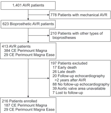
Between January 2005 and August 2018, 1,401 patients underwent AVR at our institution. Of these patients, 216 patients in whom Carpentier-Edwards Perimount Magna or Magna Ease valves were placed and who had echocar- diographic evaluations before discharge and during fol- low-up between 3 months and 2 years after surgery were enrolled (Fig. 1). The study protocol was reviewed by the institutional review board, and was approved as a mini- mal-risk retrospective study (IRB approval no., 1912-030- 1086) that did not require individual consent.
Operative data
All operations were performed using aortic and bicaval cannulation via median sternotomy by 1 of 4 surgeons at our institution. Prosthetic valves were implanted after re- moval of the AV leaflet and decalcification of the aortic annulus. The suture technique was identical in almost all study patients; non-everting mattress sutures buttress-re- inforced with polytetrafluoroethylene as a tubule (so-called
“spaghetti”) were used. One surgeon occasionally used sev- eral sutures with polytetrafluoroethylene as a pledget in- stead of a tubule. Another surgeon recently adopted a con- tinuous suture technique, which was used in 11 study patients (5.1%).
Echocardiographic evaluation
Two-dimensional echocardiography and Doppler col- or-flow imaging were performed in the early postoperative period (8.5±5.1 days) and 12.0±6.6 months after surgery.
The EOA was calculated as previously described [7]. Brief- ly, the left ventricular outflow tract (LVOT) diameter was measured in mid-systole from a parasternal long-axis view of the LVOT, with the inner to inner edge technique. The cross-sectional area (CSA) of the LVOT was calculated from the LVOT diameter by assuming it to be circular as follows: LVOT CSA=π×(LVOT diameter/2)
2. The LVOT ve- locity time integral (VTI) was obtained using the pulsed- wave Doppler signal. From these measurements, the EOA was calculated using the continuity equation as follows:
EOA=(LVOT CSA×LVOT VTI)/(VTI obtained from the continuous-wave Doppler signal) [7]. The VTI values ob- tained by pulsed-wave and continuous-wave Doppler were measured and averaged over 5 to 10 consecutive beats in
patients with atrial fibrillation. The effective orifice area index (EOAI, cm
2/m
2) was calculated by dividing the EOA by the body surface area of the patient, and moderate and severe PPM was defined as EOAI ≤0.85 cm
2/m
2and EOAI
≤0.65 cm
2/m
2, respectively [1].
Anti-thrombotic management
Warfarin anticoagulation with a target international nor- malized ratio of 2.0–2.5 was maintained for 3–6 months and throughout the patient’s life after surgery in patients with sinus rhythm and in those with atrial fibrillation, re- spectively. However, since late 2014, antiplatelet therapy in- stead of warfarin anticoagulation has been used for pa- tients with normal sinus rhythm.
Statistical analysis
Statistical analyses were performed using IBM SPSS ver.
25.0 (IBM Corp., Armonk, NY, USA). Data are expressed as means±standard deviation or proportions, as appropri- ate. Comparisons of paired data from continuous variables were performed using the paired t-test, and p-values <0.05 were considered to indicate statistical significance.
1,401 AVR patients
623 Bioprosthetic AVR patients
778 Patients with mechanical AVR
210 Patients with other types of bioprostheses
197 Patients excluded 17 Early death 26 Late death
20 Follow-up echocardiography
>2 years after AVR
88 No follow-up echocardiography 39 Aortic valve area unavailable 7 Lost to follow-up
413 AVR patients
384 CE Perimount Magna 29 CE Perimount Magna Ease
216 Patients enrolled 187 CE Perimount Magna 29 CE Perimount Magna Ease
Fig. 1. Flowchart of patient selection. Patients who underwent
AVR with CE Magna and Magna Ease between January 2005 to
August 2018 with available postoperative follow-up echocardiog-
raphy were selected. AVR, aortic valve replacement.
Haeju Lee, et al. Hemodynamic Performance of Pericardial Bioprostheses in the Aortic Position KJTCVS
Results
Patient data
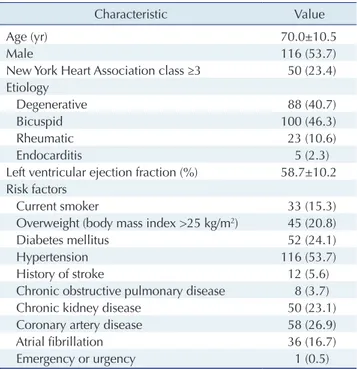
Patients’ mean age at surgery was 70.0±10.5 years, and 116 patients (53.7%) were male. Carpentier-Edwards Peri- mount Magna and Magna Ease valves were used in 187 and 29 patients, respectively. The mean left ventricular ejection fraction at surgery was 58.7%±10.2%. Bicuspid AV (n=100, 46.3%) was the most common etiology (Table 1).
Valves sized 19 mm, 21 mm, 23 mm, and 25 mm were implanted in 32, 56, 99, and 29 patients, respectively. Con- comitant procedures included arrhythmia surgery (n=20), aorta surgery (n=38), and coronary artery bypass grafting (n=29) (Table 2).
Early postoperative hemodynamic data
The transvalvular mean pressure gradient (MPG) and the EOA were measured in 216 and 181 patients, respec- tively. Overall, the transvalvular MPG on early postopera- tive echocardiography was 13.5±4.9 mm Hg, and the trans- valvular MPG values on early postoperative echocardiography for 19-mm, 21-mm, 23-mm, and 25-mm valves were 15.8±
4.8, 14.7±5.3, 12.4±4.3, and 12.2±5.0 mm Hg, respectively.
The EOAs for those valves were 1.17±0.35, 1.39±0.38, 1.63±
0.42, and 1.73±0.38 cm
2, respectively. These values were smaller than those suggested by the manufacturer (Table 3).
Hemodynamic data on follow-up
echocardiography and incidence of prosthesis- patient mismatch
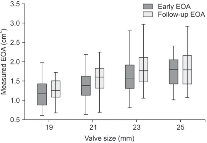
On follow-up echocardiographic evaluations, the overall transvalvular MPG was 11.4±4.2 mm Hg. The transvalvu- lar MPG for 19-mm, 21-mm, 23-mm, and 25-mm valves were 13.3±4.4, 12.6±4.2, 10.5±3.9, and 10.2±3.7 mm Hg, re- spectively. The EOAs for those valves were 1.25±0.26, 1.54±0.42, 1.81±0.41, and 1.87±0.33 cm
2, respectively (Table 4). The MPGs on follow-up echocardiography were signifi- cantly lower than those on early postoperative echocardi- ography (p<0.001) (Fig. 2). The EOAs on follow-up echo- cardiography were also significantly higher than the early Table 1. Preoperative characteristics and risk factors (N=216)
Characteristic Value
Age (yr) 70.0±10.5
Male 116 (53.7)
New York Heart Association class ≥3 50 (23.4) Etiology
Degenerative 88 (40.7)
Bicuspid 100 (46.3)
Rheumatic 23 (10.6)
Endocarditis 5 (2.3)
Left ventricular ejection fraction (%) 58.7±10.2 Risk factors
Current smoker 33 (15.3)
Overweight (body mass index >25 kg/m
2) 45 (20.8)
Diabetes mellitus 52 (24.1)
Hypertension 116 (53.7)
History of stroke 12 (5.6)
Chronic obstructive pulmonary disease 8 (3.7)
Chronic kidney disease 50 (23.1)
Coronary artery disease 58 (26.9)
Atrial fibrillation 36 (16.7)
Emergency or urgency 1 (0.5)
Values are presented as mean±standard deviation or number (%).
Table 2. Operative data (N=216)
Variable Value
Cardiopulmonary bypass time (min) 190.0±75.1 Aortic cross-clamping time (min) 121.5±50.7 Concomitant procedures
Arrhythmia surgery 20 (9.2)
Replacement of the ascending aorta 38 (17.6) Coronary artery bypass grafting 29 (13.4) Values are presented as mean±standard deviation or number (%).
Table 3. Transvalvular MPG and mEOA and rEOA of Carpentier-Edwards Magna and Magna Ease bovine pericardial valves on early postoperative (8.5±5.1 days after surgery) echocardiography
Size (mm) No. of patients for MPG MPG (mm Hg) No. of patients for mEOA mEOA (cm
2) rEOA (cm
2)
19 32 15.8±4.8 30 1.17±0.35 1.58
21 56 14.7±5.3 43 1.39±0.38 1.90
23 99 12.4±4.3 84 1.63±0.42 2.07
25 29 12.2±5.0 24 1.73±0.38 2.33
Values are presented as mean±standard deviation, unless otherwise stated.
MPG, mean pressure gradient; mEOA, measured effective orifice area; rEOA, reference effective orifice area.
KJTCVS https://doi.org/10.5090/kjtcs.19.099
postoperative values when those were compared in 181 pa- tients who had both early postoperative and follow-up data for EOA (p<0.001) (Fig. 3). However, the EOAs on fol- low-up echocardiography were still smaller than those sug- gested by the manufacturer for each size.
No patients had PPM when it was calculated based on the reference EOA. However, moderate and severe PPM was present in 56 patients (11.8%) and 9 patients (1.9%), re- spectively, when applying the measured values.
Discussion
The present study demonstrated 2 main findings. First, the currently available bovine pericardial valves had satis- factory hemodynamic performance with low rates of PPM.
Second, the actual EOAs were smaller than the values sup- plied by the manufacturer.
The aim of AVR is to relieve stenosis or regurgitation and to implant AV prostheses that reach physiological trans- valvular gradients. However, as parts of the sewing ring and the stent are positioned within the blood flow, a resid- ual gradient may remain, especially for small valves [1-3].
PPM occurs when the EOA of the prosthesis is smaller than the patient’s physiological needs, generating higher transvalvular gradients. PPM causes worse hemodynamic performance, which directly affects the clinical outcomes of AVR patients, resulting in less regression of left ventric- ular hypertrophy, higher incidence of cardiac events, and decreased long-term survival [1,4,5]. Thus, the most im- portant goal in performing AVR is to implant a valve size that corresponds to the patient’s needs [1-3].
Carpentier-Edwards Perimount Magna and Magna Ease are stented bovine pericardial bioprostheses designed to allow for complete supra-annular placement. Supra-annu- lar placement allows the insertion of larger prostheses in the same aortic annular diameter. In addition, they have a smaller sewing ring, which is intended to increase the EOA or even to allow possible upsizing of the implanted valve, thereby improving hemodynamic status [3,8,9].
While each manufacturer provides an expected EOA for each valve size and surgeons usually follow this guidance, in vivo Doppler echocardiographic results have demon- strated variation of EOA in patients with the same prosthe- sis size, and the in vivo reference values tend to be smaller than the provided values [3,6,10]. Therefore, this study was conducted to evaluate the hemodynamic performance of these stented bovine pericardial valves and to provide in- Table 4. Transvalvular MPG and mEOA and rEOA of Carpentier-
Edwards Magna and Magna Ease bovine pericardial valves on follow- up echocardiography performed at 12.0±6.6 months after surgery
Size (mm)
No. of patients
a)MPG (mm Hg)
mEOA (cm
2)
rEOA (cm
2)
19 32 13.3±4.4 1.25±0.26 1.58
21 56 12.6±4.2 1.54±0.42 1.90
23 99 10.5±3.9 1.81±0.41 2.07
25 29 10.2±3.7 1.87±0.33 2.33
Values are presented as mean±standard deviation, unless otherwise stated.
MPG, mean pressure gradient; mEOA, measured effective orifice area;
rEOA, reference effective orifice area.
a)