• 검색 결과가 없습니다.

Renal Actinomycosis Initially Misdiagnosed as Renal Cell Carcinoma with Renal Vein Thrombosis

N/A
N/A
Protected

Academic year: 2021

Share "Renal Actinomycosis Initially Misdiagnosed as Renal Cell Carcinoma with Renal Vein Thrombosis"

Copied!
3
0
0

로드 중.... (전체 텍스트 보기)

전체 글

(1)

73

Case Report

ISSN 2465-8243(Print) / ISSN: 2465-8510(Online) http://dx.doi.org/10.14777/uti.2016.11.2.73 Urogenit Tract Infect 2016;11(2):73-75

Renal Actinomycosis Initially Misdiagnosed as Renal Cell Carcinoma with Renal Vein Thrombosis

Jong Wook Kim, Hyoung Kook Jeong, Jong Jin Park, Ji Yun Chae, Hong Seok Park, Du Geon Moon, Mi Mi Oh Department of Urology, Korea University Guro Hospital, Seoul, Korea

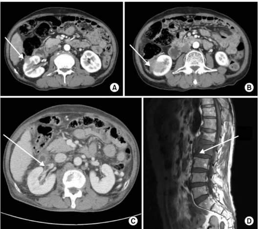
This case report describes an uncommon case of renal actinomycosis in a 63-year-old man. The patient underwent radical nephrectomy for suspicious renal cell carcinoma with renal vein thrombosis and spinal metastasis. The postoperative diagnosis of renal and spinal actinomycosis was established in accordance with the results from histological examination. Three years after surgery, the patient did not show any symptoms of recurrence.

Keywords: Actinomycosis; Carcinoma, renal cell; Renal vein thrombus

Copyright 2016, Korean Association of Urogenital Tract Infection and Inflammation. All rights reserved.

This is an open access article distributed under the terms of the Creative Commons Attribution Non-Commercial License (http://creativecommons.org/licenses/by-nc/4.0) which permits unrestricted non-commercial use, distribution, and reproduction in any medium, provided the original work is properly cited.

Received: 28 March, 2016 Revised: 13 June, 2016 Accepted: 8 July, 2016

Correspondence to: Mi Mi Oh

http://orcid.org/0000-0002-1232-2598 Department of Urology, Korea University Guro Hospital, 148 Gurodong-ro, Guro-gu, Seoul 08308, Korea Tel: +82-2-2626-1310, Fax: +82-2-2626-1321 E-mail: mamah@hanmail.net

Actinomycosis is an uncommon suppurative granulo- matous inflammation caused by the Actinomyces species, which are microaerophilic, anaerobic, gram-positive, rod-shaped bacteria [1]. Cases of abdomino-pelvic actinom- ycosis account for 10-20% of all actinomycosis cases [2].

Abdomino-pelvic actinomycosis often manifests as renal or perirenal masses, often mimicking a neoplasm [3,4]. Few reports have been published on the misdiagnosis of renal actinomycosis as renal cell carcinoma with renal vein thrombosis [5].

Herein, we report a case of renal actinomycosis initially misdiagnosed as renal cell carcinoma with renal vein thrombosis.

CASE REPORT

A 63-year-old man experiencing right flank pain and unexplained weight loss was referred to our hospital. The patient was a smoker and chronic alcoholic. He has a history of type 2 diabetes mellitus, liver cirrhosis, and hypertension.

His weight loss went from 60 kg to 54 kg in a 1-month period.

His blood pressure, pulse rate, respiratory rate, and body

temperature were 120/80 mmHg, 88 beats/min, 20 breaths/min, and 36.9

o

C, respectively. A complete blood count was performed: hemoglobin level of 11.3 g/dl, white blood cell count of 13,500 cells/l after the antibiotics treatment from another hospital, and platelet count of 217,000 cells/l. His blood urea nitrogen level was 18.5 mg/dl, and his creatinine level was 0.7 mg/dl. Urine analysis revealed microscopic hematuria and pyuria. The liver function test was within the normal range.

Abdomino-pelvic computed tomography (CT) revealed two contrast-enhanced, exophytic renal masses—1.5 cm and 3.7 cm in size (Fig. 1A, B)—with renal vein thrombosis (Fig. 1C). The masses had unclear margins with possible direct invasion to the ascending colon (Fig. 1B). A magnetic resonance imaging revealed a 2-cm soft-tissue mass on the anterior portion of the L2 vertebral body with a cortical disruption (Fig. 1D).

The clinical diagnosis was renal cell carcinoma of the right kidney with renal vein thrombosis and direct invasion to the adjacent ascending colon, as well as suspicious metastasis to the L2 vertebra.

Radical nephrectomy was performed due to the increased

(2)

74 Jong Wook Kim, et al. Renal Actinomycosis Misdiagnosed as RCC

Urogenit Tract Infect Vol. 11, No. 2, August 2016

Fig. 1. An abdomino-pelvic computed tomography scan showed two exophytic renal masses—1.5 cm and 3.7 cm in size (A, B)—with renal vein thrombosis (C, arrow). The masses showed unclear margins with possible direct invasion to the ascending colon (arrows of A, B). (D) A magnetic resonance imaging scan showed a 2-cm soft-tissue mass on the anterior portion of the L2 vertebral body with cortical disruption (arrow).

Fig. 2. A photomicrograph showed filamentous bacteria in a sulfur granule (Gomori methenamine-silver staining, ×400).

risk of infarction as a result of renal vein thrombosis. Meti- culous dissection was performed via a flank incision to dissect the kidney, which was adherent to the ascending colon.

Histopathological examination revealed sulfur granules containing Actinomyces species with neutrophil infiltration in the adjacent tissue (Fig. 2). A needle biopsy of the metastatic lesion in the L2 vertebra revealed actinomycosis.

The final diagnosis was actinomycosis of the kidney and spine. Parenteral ampicillin was prescribed for 4 weeks, followed by oral sultamicillin. Three years after the surgery, the patient, according to an abdominal CT, did not show any symptoms of recurrence in the retroperitoneal space and spine lesion.

DISCUSSION

The common forms of actinomycosis include cervico- facial, thoracopulmonary, and abdomino-pelvic actinomy- coses. Abdomino-pelvic actinomycosis accounts for approximately 10-20% of all reported cases, among which, genitourinary involvement is sporadic and mostly manifests as renal or perirenal masses [3,4]. Diagnosis of actinomycosis

is quite difficult for clinicians as a result of its indolent

and nonspecific presentation. Actinomycosis may cause an

abdominal tumor mimicking malignancy; therefore, it is

important for the lesion to be differentiated from abdominal

tumors and for the treatment strategy to be individualized

[6]. Preoperative diagnosis is usually difficult with the

majority of cases being diagnosed after the histological and

(3)

Jong Wook Kim, et al. Renal Actinomycosis Misdiagnosed as RCC 75

Urogenit Tract Infect Vol. 11, No. 2, August 2016

bacteriological examinations of the resected specimen [6].

Renal actinomycosis is commonly represented as a chronic process of inflammation. The symptoms are usually nonspecific. Abdominal or flank pain is the most common symptom, along with weight loss, fever, fatigue, and night sweats. Hematuria is relatively an uncommon symptom.

Lower urinary tract symptoms may also be present. In this case, the patient presented with flank pain, fatigue, and weight loss; however, it is worth nothing that these are also common symptoms of renal cell carcinoma.

Abdominopelvic CT is the most reliable imaging modality to identify the location of lesions and to provide sufficient information on the anatomical extent of renal actinomycosis.

However, the findings from the CT scan are nonspecific and may vary with the stage of disease and regions involved.

Most patients present with a solid renal mass of variable contrast enhancement; some of these patients may have initially been diagnosed with a renal neoplasm, especially a malignant lymphoma, rather than inflammation [7].

Actinomycosis infection of the bone is mainly due to adjacent tissue infection or as a result of hematogenous spread of infection; however, it can also be seen in some fractures [8]. In this case, renal actinomycosis had spread hematogeneously to the spine, which contributed to the misdiagnosis of renal cell carcinoma with metastasis to the spine.

In selected cases, the treatment of choice included the use of antimicrobial agents with a high-dose of penicillin over an extended period of time—18 to 24 million units/day for 2 to 6 weeks, followed by an oral therapy with penicillin or amoxicillin for an addition 6 to 12 months; and this has been reported to completely eradicate actinomycosis [8].

This case report examined a case of renal and spinal actinomycosis, which was initially misdiagnosed as renal

cell carcinoma with renal vein thrombosis. A diagnosis of renal actinomycosis is difficult due to its nonspecific clinical presentations and the limitations of imaging modalities.

However, actinomycosis could be suspected when patients present with flank pain, fever, weight loss, and leukocytosis associated with characteristic findings on a CT scan.

CONFLICT OF INTEREST

No potential conflict of interest relevant to this article was reported.

REFERENCES

1. Mueller MC, Ihrler S, Degenhart C, Bogner JR. Abdominal actinomycosis. Infection 2008;36:191.

2. Das N, Lee J, Madden M, Elliot CS, Bateson P, Gilliland R. A rare case of abdominal actinomycosis presenting as an inflam- matory pseudotumour. Int J Colorectal Dis 2006;21:483-4.

3. Lee IJ, Ha HK, Park CM, Kim JK, Kim JH, Kim TK, et al.

Abdominopelvic actinomycosis involving the gastrointestinal tract: CT features. Radiology 2001;220:76-80.

4. Wagenlehner FM, Mohren B, Naber KG, Mannl HF. Abdominal actinomycosis. Clin Microbiol Infect 2003;9:881-5.

5. Chang DS, Jang WI, Jung JY, Chung S, Choi DE, Na KR, et al.

Renal actinomycosis with concomitant renal vein thrombosis.

Clin Nephrol 2012;77:156-60.

6. Hayashi M, Asakuma M, Tsunemi S, Inoue Y, Shimizu T, Komeda K, et al. Surgical treatment for abdominal actinomy- cosis: a report of two cases. World J Gastrointest Surg 2010;2:405-8.

7. Lin CD, Tai HC, Wang CC, Yu CT, Chang SJ, Hsieh CH, et al.

Renal actinomycosis—an unusual cause of a renal abscess. Urol Sci 2012;23:129-32.

8. Acevedo F, Baudrand R, Letelier LM, Gaete P. Actinomycosis: a

great pretender. Case reports of unusual presentations and a

review of the literature. Int J Infect Dis 2008;12:358-62.

수치

Fig. 1. An abdomino-pelvic computed  tomography scan showed two exophytic  renal masses—1.5 cm and 3.7 cm in size  (A, B)—with renal vein thrombosis (C,  arrow)

참조

관련 문서

Patients with breast cancer; ovarian cancer; renal cell carcinoma; pancreatic neuroendocrine cancer; colorectal cancer; head and neck cancer; non-small cell lung

Intermittent hemodialysis(IDH) and Continuous renal replacement therapy (CRRT) has been used widely for treating critically ill patients with acute renal failure, We

(Method) The study subjects were 25 cancer patients out of 796 end-stage renal disease (ESRD) patients maintained on hemodialysis or peritoneal dialysis at Chosun

KEY WORDS : Computed tomography, Dual energy, Renal stone, Uric acid stone, Non-uric acid stone,

Differential Expression of Desmoglein1, Desmoglein3, Epithelial Membrane Antigen, Ber-EP4 and CD10 in Basal Cell Carcinoma and Squamous Cell Carcinoma

PCT, serum procalcitonin; ESRD, end-stage renal disease; DMN, diabetic nephropathy; HTN, hypertensive nephropathy; HD, hemodialysis; PD, peritoneal

Haemorragic fever with renal syndrome (HFRS) caused by Hantanvirus has been one of the principal acute febrile disease in Korea. Hantaviruses are carried by numerous

Undiagnosed malignancy in patients with deep vein thrombosis: incidence, risk indicators, and diagnosis.. Monreal M, Fernandez-Llamazares J, Perandreu