Effects of Hepatocyte Growth Factor on Collagen Synthesis and Matrix Metalloproteinase Production in Keloids
Keloids are pathologic proliferations of the dermal layer of the skin resulting from excessive collagen production and deposition. Hepatocyte growth factor (HGF) increases the expression of matrix metalloproteinase (MMP)-1 and suppresses collagen synthesis to modulate extracellular matrix turnover. To investigate the anti-fibrotic effects of HGF, we examine the mRNA expression of collagen types I and III and matrix metalloproteinase (MMP-1, MMP-3) on human dermal fibroblast (HDF) cell lines and keloid fibroblasts (KFs, n = 5) after adding various amount of HGF protein. We also evaluated the enzymatic activity of MMP-2, MMP-9 by zymograghy. In HDFs treated with TGF-β1 and HGF protein simultaneously, both type I and III collagen mRNA expression significantly decreased (P <
0.05). Expression of MMP-1, MMP-3 mRNA also decreased. However, the mRNA expression of MMP-1, MMP-3 significantly increased in KFs with increasing amount of HGF in dose dependent manner (P < 0.05). The enzymatic activities of MMP-2 increased with increasing HGF protein in a dose-dependent manner. However, the enzymatic activity of MMP-9 did not change. These results suggest that the anti-fibrotic effects of HGF may have therapeutic effects on keloids by reversing pathologic fibrosis.
Key Words: Hepatocyte Growth Factor; Keloid Fibroblast; Dermal Fibroblast; Collagen;
Matrix Metalloproteinase Won Jai Lee, Sang Eun Park
and Dong Kyun Rah
Institute for Human Tissue Restoration, Department of Plastic and Reconstructive Surgery, Yonsei University Health System, Severance Hospital, Seoul, Korea
Received: 8 April 2011 Accepted: 21 June 2011 Address for Correspondence:
Won Jai Lee, MD
Institute for Human Tissue Restoration, Department of Plastic and Reconstructive Surgery, Yonsei University Health System, Severance Hospital, 262 Seongsan-ro, Seodaemun-gu, Seoul 120-752, Korea
Tel: +82.2-2228-2219, Fax: +82.2-393-6947 E-mail: [email protected]
This work was supported by the National Research Foundation of Korea (NRF) grant funded by the Republic of Korea Government (MEST) (No. 2009-0073055). All authors have no conflicts of interest or financial arrangements that could potentially influence the described research.
DOI: 10.3346/jkms.2011.26.8.1081 • J Korean Med Sci 2011; 26: 1081-1086 Surgery
INTRODUCTION
Keloids and hypertrophic scars are pathologic conditions result- ing from excessive collagen deposition in the dermis due to skin injuries including surgery and trauma. They can lead to func- tional problems such as limited joint motion and aesthetic and social issues for patients. At present, the demand for keloid treat- ment is increasing.
Keloid fibroblasts (KFs) are similar to human normal dermal fibroblasts (HDFs) in size and shape, but show a great capacity to proliferate and produce high levels of extracellular matrix such as collagen, fibronectin, elastin, and proteoglycans (1-5). Keloids show an abnormal balance between proliferative and apoptotic cell death (6) and an excessive synthesis of extracellular matrix due to a failure to maintain homeostasis in the wound healing process. Many investigators have found that the excessive extra- cellular matrix (ECM) production is related to an increase or de- crease of matrix metalloproteinases (MMP-1, -2, -3, -9, -13) and an increase of tissue inhibitors of metalloproteinases (TIMPs) (7-10).
Gelatinases (MMP-2, 9) digest denatured collagens (types I and III) fibrils, type IV collagen, fibronectin and elastin; addi- tionally, these enzymes can potentiate the degradation of ECM
components by activating collagenase-3 (MMP-13) and neutro- phil collagenase (MMP-8). Therefore, gelatinases are crucial in physiological processes such as tissue remodeling, and in path- ological processes such as wound healing and the invasion of malignant tumors (11-13). MMP-9 activity is crucial during the epithelization process and early repair events, whereas the ge- latinolytic activity of MMP-2 is important during the prolonged remodeling phase (14-16). The gelatinases act on cleaved colla- gen more effectively than other MMPs (11).
Hepatocyte growth factor (HGF), initially discovered to be a potent mitogen for hepatocytes (17, 18) and also known as a “scat- ter factor”, acts as a multifunctional mediator on many kinds of cells. It is secreted by mesenchymal cells and regulates cell growth, cell motility, apoptotic cell death (19), and the morphogenesis of hepatocytes, fibroblasts, and keratinocytes (20) primarily by activating a tyrosine kinase signaling cascade after binding to the proto-oncogenic c-Met receptor (21-23). It is secreted as a single, inactive polypeptide and is cleaved by serine proteases into a 69-kDa α-chain and a 34-kDa β-chain.
Recent studies have found a reverse anti-fibrotic effect in which HGF prevents fibrosis after damage to the liver (24), lungs, and kidneys (20, 25). HGF not only protects against fibrosis through inhibiting ECM deposition such as collagen but also reduces the
previously formed ECM by the induction of MMP-1 (26). How- ever, these studies show these anti-fibrotic effects only in ani- mal models and with HGF gene therapy, and the actions of HGF on human keloid fibroblasts have yet to be studied.
Some investigators have studied an anti-fibrogenic mecha- nism of HGF in which increasing MMP expression promotes collagen degradation (26, 27). Adding HGF protein to fibroblast cultures of scleroderma patients significantly decreased colla- gen production and increased MMP-1 expression and activity (26). This means that HGF may have therapeutic effect on ke- loids that are similar to scleroderma with pathologic dermal fi- brosis.
In this study, we investigated the mRNA levels of type I and III collagen synthesis, MMP-1, 3 expression, and MMP-2, 9 en- zymatic activities in HDFs and KFs after administration of vari- ous amounts of HGF protein with or without TGF-β1.
MATERIALS AND METHODS
Keloid-derived fibroblast cultures and the human dermal fibroblast cell line
Keloid-derived fibroblasts (KFs, n = 5) were obtained from tissues excised during surgical procedures. Cells were obtained from the central dermal layer of active stage keloid patients (Table 1) after having obtained informed consent according to a protocol approved by the Yonsei University College of Medicine Institu- tional Review Board. All experiments involving humans were performed in adherence to the Helsinki Guidelines. Keloids were identified by trained clinicians and pathologists. The sep- arated cells were cultured in Dulbecco’s modified Eagle’s medi- um (DMEM; GIBCO, Grand Island, NY, USA) supplemented with heat-inactivated 10% fetal bovine serum (FBS) and peni- cillin (30 U/mL), streptomycin (300 mg/mL), and actinomysin.
The culture medium was changed in 2-3 day intervals. All cells used in this study were taken before passage 5. The human nor- mal dermal fibroblasts (HDFs) cell line was purchased from the American Type Culture Collection (ATCC, Manassas, VA, USA).
The HDFs were cultured in DMEM (Gibco BRL) supplemented with 10% FBS (Gibco BRL), penicillin (30 IU/mL), and strepto- mycin (300 μg/mL).
Hepatocyte growth factor (HGF) and TGF-ϐ1 treatment The HDFs and KFs were treated with different amounts of HGF
protein (1, 10, 20, and 40 ng). Three days after adding the HGF protein, the mRNA levels of collagen type I, III, MMP-1, 3 were examined by RT-PCR. Additionally, MMP-2 and -9 enzymatic activities were examined by zymography. TGF-β1 (5 ng/mL) was added simultaneously with the HGF protein.
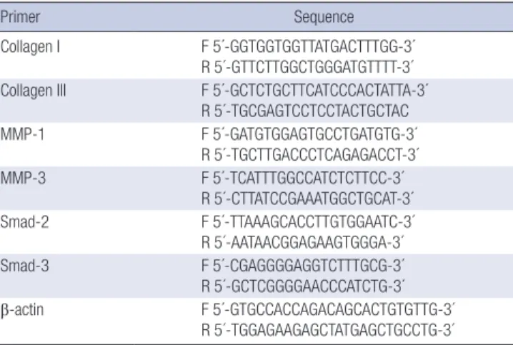
Reverse transcriptase-polymerase chain reaction (RT-PCR) Total RNA was extracted from samples using a Trizol reagent (Invitrogen Corp., Carlsbad, CA, USA) according to the manu- facturer’s protocol. One microgram of total RNA was converted into the complementary DNA using a First Strand cDNA Syn- thesis kit (Promega Corp., Madison, WI, USA) with random prim- ers. A polymerase chain reaction was performed using 2× Taq Premix 2 (Solgent Co., Ltd., Seoul, Korea) and the synthetic gene- specific primers for collagen types I and III, MMP-1, MMP-3 and β-actin (Table 2). All target sequences were separately amplified for 35 cycles. Samples of each reaction product were separated by 2% agarose gel electrophoresis, visualized by ethidium bro- mide staining, and photographed with 290-nm ultraviolet illu- mination. The density of each band was measured by Image J.
Relative expression of the mRNA of collagen types I and III, MMP- 1, and MMP-3 were normalized to the expression of an internal control (β-actin).
MMP zymography
The enzyme activities of MMP-2 and MMP-9 were measured us- ing zymography. Culture supernatants were collected and ana- lyzed by gel substrate zymography. Briefly, 10 mL of non-reduc- ing sample buffer (125 mM Tris-HCl [pH 6.8], 10%[v/v] glycerol, 0.1%[w/v] BPB) was added to each sample. Samples were then loaded onto 12.5% SDS-PAGE gels containing 0.2% gelatin (Sig- ma, St. Louis, MO, USA). The gels were incubated with renatur- ing buffer (2.5% Triton X-100, 50 mM Tris-HCl, pH 7.5, and 0.1 M NaCl) for 1 hour at room temperature, and then with devel- oping buffer (50 mM Tris-pH (pH 7.5), 10 mM CaCl2, and 0.02%
NaN3) for 18 hr at 37°C. The gels were then stained with 1% Coo-
Table 1. Profiles of the keloid scar fibroblasts used in the study
Case Sex Race Age (yr) Location of keloid
1 F Korean 28 Earlobe
2 F Korean 28 Shoulder
3 F Korean 27 Earlobe
4 F Korean 20 Earlobe
5 F Korean 23 Earlobe
Table 2. List of primers used in our study
Primer Sequence
Collagen I F 5´-GGTGGTGGTTATGACTTTGG-3´
R 5´-GTTCTTGGCTGGGATGTTTT-3´
Collagen III F 5´-GCTCTGCTTCATCCCACTATTA-3´
R 5´-TGCGAGTCCTCCTACTGCTAC
MMP-1 F 5´-GATGTGGAGTGCCTGATGTG-3´
R 5´-TGCTTGACCCTCAGAGACCT-3´
MMP-3 F 5´-TCATTTGGCCATCTCTTCC-3´
R 5´-CTTATCCGAAATGGCTGCAT-3´
Smad-2 F 5´-TTAAAGCACCTTGTGGAATC-3´
R 5´-AATAACGGAGAAGTGGGA-3´
Smad-3 F 5´-CGAGGGGAGGTCTTTGCG-3´
R 5´-GCTCGGGGAACCCATCTG-3´
β-actin F 5´-GTGCCACCAGACAGCACTGTGTTG-3´
R 5´-TGGAGAAGAGCTATGAGCTGCCTG-3´
massie brilliant blue R-250, and gelatinolytic activity was detect- ed as unstained bands on a blue background.
Statistics
Results are expressed as mean ± standard error of the mean (SEM). Statistical comparisons across KFs and HDF cell lines treated with different amounts of HGF were made using paired t-tests. P values less than 0.05 were considered significant.
Ethics statement
This study was approved by the institutional review board of Yon- sei Medical Center (IRB No.: 4-2011-0072). Informed consent was acquired from all patients.
RESULTS
The effects of hepatocyte growth factor on the mRNA expression of collagen types I and III in the HDFs, TGF-ϐ1 stimulated HDFs, and keloid fibroblasts
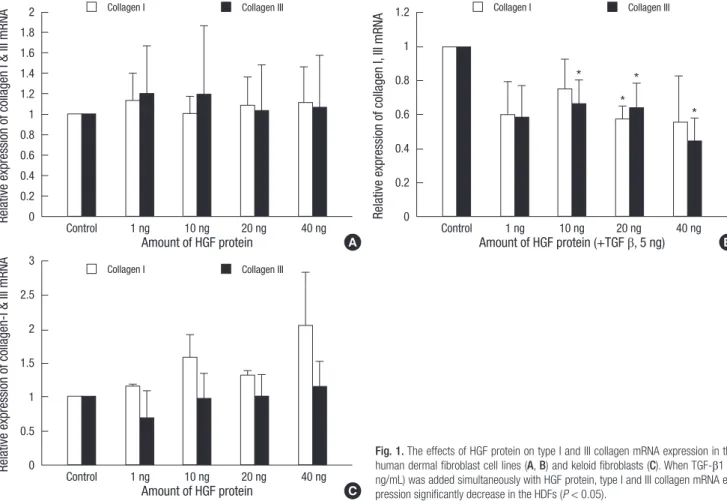
The mRNA expression of collagen types I and III were examined by RT-PCR. We found no significant differences in HDFs after adding HGF alone (Fig. 1A). However, when TGF-β1 (5 ng/mL) was added simultaneously with the HGF protein, collagen types I and III mRNA expression decreased. In HDFs treated with 40
ng HGF protein, this decrease was 57% for type I and 64% for type III collagen (P < 0.05, Fig. 1B). On the other hand, the HGF protein caused no significant change in type I collagen mRNA expression in KFs (Fig. 1C).
The effects of hepatocyte growth factor on the mRNA expression of MMP-1 and 3 in the HDFs, TGF-ϐ1 stimulated HDFs and keloid fibroblasts
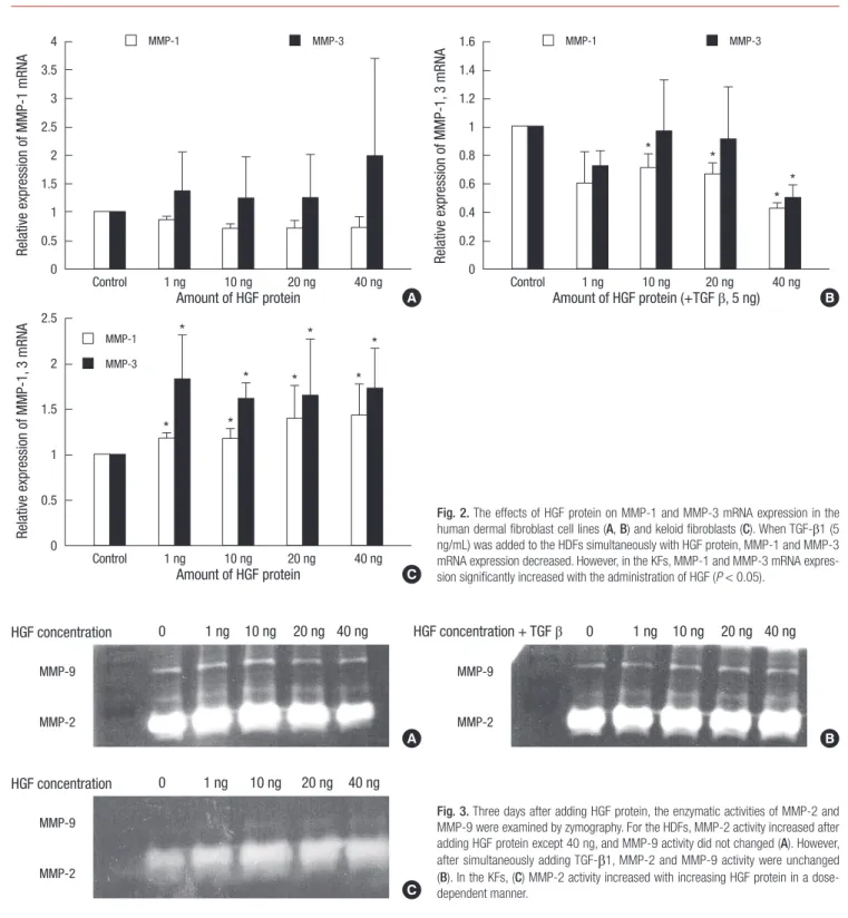
The expression of MMP-1 and MMP-3 mRNA was examined by RT-PCR. We found no significant differences in the expression of MMP-1 and MMP-3 mRNA when the HGF protein was treat- ed alone. When TGF-β1 (5 ng/mL) was administered simulta- neously with the HGF protein, both MMP-1 and MMP-3 mRNA expression decreased. This decrease was approximately 43% for MMP-1 and 49% for MMP-3 after adding 40 ng of HGF protein (Fig. 2A, B). However, mRNA expressions of MMP-1 and MMP- 3 in the KFs were increased after the administration of HGF pro- tein alone with statistical significance (P < 0.05, Fig. 2C).
The effects of hepatocyte growth factor on MMP-2, MMP-9 enzymatic activities
The HDFs and KFs were treated with various amounts of HGF protein (1, 10, 20, and 40 ng). Three days after the administration of HGF protein, MMP-2 and MMP-9 enzymatic activities were
Collagen I
Collagen I
Collagen I Collagen III
Collagen III
Collagen III
Relative expression of collagen I & III mRNA
Amount of HGF protein
Control 1 ng 10 ng 20 ng 40 ng
2 1.8 1.6 1.4 1.2 1 0.8 0.6 0.4 0.2 0
A
Relative expression of collagen-I & III mRNA
Amount of HGF protein
Control 1 ng 10 ng 20 ng 40 ng
3
2.5
2
1.5
1
0.5
0
C
Relative expression of collagen I, III mRNA
Amount of HGF protein (+TGF β, 5 ng)
Control 1 ng 10 ng 20 ng 40 ng
1.2
1
0.8
0.6
0.4
0.2
0
B
*
*
*
*
Fig. 1. The effects of HGF protein on type I and III collagen mRNA expression in the human dermal fibroblast cell lines (A, B) and keloid fibroblasts (C). When TGF-β1 (5 ng/mL) was added simultaneously with HGF protein, type I and III collagen mRNA ex- pression significantly decrease in the HDFs (P < 0.05).
examined by zymography.
For the HDFs, we found that MMP-2 enzymatic activity in- creased after adding 1, 10, and 20 ng of HGF protein (Fig. 3A).
However, the enzymatic activity of MMP-9 did not change. When TGF-β1 (5 ng/mL) was added simultaneously with HGF protein, the enzymatic activities of MMP-2 and MMP-9 were unchanged (Fig. 3B). For the KFs, we found that MMP-2 activity increased with increasing amounts of HGF protein in a dose-dependent manner (Fig. 3C). However, the enzymatic activity of MMP-9 ei- ther did not change or showed only a slight increase.
DISCUSSION
This study investigated the anti-fibrogenic effects of hepatocyte growth factor (HGF) on the human dermal fibroblasts (HDFs) cell line and keloid fibroblasts (KFs). Fujiwara et al. (28) found that the production of type 1 collagen, MMP-1, and MMP-2 was 3- fold, 6-fold, and 2.4-fold higher, respectively, in keloid fibro- blasts compared to normal dermal fibroblasts. In this study, KFs showed excessive collagen synthesis and breakdown of the ex- tracellular matrix (ECM) due to increased MMP-1 and MMP-2
MMP-1
MMP-1
MMP-1 MMP-3
MMP-3
MMP-3
Relative expression of MMP-1 mRNA
Amount of HGF protein
Control 1 ng 10 ng 20 ng 40 ng
4 3.5 3 2.5 2 1.5 1 0.5 0
A
Relative expression of MMP-1, 3 mRNA
Amount of HGF protein
Control 1 ng 10 ng 20 ng 40 ng
2.5
2
1.5
1
0.5
0
C
Relative expression of MMP-1, 3 mRNA
Amount of HGF protein (+TGF β, 5 ng)
Control 1 ng 10 ng 20 ng 40 ng
1.6 1.4 1.2 1 0.8 0.6 0.4 0.2 0
B
*
*
*
*
*
*
* *
*
*
*
*
Fig. 2. The effects of HGF protein on MMP-1 and MMP-3 mRNA expression in the human dermal fibroblast cell lines (A, B) and keloid fibroblasts (C). When TGF-β1 (5 ng/mL) was added to the HDFs simultaneously with HGF protein, MMP-1 and MMP-3 mRNA expression decreased. However, in the KFs, MMP-1 and MMP-3 mRNA expres- sion significantly increased with the administration of HGF (P < 0.05).
Fig. 3. Three days after adding HGF protein, the enzymatic activities of MMP-2 and MMP-9 were examined by zymography. For the HDFs, MMP-2 activity increased after adding HGF protein except 40 ng, and MMP-9 activity did not changed (A). However, after simultaneously adding TGF-β1, MMP-2 and MMP-9 activity were unchanged (B). In the KFs, (C) MMP-2 activity increased with increasing HGF protein in a dose- dependent manner.
HGF concentration MMP-9
MMP-2
0 1 ng 10 ng 20 ng 40 ng
C
HGF concentration HGF concentration + TGF β
MMP-9 MMP-9
MMP-2 MMP-2
0 1 ng 10 ng 20 ng 40 ng 0 1 ng 10 ng 20 ng 40 ng
A B
activity. TGF-β1 played a role in modulating type 1 collagen, MMP- 1, and MMP-2. Since these keloids are characterized by an ac- cumulation of collagen, it appears that the down regulation of collagen I and III expression and the upregulation of MMP-1 and MMP-2 are able to ablate the excess collagen formed by the in- crease in synthetic activity.
In our study, the expressions of collagen I and III mRNA induc- ed by exogenous TGF-β1 were suppressed by adding HGF pro- tein on the HDFs. However, these expressions were not changed by adding HGF protein alone. This means that HGF does not influence or increase ECM production of normal dermal fibro- blasts, and suppresses the increased expression of collagen I and III mRNA induced by exogenous treatment with TGF-β. This suggests a high potential for HGF as therapeutics in abnormal fibrotic disease because HGF do not affect basal collagen levels in normal fibroblasts.
Applying HGF proteins to KFs did not significantly change the expression of collagen I and III mRNA (Fig. 1). In previous stud- ies, collagen I and III mRNA expression was found to be more enhanced in KFs than HDFs, so that we expected the addition of HGF protein to cultured KFs to decrease collagen I and III mRNA expression since TGF-β is antagonized by HGF (20). How- ever, contrary to our hypothesis, this did not happen. This seems to be caused by a different profibrogenic reaction with TGF-β in KFs compared with HDFs. More TGF-β is secreted in KFs than in normal fibroblasts and it plays a predominant role in the pro- fibrogenic reaction; the profibrogenic reaction of TGF-β in KFs is more sensitive and persistent than in HDFs. Thus, the anti-fi- brogenic reaction of HGF seems to be different in KFs from that in HDFs. Therefore, it appears that HGF protein decreases the expression of type I and III collagen mRNA in profibrogenic con- ditions (HDFs adding TGF-β), but does not influence these ex- pression in keloid-like conditions (high TGF-β concentration).
However, Jinnin et al. (26) reported that HGF decreased the de- position of type I collagen protein by C-met overexpression and MMP-1 induced collagenolysis. Therefore, further investigation of the effects of HGF on collagen metabolism of keloid-derived fibroblast would be warranted.
Applying TGF-β (5 ng) and HGF to the cultured HDFs signifi- cantly decreased the expression of MMP-1 and MMP-3 mRNA, but adding HGF alone showed no significant differences. Also, when TGF-β and HGF are both applied to cultured HDFs, the expression of collagen type I and type III mRNA decreases. Be- cause healing of wounds such as lacerations, requires early pro- fibrogenic conditions similar to secreted TGF-β in HDFs, HGF treatments may impair the wound healing process due to a de- crease in extracellular matrix synthesis and degradation rate. In addition, we found no changes in MMP-9 activity in the HDFs, which is further evidence that HGF protein therapy will have lit- tle benefit in the wound healing process. However, other study shows that continuous high HGF protein expression by the in-
troduction of the HGF gene promotes dermal regeneration, pos- sibly not only by suppres sing the overgrowth of collagen due to its antifibrotic effect but also by promoting regeneration (22).
Therefore we think that repetitive high doses of HGF could en- hance effectively an acute wound healing. But, this approach has disadvantages, including the requirement for large amounts of HGF, the short half-life, the potential toxicity, and difficulty with appropriate delivery of HGF in the wound. Further studies are necessary for changes of collage and MMPs expression us- ing HGF gene therapy.
Adding HGF to cultured KFs increased MMP-1 and MMP-3 mRNA expression (Fig. 2C). The enzymatic activity of MMP-2 also increased with increasing HGF protein concentration (Fig.
3). In contrast, we saw no detectable changes in MMP-9 activity in the KFs. The increase in MMP-2 activity is important in induc- ing the degradation of excessive ECM in keloids, which seems to enhance MMP-1 and MMP-3 expression, indicating that HGF may have therapeutic effects on keloids by reversing pathologic fibrosis. The anti-fibrogenic effects of HGF generally increased in a dose-dependent manner, and so the appropriate treatment dose remains to be determined. In addition, further studies are necessary to develop efficient continuous release vehicles for HGF such as gene therapy that has its very short half-life.
In conclusion, contrary to previously published reports which found that HGF promotes wound healing, this study showed that HGF might inhibit the profibrogenic effects in wound heal- ing. In addition, we found that HGF may have a therapeutic ef- fect on keloids through enhancing MMP-1 and MMP-3 mRNA expression and MMP-2 enzymatic activity.
REFERENCES
1. Babu M, Diegelmann R, Oliver N. Fibronectin is overproduced by keloid fibroblasts during abnormal wound healing. Mol Cell Biol 1989; 9: 1642-50.
2. Bettinger DA, Yager DR, Diegelmann RF, Cohen IK. The effect of TGF- beta on keloid fibroblast proliferation and collagen synthesis. Plast Re- constr Surg 1996; 98: 827-33.
3. Calderon M, Lawrence WT, Banes AJ. Increased proliferation in keloid fibroblasts wounded in vitro. J Surg Res 1996; 61: 343-7.
4. Kischer CW, Hendrix MJ. Fibronectin (FN) in hypertrophic scars and keloids. Cell Tissue Res 1983; 231: 29-37.
5. Russell SB, Trupin JS, Kennedy RZ, Russell JD, Davidson JM. Glucocor- ticoid regulation of elastin synthesis in human fibroblasts: down-regula- tion in fibroblasts from normal dermis but not from keloids. J Invest Der- matol 1995; 104: 241-5.
6. Akasaka Y, Fujita K, Ishikawa Y, Asuwa N, Inuzuka K, Ishihara M, Ito M, Masuda T, Akishima Y, Zhang L, Ito K, Ishii T. Detection of apoptosis in keloids and a comparative study on apoptosis between keloids, hypertro- phic scars, normal healed flat scars, and dermatofibroma. Wound Repair Regen 2001; 9: 501-6.
7. Reynolds JJ. Collagenases and tissue inhibitors of metalloproteinases: a functional balance in tissue degradation. Oral Dis 1996; 2: 70-6.
8. Visse R, Nagase H. Matrix metalloproteinases and tissue inhibitors of metalloproteinases: structure, function, and biochemistry. Circ Res 2003;
92: 827-39.
9. Brew K, Dinakarpandian D, Nagase H. Tissue inhibitors of metallopro- teinases: evolution, structure and function. Biochim Biophys Acta 2000;
1477: 267-83.
10. Kischer CW, Thies AC, Chvapil M. Perivascular myofibroblasts and mi- crovascular occlusion in hypertrophic scars and keloids. Hum Pathol 1982; 13: 819-24.
11. Parks WC. Matrix metalloproteinases in repair. Wound Repair Regen 1999; 7: 423-32.
12. Vihinen P, Ala-aho R, Kähäri VM. Matrix metalloproteinases as thera- peutic targets in cancer. Curr Cancer Drug Targets 2005; 5: 203-20.
13. Santos MC, Souza AP, Gerlach RF, Tabchoury CM, Line SR. Inhibition of human gelatinases (matrix metalloproteinase-2 and matrix metallo- proteinase-9) activity by zinc oxide: a possible mechanism to enhance wound healing. Br J Dermatol 2001; 145: 854-5.
14. Gillard JA, Reed MW, Buttle D, Cross SS, Brown NJ. Matrix metallopro- teinase activity and immunohistochemical profile of matrix metallopro- teinase-2 and -9 and tissue inhibitor of metalloproteinase-1 during hu- man dermal wound healing. Wound Repair Regen 2004; 12: 295-304.
15. Inkinen K, Turakainen H, Wolff H, Ravanti L, Kähäri VM, Ahonen J. Ex- pression and activity of matrix metalloproteinase-2 and -9 in experimen- tal granulation tissue. APMIS 2000; 108: 318-28.
16. Agren MS. Gelatinase activity during wound healing. Br J Dermatol 1994;
131: 634-40.
17. Nakamura T, Nawa K, Ichihara A. Partial purification and characteriza- tion of hepatocyte growth factor from serum of hepatectomized rats. Bio- chem Biophys Res Commun 1984; 122: 1450-9.
18. Asami O, Ihara I, Shimidzu N, Shimizu S, Tomita Y, Ichihara A, Nakamu- ra T. Purification and characterization of hepatocyte growth factor from injured liver of carbon tetrachloride-treated rats. J Biochem 1991; 109:
8-13.
19. Bardelli A, Longati P, Albero D, Goruppi S, Schneider C, Ponzetto C, Co- moglio PM. HGF receptor associates with the anti-apoptotic protein BAG- 1 and prevents cell death. EMBO J 1996; 15: 6205-12.
20. Sato C, Tsuboi R, Shi CM, Rubin JS, Ogawa H. Comparative study of he- patocyte growth factor/scatter factor and keratinocyte growth factor ef- fects on human keratinocytes. J Invest Dermatol 1995; 104: 958-63.
21. Ono I. The effects of basic fibroblast growth factor (bFGF) on the break- ing strength of acute incisional wounds. J Dermatol Sci 2002; 29: 104-13.
22. Ono I, Yamashita T, Hida T, Jin HY, Ito Y, Hamada H, Akasaka Y, Ishii T, Jimbow K. Local administration of hepatocyte growth factor gene en- hances the regeneration of dermis in acute incisional wounds. J Surg Res 2004; 120: 47-55.
23. Matsumoto K, Nakamura T. Emerging multipotent aspects of hepatocyte growth factor. J Biochem 1996; 119: 591-600.
24. Ueki T, Kaneda Y, Tsutsui H, Nakanishi K, Sawa Y, Morishita R, Matsumoto K, Nakamura T, Takahashi H, Okamoto E, Fujimoto J. Hepatocyte growth factor gene therapy of liver cirrhosis in rats. Nat Med 1999; 5: 226-30.
25. Patijn GA, Lieber A, Schowalter DB, Schwall R, Kay MA. Hepatocyte growth factor induces hepatocyte proliferation in vivo and allows for ef- ficient retroviral-mediated gene transfer in mice. Hepatology 1998; 28:
707-16.
26. Jinnin M, Ihn H, Mimura Y, Asano Y, Yamane K, Tamaki K. Effects of he- patocyte growth factor on the expression of type I collagen and matrix metalloproteinase-1 in normal and scleroderma dermal fibroblasts. J Invest Dermatol 2005; 124: 324-30.
27. Esposito C, Parrilla B, Cornacchia F, Grosjean F, Mangione F, Serpieri N, Valentino R, Villa L, Arra M, Esposito V, Dal Canton A. The antifibrogen- ic effect of hepatocyte growth factor (HGF) on renal tubular (HK-2) cells is dependent on cell growth. Growth Factors 2009; 27: 173-80.
28. Fujiwara M, Muragaki Y, Ooshima A. Keloid-derived fibroblasts show in- creased secretion of factors involved in collagen turnover and depend on matrix metalloproteinase for migration. Br J Dermatol 2005; 153: 295-300.
AUTHOR SUMMARY
Effects of Hepatocyte Growth Factor on Collagen Synthesis and Matrix Metalloproteinase Production in Keloids
Won Jai Lee, Sang Eun Park and Dong Kyun Rah
The purpose of this study is to investigate the effects of hepatocyte growth factor (HGF) in the human dermal fibroblasts (HDFs) and keloid fibroblasts (KFs). In HDFs, treatment with TGF-β1 and HGF decreased type I and III collagen mRNA expression. However, the expressions of MMP-1, MMP-3 mRNA were also decreased. In contrast, the expressions of MMP-1 and MMP-3 mRNA were increased in KFs by HGF. Also, the enzymatic activity of MMP-2 was increased by HGF. These results suggest that the anti-fibrotic effects of HGF may have therapeutic effects on keloids by reversing pathologic fibrosis.