INTRODUCTION
Dendritic cells (DCs) are antigen-presenting cells (APC) with a key role in the immune system as initiators and stimu- lators of naive T-cell responses against microbial pathogens and tumors (1). Since the recognition of DCs in lymphoid organs in 1973 (2), research on these cells has increased because they might be useful for antitumor immunotherapy (1, 3, 4). Furthermore, the central role of DCs in the initia- tion of immune responses and new methods for the genera- tion of large numbers of pure DCs by culturing progenitor cells in the presence of granulocyte-macrophage colony-stimu- lating factor (GM-CSF), tumor necrosis factor (TNF)- and interleukin (IL)-4, creates possibilities for the development of novel immunotherapeutic strategies against tumors and other diseases (5-8).
DCs are present in small numbers in most tissues, includ- ing skin, lung, liver, spleen, blood, lymphoid organs, and bone marrow (9). Morphologically, they are large cells with elongated stellate dendrites. These cells have been shown to internalize and process exogenous or endogenous antigens efficiently, and to present soluble antigens as peptides in conjunction with MHC classes I and II (10, 11). In addition,
DCs have the unique ability to cause clustering of naive T- cells. They also respond to antigen by rapid upregulation of the expression of MHC factors and co-stimulatory molecules, the production of cytokines, and migration toward lym- phatic organs (12, 13). DCs, helper T-cell-dependent anti- body responses, as well as the generation of primary and secondary cytotoxic T-cell responses to tumor-associated antigens, have proven to be effective immunogens when pulsed with tumor-associated antigens (9, 14). However, T- cell defined epitopes that can be presented by DCs have not been identified for most tumors. One study suggested that immunization with undefined tumor antigens might be more effective in eliciting antitumor immunity (15). Although the use of a single antigen-derived epitope has been shown to effect antitumor immunity in murine models, the presen- tation of multiple antigen-derived epitopes may enhance antitumor immunity. Johnston et al. demonstrated that the increased immunogenicity of tumor cells expressing the B7.1 gene was caused by the expansion of the antigenic repertoire of the tumor (16). Because of the extensive diversity of MHC antigens, designing common peptides for T-cell recognition for different individuals with the same disease may be diffi- cult to prove (17). To circumvent this problem and to pro-
*
*
Division of Hematology/Oncology, Department of Medicine, Asan Medical Center, College of Medicine, University of Ulsan, and Asan Institute for Life Science*, Seoul, Korea
Sang-Hee Kim, M.D.
Asan Institute for Life Science Division of Hematology/Oncology, Department of Medicine, College of Medicine, University of Ulsan, 388-1 Poongnap-dong, Songpa-gu, Seoul 138-736, Korea
Tel : +82.2-3010-4157, Fax : +82.2-3010-4182 E-mail : [email protected]
*Presented at the 41st Annual Meeting of the Ameri- can Society of Hematology, New Orleans LA, U.S.A., December 3-7, 1999.
372
Dendritic cells (DCs) are potent antigen-presenting cells for the induction and activation of cytotoxic T lymphocytes. We tested whether bone marrow-derived DCs are capable of inducing protective immunity against a murine lymphoma (A20). DCs were grown from tumor-bearing BALB/c mice by culturing bone mar- row cells. BALB/c mice were injected (sc) with A20 cells on day 0. Intraperitoneal immunization with DCs mixed with lethally irradiated A20 cells were started when the tumor reached ca. 4-5 mm in diameter (Group A) or on day -7 (Group B). Booster immunizations were given every 3-4 days for four weeks. By 31 days in group A, there was a significant reduction in tumor growth in the mice immunized with DCs mixed with irradiated A20 cells as compared with the control groups (p
=0.016). In group B, tumor growth was completely inhibited and there was no tumor growth following extended observations after completion of immunization.
Thus, DCs mixed with irradiated tumor cells can induce an antitumor effect. This provides a rationale for the use of DCs mixed with irradiated tumor cells in immunotherapy for minimal residual disease of lymphomas.
Key Words : Lymphoma; Vaccination; Dendritic cells; Neoplasm, Residual
Received : 28 October 2002 Accepted : 18 February 2003
vide DCs with presentable tumor-associated peptides, several methods have been developed to isolate immunogenic pep- tides from tumor cells. However, there is no defined stan- dard method. Despite the advances in chemotherapy, includ- ing high-dose therapy with autologous stem cell transplan- tation for lymphomas, relapse of the underlying disease remains a significant obstacle (18). Recent advances in can- cer vaccine development now make it possible to consider combining active specific immunotherapy as a strategy for the elimination of minimal residual disease.
The aim of this study was to assess the ability of bone marrow-derived DCs to induce therapeutic and protective immunity against a murine lymphoma model. Using immu- nization with DCs mixed with lethally irradiated target tumor cells, we investigated whether tumor cells, as a source of undefined tumor antigens, could intensify the antitumor immune response of DCs, and evaluated whether the sup- pression of tumor growth was correlated with T-cell function.
MATERIALS AND METHODS Mice
Female BALB/c AnN (BALB/c) mice (6-8 weeks old) were purchased from Jackson Laboratories (Bar Harbor, ME, U.S.A.) and housed in specific pathogen-free units of the Animal Resources Center at Asan Institute for Life Science.
Mice were maintained and treated according to National Institutes of Health guidelines. All aspects of the studies requiring animal experimentation were approved by the Asan Institute of Life Science Animal Care and Use Com- mittee.
Cell Lines
A20 lymphoma cells were purchased from American Type Culture Collection (ATCC: Rockville, MD, U.S.A.). This is a BALB/c B lymphoma cell line derived from a spontaneous reticulum cell neoplasm found in an old BALB/c mouse (19, 20). Cells were maintained in complete RPMI 1640 medium (GIBCO BRL, Aithersburg, MD, U.S.A.) supple- mented with 100 IU/mL penicillin (Sigma, St Louis, MO, U.S.A.), 0.1 mg/mL streptomycin (Sigma), 10-5M -mer- captoethanol (Sigma), and 10% fetal bovine serum (FBS:
GIBCO BRL).
Tumor-Bearing Bone Marrow Cell Culture and Isola- tion of Dendritic Cells
A20 tumor cells (2 106) were injected subcutaneously (sc) into the shaved back of BALB/c mice. Animals were sacrificed three weeks after the injection, when the tumor reached 5-10 mm in diameter. Bone marrow cells were
obtained from the femurs and tibias of BALB/c mice as des- cribed (5). After three washes in RPMI 1640, mononuclear cells were obtained and allowed to adhere to a tissue culture flask for 3 hr at 37 . The adherent cells were then removed and non-adherent cells were placed in 100 mm diameter tissue culture dishes at a concentration of 1 105/mL in the medium supplemented with 20 ng/mL murine recombinant (r) GM-CSF (R & D Systems, Minneapolis, MN, U.S.A.), 10 ng/mL murine IL-4 (R & D) and 2.5 ng/mL murine TNF- (R & D). Culture dishes were fed once every three days. On day 11, non-adherent cells were harvested and used for assays and immunization.
Evaluation of Cell Yield and FACS Analysis
Cultured cells were washed once and an aliquot volume was mixed 1:1 in Trypan blue solution (Sigma). Trypan blue negative, and large cells were counted as viable under the microscope in a Neubauer chamber and cells were identified by their distinctive morphology. Cells (1 106) were incu- bated with the corresponding antibody for 30 min at 4 . The cells were then washed and fixed in 1% paraformalde- hyde. Flow cytometric analysis was performed using a FAC- Scan (Becton Dickinson, Mountain View, CA, U.S.A.). DCs were phenotyped with antibodies to the following markers:
isotype controls for hamster IgG; rat IgG2a; DC markers DEC-205 (NLDC-145) and CD11C; co-stimulatory/adhe- sion molecules CD80 (B7-1) and CD86 (B7-2); macrophage markers CD14 and F4/80, and granulocyte marker Gr-1 (Pharmingen, Hamburg, Germany).
Preparation of Splenocytes
Spleen cells were obtained from the same mice, and sus- pended and red cells were lysed with ammonium chloride.
To obtain purified T-cells, spleen cells were incubated in tissue culture flasks for 1 hr in medium as above, and non-adherent cells were collected and filtered through a nylon wool column.
Mixed Leukocyte Reaction (MLR)
DCs (5 103) from BALB/c AnN mice were irradiated (1,500 rads) to stimulate the proliferation of DCs and were added to T-cells (5 104) from BALB/c mice. A20 cells (5 104 lethally irradiated at 10,000 rad) were co-cultured in 96-well plates for each immunized group. The cells were co-cultured for three days at 37 (under 5% CO2). Cells were divided into three groups: DCs mixed with T-cells, lethally irradiated A20 cells plus T-cells, and T-cells only.
Each group of cells was incubated for 18 hr with 1 ci of [3H] thymidine (Amersham, Arlington Heights, IL, U.S.A.) at the end of the three-day culture, and isotope incorporation was determined using a liquid scintillation counter. Results were tested three times in triplicates.
Cytotoxic T Lymphocyte (CTL) Assay
Cytotoxic activity of the stimulated cells was measured using a 6 hr 51Cr release cytotoxic assay (21). For the immu- nization group, DCs (4 105) from BALB/c mice were irra- diated (1,500 rads), added to T-cells (4 105) from BALB/c mice, and 4 105of lethally irradiated (10,000 rads) A20 cells, and co-cultured in 12-well plates for six days at 37 (under 5 % CO2). Control groups were cultured DCs mixed with T-cells, T-cells mixed with lethally irradiated A20 cells, or T-cells alone. Target cells (A20 cells) were labeled for two hours with 51Cr at the end of the three day culture, then washed three times and 2 104of the target cells were mixed with each group of effector cells. After incubation of six hours, supernatants were harvested, and the amount of
51Cr release was measured using a Packard Parias gamma spectrometer (Packard Instruments, Meridian, CT, U.S.A.).
Results were tested three times in triplicate. The maximum and spontaneous release and the percentage of specific release were determined as described (21).
Post-Tumor Induction Immunization
For the evaluation of the effect of immunization on the growth of the established tumor, mice were injected (sc) with 2 106A20 cells. Intraperitoneal immunizations with 2 4
105DCs mixed with lethally irradiated (10,000 rads) 2 106A20 cells were started when the tumors reached 5-10 mm in diameter.
Mice in control groups were given intraperitoneal inocu- lations of phosphate-buffered saline solution (PBS), or 2-4 105DCs alone. Each group contained five mice and booster immunizations were performed once every three days for four weeks.
Mice were examined daily, and tumor volumes were record- ed daily with calipers.
Immunization Before Tumor Implantation
Intraperitoneal immunizations with 2-4 105DCs mixed with 2 106lethally irradiated (10,000 rads) A20 cells were started on day -7. The mice in control groups were given intraperitoneal injections of PBS only. Booster immuniza- tions were performed once every three days for four weeks.
On day 0, the mice were injected sc with 5 104of A20 cells, and tumor volumes were recorded daily using calipers.
Evaluation of the Effect of Immunization on T-Cells
Splenocytes were obtained from each group of mice after the final post-tumor induction immunization. Viable T-cells were counted using trypan blue vital stain, and FACScan analysis was performed to compare the ratio of CD4+/CD8+
cells using anti-CD4/FITC, anti-clone MT310+/ anti-CD8/PE,
and anti-clone DK 205 antibodies (DAKO, Denmark). Esti- mation of the induction activity of T-cells after DC immuni- zation was performed on 1 105T-cells in a [3H] thymidine uptake test.
For the evaluation of the efficacy of immunization associ- ated with alteration of the Th1/Th2 profile of the T-cells in tumor-bearing mice, 1 106of splenic T-cells were cultured for three days at 37 (5% CO2) and the concentrations of IL-2 and IL-4 proteins in supernatants were measured with ELISA kits (R&D).
Apoptosis Assay
For the evaluation of the effect of immunization on tumor tissue, tumor tissues were obtained from each group of mice after the final post-tumor induction immunization.
Demonstration of apoptosis in the tumors was performed by the TdT-mediated biotinylated-dUTP nick end labeling (TUNEL) method using the ApopTaq in situ apoptosis detec- tion kit (Oncor, MD, U.S.A.) with slight modifications. Nuclei of tumor sections were stripped from proteins by incubation with 20 g/mL proteinase K for 15 min at room tempera- ture. The sections were then washed in distilled water and immersed in 3% H2O2in methanol for 5 min to quench the endogenous peroxidase activity. The tumor sections were immersed in the kit’s equilibration buffer for 10 min. Ter- minal deoxynucleotidyl transferase (TdT) and dUTP-digoxi- genin were added to the sections, and the slices were incu- bated in a humidified chamber at 37 for 1 hr. After wash- ing in PBS, the sections were incubated with an anti-digoxi- genin-peroxidase solution for 30 min. The slices were stained with DAB/H2O2solution (0.05% diaminobenzidine tetra- chloride and 0.02% H2O2in 50 mM Tris-HCl buffer), and then counterstained with hematoxylin and eosin (22).
Statistical Methods
Different experimental groups within the study were com- pared using the non-parametric Kruskal-Wallis, Friedman, or Mann-Whitney tests. A p value <0.05 was considered significant and statistical analysis was done using the SAS program.
RESULTS Development of BM-DCs
Clusters of round granulocytes developed around the third day, and increasing numbers of macrophages adhered to the plastic bottom of the well. By the fourth to the sixth day, isolated aggregates of matured DCs were visible. Around the seventh to the ninth day, DCs were enlarged and numerous typical long and small dendritic processes were visible.
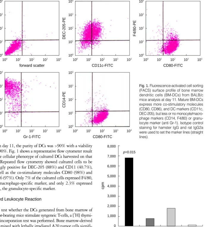
On day 11, the purity of DCs was >90% with a viability of >90%. Fig. 1 shows a representative flow cytometer result of the cellular phenotype of cultured DCs harvested on that day. Repeated flow cytometry showed cultured cells to be strongly positive for DEC-205 (88%) and CD11 (40.7%), as well as the co-stimulatory molecules CD80 (98%) and CD86 (97%). Only 7% of the cultured cells expressed F4/80, the macrophage-specific marker, and only 2.3% expressed Gr-1, the granulocyte-specific marker.
Mixed Leukocyte Reaction
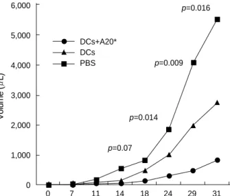
To test whether the DCs generated from bone marrow of tumor-bearing mice stimulate syngeneic T-cells, a [3H] thymi- dine incorporation test was performed. Bone marrow-derived DCs mixed with lethally irradiated A20 tumor cells signifi- cantly stimulated syngeneic T-cells, as compared with con- trol groups (Fig. 2). Microscopic examination revealed that DCs mixed with lethally irradiated A20 tumor cells stimu- lated and induced cluster formation and aggregation of T- cells.
Cytotoxic T Lymphocyte (CTL) Assay
Estimation of the induction and functional activity of cytotoxic T lymphocytes after DC immunization was per- formed by a 6 hr 51Cr release cytotoxic assay. DCs were cul-
tured with T-cells at a ratio of 1:10 for five days and the effector: target ratios were 20:1 in triplicate. DCs generated
cpm
8,000 7,000 6,000 5,000 4,000 3,000 2,000 1,000 0
DCs+ DCs A20* T cells
A20* only
p=0.015
Fig. 2. Dendritic cell (DCs) generation from bone marrow (BM) of tumor-bearing mice mixed with lethally irradiated tumor cells stimulate syngeneic T-cells. BM-derived DCs (5 103) mixed with 5 104lethally irradiated (10,000 rads) A20 cells showed a significantly increased stimulation of syngeneic T-cells (5 104: p=0.015).
* irradiated A20 cells.
Fig. 1.Fluorescence-activated cell sorting (FACS) surface profile of bone marrow dendritic cells (BM-DCs) from BALB/c mice analysis at day 11. Mature BM-DCs express more co-stimulatory molecules (CD80, CD86), and DC markers (CD11c, DEC-205), but less or no monocyte/macro- phage markers (CD14, F4/80) or granu- locyte marker (anti Gr-1). Isotype control staining for hamster IgG and rat IgG2a were used to set the marker lines (straight lines).
104
103
102
101
100
side scatter
forward scatter
100 101 102 103 104 104
103
102
101
100
DEC-205-PE
CD11c-FITC
100 101 102 103 104 104
103
102
101
100
F4/80-PE
CD80-FITC
100 101 102 103 104
104
103
102
101
100
CD86-PE
Gr-1-FITC
100 101 102 103 104 104
103
102
101
100
CD14-PE
CD80-FITC
100 101 102 103 104
from the bone marrow of tumor-bearing mice mixed with lethally irradiated tumor cells stimulated the CTL as mea- sured by this assay (Fig. 3).
Effect of Immunization on the Growth of Established Tumors-Immunization After Tumor Induction
To study the effects of immunization on the growth of established tumors, intraperitoneal immunizations with DCs mixed with lethally irradiated tumor cells were started when the tumors reached ca. 5-10 mm in diameter, and continued once every three days for four weeks. A significant reduction in the primary tumor growth of A20 lymphomas was noted
Cytotoxicity %
40 35 30 25 20 15 10 5 0
DCs+ DCs A20* T cells
A20* only
p=0.45
Fig. 3. Dendritic cell (DC) generation from bone marrow (BM) of tumor-bearing mice mixed with lethally irradiated tumor cells sti- mulate CTL. BM-derived DCs (4 104) mixed with 4 105lethally irradiated (10,000 rads) A20 cells showed increased stimulation of CTL (4 105).
* irradiated A20 cells.
Volume (L)
6,000
5,000
4,000
3,000
2,000
1,000
0
0 7 11 14 18 24 29 31
p=0.07 p=0.014
p=0.009 p=0.016
DCs+A20*
DCs PBS
Fig. 4. The effect of immunization with dendritic cells (DCs) on the growth of established tumors. BALB/c mice were injected (sc) with 2 106A20 lymphoma cells. Intraperitoneal immunization with 2-4 105DCs mixed with 2 106lethally irradiated A20 cells, 2-4 105DCs only or with phosphate-buffered saline solution (PBS) alone, were started when the tumors reached 5-10 mm in diameter. Each group contained five mice. Mean tumor sizes in each group are given. p values for differences between the groups of mice immunized with different methods are shown above the data points.
day
Tumor Volume (L)
900 800 700 600 500 400 300 200 100 0
-7 0 7 11 17 22 24 28
DCs+A20*
PBS
Fig. 5. The effect of immunization with dendritic cells (DCs) before tumor implantation on subsequent tumor growth. Intraperitoneal immunization of BALB/c mice with 2-4 105DCs mixed with lethally irradiated (10,000 rads) 2 106A20 cells or phosphate- buffered saline solution (PBS) were started before tumor induc- tion on day 7. Mice were injected (sc) with 5 104A20 lymphoma cells on day 0. Immunization before tumor injection prevents tumor growth.
day
A. 2-4 106DC 18/47 17951 1935 1536/1150
+ 2 106A20 cells
B. 2-4 106DC 16/35 26324 2301 2229/1144
C. PBS 9/27 4045 796 130/81
Normal control 13/31 128 53 116/70
BALB/c mice were injected (sc) with 2 106A20 lymphoma cells. Intra- peritoneal immunization with 2-4 105dendritic cells (DCs) mixed with 2 106lethally irradiated (10,000 rads) A20 cells, 2-4 105DCs only, or phosphate-buffered saline solution (PBS) alone, were started when the tumors reached c. 5-10 mmin diameter. For evaluation of the effect of immunization on T-cells, [3H] thymidine uptake test, FACS scan analy- sis of T-cells and IL-2/IL-4 assays (using ELISA) with T-cells from spleno- cytes of each group were used.
CD4/CD8 3[H] thymidine IL2/IL4 (%) uptake test (cpm) (pg/mL) Immunization group
Table 1.The effect of immunization with dendritic cells on T- cells
at day 31, and there were significant differences in the rates of growth of lymphomas between groups (Fig. 4).
Effect of Immunization on the Growth of Established Tumors-Immunization Before Tumor Induction
To test whether immunization with DCs mixed with lethally irradiated tumor cells was capable of inducing protec- tive antitumor immunity, immunization was started before
tumor induction. As shown in Fig. 5, immunization before tumor injection could completely prevent tumor growth and there was no growth after the completion of immuniza- tion. By contrast, the mice of control groups showed progres- sive tumor growth.
Effect of Immunization on T-Cells
Spleens were obtained from each group of mice after the
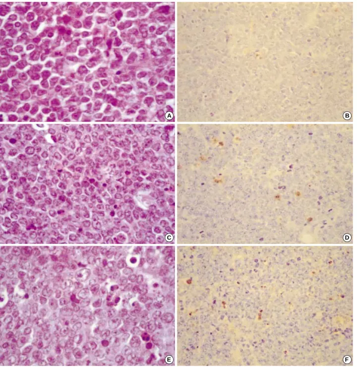
Fig. 6. Microscopic findings of A20 lymphomas treated with PBS (A, B), DC (C, D), and DC mixed with irradiated A20 cells (E, F). Some of the apoptotic cells showed fragmentation of the condensed nucleus into multiple apoptotic bodies. (A, C, E: Hematoxylin and eosin staining 400). Apoptotic cells are identified by dark brown nuclear staining using the TUNEL method. (B, D, F: 200).
A B
C D
E F
final post-tumor induction immunization. As summarized in Table 1, the ratio of CD4+ to CD8+ cells was highest in the group immunized with DCs mixed with lethally irradi- ated tumor cells. The mice of both immunized groups and the tumor-bearing control group had higher [3H] thymidine uptakes than normal controls. Mice of the immunized groups showed considerably higher levels of IL-2 and IL-4 than mice of the non-immunized group.
Evaluation of Apoptosis by In Situ End-Labeling
Tumor tissues were obtained from each group of mice after the final post-tumor induction immunization. Micro- scopically, A20 lymphomas treated with PBS (A, B), DC (C, D) and DC mixed with irradiated A20 cells (E, F), showed some apoptosis with fragmentation of the condensed nucle- us in multiple apoptotic bodies (Fig. 6A, C, E), and the apop- totic cells were identified by dark brown nuclear staining using the TUNEL method (Fig. 6B, D, F). The apoptosis indices were 0.5%, 4%, and 6% for B, D and E, respectively.
DISCUSSION
In this study, we used bone marrow-derived DCs to induce a therapeutic and protective immune response against a murine lymphoma. DCs mixed with lethally irradiated lymphoma cells as a source of tumor antigens stimulated an effective anti-lymphoma immune response. This was associated with an increase in tumor-specific cytotoxic-T cell responses.
Although the exact mechanism of antitumor immunity was not defined, it is possible that the lethal irradiation of the A20 lymphoma cells resulted in rapid cell death with the release of cellular antigens that were then processed by the adjacent DCs (23), which then initiated the stimulation of tumor-specific cytotoxic T-cells.
Bone marrow-derived myeloid lineage DCs provide criti- cal antigen-presenting cell activity for initiating specific T lymphocyte activation and proliferation; thus many studies have used bone marrow-derived DC for immunotherapy (24-26). Several studies applied a combination of cytokines including GM-CSF, TNF- and IL-4 to harvest a large num- ber of purified DCs from bone marrow cells (6-8). We also obtained DCs with morphologically typical dendrites using GM-CSF, TNF- and IL-4. In this study, we reduced the dose of GM-CSF and increased that of TNF- at a later stage of ex vivo culture. This resulted in minimal contamination with granulocytes and macrophages, and we obtained func- tionally active mature DCs. Our FACS analysis using the DC markers CD11c and DEC-205, and the co-stimulatory molecules CD80 and CD86 showed higher level of DCs at day 11 of culture. However, the macrophage-specific mark- ers F4/80 and CD14, and granulocyte-specific marker Gr-1 were expressed at much lower levels. We also found that
DCs mixed with lethally irradiated A20 tumor cells stimu- lated T-cells and induced cluster formation and aggregation of T-cells.
We performed [3H] thymidine incorporation assays to show that DCs generated from the bone marrow of the tumor- bearing mice significantly stimulated syngeneic T-cells as compared with control groups. The cytotoxic activity of the T lymphocytes measured by 51Cr release was stimulated in the mice immunized with the DCs mixed with lethally irradiated tumor cells. If we were to increase the ratio of the effector cells, this might stimulate the CTL significantly.
Immunization before tumor implantation resulted in com- plete prevention of tumor growth, and there was no tumor growth after completion of the immunization. We observed that these mice were alive without any tumor growth for more than 12 weeks. This result suggests the feasibility of active tumor-specific immunotherapy with autologous den- dritic cell transplantation as a strategy for the elimination of minimal residual disease (MRD) of lymphomas. Our results with syngeneic bone marrow-derived DCs for immunother- apy suggest that effective ex vivo expansion and maturation of DC from autologous stem cells could be an important and useful tool for immunotherapy.
In many model systems, the balance between the cytokines induced from Th1 and Th2 effector cells plays an important role in the regulation of immune responses. A Th1-positive cell response is thought involved in cellular and tumor immu- nity, and Th2-positive cells are associated with the suppres- sion of cytolytic activity (27, 28). In this study, we tried to evaluate the possible role of Th1/Th2 cell balance in the immune response to DCs sensitized with irradiated tumor cells. Tumor progression in control mice was associated with a decreased absolute level of Th1-cell induced cytokine (IL- 2) and Th2-cell induced cytokine (IL-4), whereas effective immunization blocks tumor progression, which in turn was closely associated with increased absolute levels of Th1-cells (IL-2). However, we found no significant differences in Th1/Th2 cell ratios between the groups. The induction of antitumor immunity by bone marrow-derived DCs requires the presentation of MHC class-II restricted molecules and activation of CD4+ T-cells (29). Our study demonstrated that the cultured DCs mixed with whole but dead tumor cells as a vaccine and induced antitumor immunity and com- plete prevention of tumor growth in the co-adjuvant treat- ment group. Recently, Kim et al. demonstrated that NK cells were required during the priming of cytotoxic T-cell response by DCs-based tumor vaccine and DCs can induce an antitumor immune response by enhancing NK cell-de- pendent CTL activation (30).
Evaluation of apoptosis by TUNEL demonstrated that the PBS-alone treated control group showed an apoptotic index of 0.5% (Fig. 6B), while in the DC-only treated group it was 4% (Fig. 6D), and in the group treated with DC mixed with irradiated A20 cells treated group it was 6%
(Fig. 6F). Programmed cell death was thus increased over tenfold in the treated group F.
We conclude that DCs mixed with irradiated tumor cells as a source of undefined tumor antigens can induce an effective antitumor immune response and completely prevent tumor growth. It also provides a rationale for the use of DCs mixed with irradiated tumor cells as an immunotherapy for MRD of lymphomas. Further studies on better methods of cultur- ing DCs, optimizing the means of antigen loading on DCs and the most efficacious vaccination schedules and dosages should be investigated. Moreover, the mechanism by which DC vaccine can provide the desired immunity remains to be determined.
REFERENCES
1. Cella M, Salluso F, Lanzavecchia A. Origin, maturation and antigen presenting function of dendritic cells. Curr Opin Immunol 1997; 9:
10-6.
2. Steinman RM, Cohn ZA. Identification of a novel cell type in peri- pheral lymphoid organs of mice. I. Morphology, quantitation, tissue distribution. J Exp Med 1973; 137: 1142-62.
3. Grabbe S, Beissert S, Schwarz T, Granstein RD. Dendritic cells as initiators of tumor immune responses. A possible strategy for tumor immunotherapy? Immunol Today 1995; 16: 117-21.
4. Girolomoni G, Ricciardi-Castagnoli P. Dendritic cells hold promise for immunotherapy. Immunol Today 1997; 18: 102-4.
5. Inaba K, Inaba M, Romani N, Aya H, Deguchi M, Ikehara S, Mura- mats S, Steinman RM. Generation of large numbers of dendritic cells from mouse bone marrow cultures supplemented with granulo- cyte/macrophage colony stimulating factor. J Exp Med 1992; 176:
1693-702.
6. Lutz MB, Kukutsch N, Ogilvie AL, Rossner S, Koch F, Romani N, Schuler G. An advanced culture method for generating large quan- tities of highly pure dendritic cells from mouse bone marrow. J Immu- nol Methods 1999; 223: 77-92.
7. Chirinos-Rojas CL, Steward MW, Partidos CD. A peptidomometic antagonist of TNF-alpha-mediated cytotoxicity identified from a phage-displayed random peptide library. J Immunol 1998; 161:
5621-6.
8. Luykx-de Bakker SA, de Gruijl TD, Scheper RJ, Wagstaff J, Pinedo HM. Dendritic cells: Novel therapeutic modality. Ann Oncol 1999;
10: 21-7.
9. Steinman RM. The dendritic cell system and its role in immunogenici- ty. Annu Rev Immunol 1991; 9: 271-96.
10. Inaba K, Metlay JP, Crowley MT, Witmer-Pack M, Steinman RM.
Dendritic cells as antigen presenting cells in vivo. Int Rev Immunol 1990; 6: 197-206.
11. Crowley M, Inaba K, Steinman RM. Dendritic cells are the princi- pal cell in mouse spleen bearing immunogenic fragments of foreign proteins. J Exp Med 1990; 172: 383-96.
12. Steinman RM, Witmer-Pack M, Inaba K. Dendritic cells: antigen presentation, assessory function and clinical relevance. Adv Exp
Med Biol 1993; 329: 1-9.
13. Inaba K, Romani N, Steinman RM. An antigen-independent contact mechanism as an early step in T cell proliferative responses to den- dritic cells. J Exp Med 1989; 170: 527-42.
14. Sornasse T, Flamand V, De Becker G, Bazin H, Tielemans F, Thiel- mans K, Urbain J, Leo O, Moser M. Antigen-pulsed dendritic cells can efficiently induce an antibody response in vivo. J Exp Med 1992;
175: 15-21.
15. Takahashi H, Cohen J, Hosmalin A, Cease KB, Houghten R, Cor- nette JL, DeLisi C, Moss B, Germain RN, Berzofsky JA. An immu- nodominant epitope of the human immunodeficiency virus envelope glycoprotein gp160 recognized by class I major histocompatibility complex molecule-restricted murine cytotoxic T lymphocytes. Proc Natl Acad Sci U.S.A. 1988; 85: 3105-9.
16. Johnston JV, Malacko AR, Mizuno MT, McGrown P, Hellstrom I, Hellsrom KE, Marquardt H, Chen L. B7-CD28 costimulation unveils the hierarchy of tumor epitopes recognized by major histo- compatibility complex class I restricted CD8+ cytolytic T lympho- cytes. J Exp Med 1996; 183: 791-800.
17. Guillaume T, Rubinstein DB, Symann M. Immune reconstitution and immunotherapy after autologous hematopoietic stem cell trans- plantation. Blood 1998; 92: 1471-90.
18. Kwak LW. Tumor vaccination strategies combined with autologous peripheral stem cell transplantation. Ann Oncol 1998; 9: S41-6.
19. Kim KJ, Kanellopoulos-Langevin C, Merwin RM, Sachs DH, Asofsky R. Establishment and characterization of BALB/c lym- phoma lines with B cell properties. J Immunol 1979; 122: 549-54.
20. Glimcher LH, Kim KJ, Green I, Paul WE. Ia antigen-bearing B cell tumor lines can present protein antigen and alloantigen in a major histocompatibility complex-restricted fashion to antigen-restrictive T cells. J Exp Med 1982; 155: 445-59.
21. Coveney E, Wheatley GH III, Lyerly HK. Active immunization using dendritic cells mixed with tumor cells inhibits the growth of primary breast cancer. Surgery 1997; 122: 228-34.
22. Kerr JF, Winterford CM, Harmon BV. Apoptosis. Its significance in cancer and cancer therapy. Cancer 1994; 73: 2013-26.
23. Gabrilovich DI, Ciernik IF, Carcone DP. Dendritic cells in anti-tumor immune responses. I. Defective antigen presentation in tumor-bear- ing hosts. Cell Immunol 1996; 170: 101-10.
24. Gabrilovich DI, Nadaf S, Corak J, Berzofsky JA, Carbone DP. Den- dritic cells in antitumor immune responses. II. Dendritic cells grown from bone marrow precursors, but not mature DC from tumor bear- ing mice, are effective antigen carriers in the therapy of established tumors. Cell Immunol 1996; 170: 111-20.
25. Hart DN. Dendritic cells: unique leukocyte populations which con- trol the primary immune response. Blood 1997; 90: 3245-87.
26. Kraal G, Breel M, Janse M, Bruin G. Langerhans cells, veiled cells and interdigitating cells in the mouse recognized by a monoclonal antibody. J Exp Med 1986; 163: 981-97.
27. Paul WE, Seder RA. Lymphocyte responses and cytokines. Cell 1994; 76: 241-51.
28. Porgador A, Synder D, Gilboa E. Induction of antitumor immunity using bone marrow generated dendritic cells. J Immunol 1996; 156:
2918-26.
29. Gavrieli Y, Sherman Y, Ben-Sasson SA. Identification of program- med cell death in situ via specific labeling of nuclear DNA fragmen- tation. J Cell Biol 1992; 119: 493-501.
30. Kim KD, Choi SC, Kim A, Choe YK, Choe IS, Lim JS. Dendritic
cell-tumor coculturing vaccine can induce antitumor immunity through both NK and CTL interaction. Int Immunopharmacol 2001;
1: 2117-29.