Prosthetic rehabilitation of the edentulous patients using O-ring attachment:A clinical report
Jae-Jin Ahn, DDS, MSD, PhD, Byung-Woan Jo, DDS, PhD, Sang-Hun Ahn, DDS, Jong-Pil Kim, DDS.*
Department of Dentistry, Fatima Hospital, Taegu, Korea.
Section of Dentistry, School of Medicine, University of Soonchunhyang, Kumi, Korea.*
I. INTRODUCTION
There are many methods and reports on im- plant-assisted rehabilitation of the edentulous mouth and maxillofacial defects. The ideal oral rehabilitation of edentulous patients seems to be an implant-supported fixed complete denture.
However this superior prosthodontic treatment is not always possible to perform. The next best choice would be the implant-supported over- denture. In addition to functional satisfaction, ex- cellent cosmetic and phonetic results can be achieved with the implant-supported overdenture.
Many different available attachments today may be used to support an implant-supported overdenture1). The most popular implant at- tachments currently used are the bar and clip, stud attachments and the magnet2).
The O-ring attachment has many advantages, such as simple and fast prosthetic technique, easy maintenance, good oral hygiene, broad indication, economical fee, and so forth.
Although the superiority of O-ring attachment has notbeen proved scientifically in comparison to the other attachments, the authors think that the O-ring attachment is the attachment of choice
for an implant-supported overdenture. This clin- ical report describes the application for the O-ring attachment of two edentulous patients and a maxillectomy patient rehabilitated with the im- plant-supported overdentures using O-ring at- tachment of the Steri-Oss� Implant System.
INDICATIONS
1. Tissue-supported overdenture restoration 2. Long span between implants that is con- traindicated for bar splinting
3. Difficulty with oral hygiene (elderly patients) 4. Financial considerations
CONTRAINDICATION
Implants greater than 10�divergent
COMPONENTS 1. Abutment 2. Analog 3. Spacer
4. Retaining ring (metal)
5. Red O-ring (rubber) : For laboratory use 6. White O-ring (rubber) : For clinical use The O-ring attachment can be processed in- to the denture by either direct or indirect tech-
대한치과보철학회지:Vol. 36, No. 4, 1998
nique. For the direct technique, the O-ring abutment is placed into the implant and the O- ring assembly is picked up in the denture at chairside. The indirect technique utilizes the O-ring analog in a laboratory working cast.
II. CLINICAL REPORT
CASE 1. A 64-year-old female patient, with full edentulous ridge of the maxilla and mandible, presented for complete denture construction.
She was rehabilitated with implant-supported overdenture in the mandible and conventional complete denture in the maxilla. The two im-
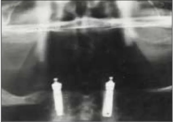
plants of the mandible were 3.8 mm in diame- ter and 18 mm in length and threaded HA- coated titanium implants (Fig. 1). Two mandibu- lar implants were exposed and abutments were connected at five months post-implantation.
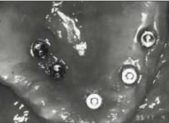
The heights of the O-ring abutments were 3 mm for right and 2 mm for left (Fig. 2). At 32-month follow-up, there was no sign of im- plant failure and prosthesis was functionally sound. Two mandibular implants were considered successful according to the Albrektsson’s crite- ria3).
CASE 2. A 47-year-old female patient pre- sented with multiple carious lesions, retained roots and generalized mobile teeth. She underwent full mouth extraction, alveoloplasty, immediate place- ment of four implants on the maxilla and three implants on the mandible (Fig. 3). All the implants were threaded HA-coated titanium implants and longer than 12 mm in length. Each implant was less than 10。 divergent. Seven months later, second surgery was done and abutments were connected. All the O-ring abutments were 2 to 4 mm in height (Fig. 4, 5). At 28-month follow-up, all the implants sat- isfied Albrektsson’s success criteria.
CASE 3. A 67-year-old female patient was re- ferred to our clinic with complaint of swelling on Fig. 1. Immediate postoperative panoramic radi-
ograph of case 1. Two implants in mandible.
Fig. 2. Inner side of the denture. Notice the met- al retaining ring and rubber ring in the denture.
Fig. 3. Postoperative panoramic radiograph of case 2, 12 months after surgery.
the maxillary right anterior vestibule for 1 year.
There was no past medical or dental history.
Physical examination revealed intraoral swelling and otherwise unremarkable. The preoperative computed tomographic examination of the max- illa showed relatively defined, lobulated and multiloculated expansile lesion in right maxilla about 3×4 cm in size. An incisional biopsy was performed and the lesion was diagnosed as fi- bromyxoma. After right partial maxillectomy, im- mediate placement of five implants on the maxilla was done simultaneously (Fig. 6). The implants were 8, 10, 14, 12, 10 mm long from right to left, and all the implants were thread-
ed HA-coated titanium implants, 4.5 mm in di- ameter. Two of five implants on the normal side were placed in the residual alveolar ridge, and the other three implants were located in the hard palate near by the defect area. The bone qual- ity and quantity of the residual maxillary bone permitted installation of a sufficient number of implants and afforded immediate loading. Healing abutments were connected at first surgery. The fabrication of the prosthesis was initiated as soon as the soft tissue healed. The patient has been doing well since her surgery. The functionally and esthetically satisfactory prosthesis has now been in use for 36 months.
Fig. 4. Intraoral photograph of the O-ring abut- ments
Fig. 5. Inner side of the denture.
Fig. 6. Postoperative panoramic radiograph of case 3, 18 months after surgery. Five implants were placed immediately after partial maxillectomy.
Fig. 7. Intraoral photograph of the O-ring abut- ments.
PROSTHETIC PROCEDURE OF CASE 3 1. One month later, remove healing abutments and placed O-ring abutments on to the implants (Fig. 7).
2. Impression was recorded over O-ring abut- ments.
3. Inserted O-ring abutment analogs in the im- pression and made a cast.
4. Placed a spacer over O-ring abutment analogs.
5. Placed red processing O-ring in the retain- ing ring then placed retaining ring over O-ring abutment analog with larger diameter opening towards tissue. Processed the denture (Fig. 8).
Removed red processing O-ring and replaced it with white O-ring.
6. Delivered the denture and checked occlusion.
III. DISCUSSION
The case one was the simplest design for im- plant-prosthetic treatment of the edentulous mandible, where two implants were placed at an adequete distance (15-20 mm) in the inter- foraminal region4). In case 2, four implants were placed on the maxilla because of the reduced bone quality. The number of implants placed on the maxilla should be more than mandible to in- crease the implant-bone contact surface. An overdenture is functionally and esthetically more
effective than fixed partial denture type since on- ly four implants are placed on the maxilla and two or three implants are placed on the mandible.
Presently the selection of implant attach- ments is based largely on clinical experience and anecdote rather than scientific findings. In gen- eral, bar and clip attachment seems to be the preferred choice of design for the implant-sup- ported overdenture.
However, we would like to compare O-ring at- tachment overdenture with bar and clip at- tachment overdenture. Although bar and clip would be better in some clinical situation, our clinical experience and results available to date provide strong support for the use of O-ring at- tachment. The overdenture with O-ring at- tachment requires no metal bar frame, therefore eliminating need of casting and fitting the bar that can be a difficult procedure. The O-ring at- tachment overdenture has also less possibility of the denture fracture than bar and clip over- denture, because O-ring overdenture does not re- quire a large room inside the denture to ac- commodate bar and clip. The fabrication of the O-ring attachment overdenture has reduced prosthetic procedure and expense, broad indica- tion and fewer risks. Also, maintenance and oral hygiene are simpler. It does not encroach on tongue space.
The O-ring attachment does not distribute torquing stress to the implants because it does not splint the implants with a bar. Instead, an early detection of mobility in the O-ring con- nected implant can be possible and it can pre- vent the neighboring implant from harmful torquing stress of mobile implant. Splinting of the implants may lead to neglecting implant mobil- ity.
The transverse splinting via bars creates pres- sure and tension in the implant bed because of the elastic deformation of the mandible during Fig. 8. Inner side of the denture.
function4).
In case 3, placement of implants and O-ring attachment overdenture provided a greater re- tention and stability of the prosthesis in the edentulous maxillectomy patient. Many clinical re- ports have documented prosthetic rehabilitation of the maxillectomy defect using bar and clip, stud attachments and the magnet5-8). The O-ring attachment is very efficient and simpler than bar and clip attachment, because it can be used in patients with lone-standing implants or in situ- ations where inadequate space for a tissue bar.
If patient does not object removable prosthe- sis, we strongly recommend O-ring attachment as the treatment of choice for the edentulous pa- tients.
IV. SUMMARY
A successful prosthesis is difficult to produce without using the implants in the severely re- sorbed alveolar ridges and maxillofacial defects.
This report describes clinical experience of the overdenture using O-ring attachment. Clinical results have revealed successful application for the O-ring attachment in the two edentulous patients and a partial maxillectomy patient.
REFERENCES
1. Petropoulos VC, Smith W, Koussvelari E.
Comparison of retention and release periods
for implant overdenture attachments. Int J Oral Maxillofac Implants 1997 ; 12 : 176-185.
2. Mensor MC Jr. Removable partial overden- tures with mechanical(precision) attachments.
Dent Clin North Am 1990 ; 34 : 669-681.
3. Albrektsson T, Zarb G, Worthington P, et al.
The long-term efficacy of currently used den- tal implants : a review and proposed criteria of success. Int J Oral Maxillofac Implants 1986 ; 1 : 11-25.
4. Spiekermann H, Donath K, Hassell TM, et al.
Implantology. New york : Thieme Medical Publishers, 1995 : 150-179.
5. Block MS, Guerra LR, Kent JN, et al.
Hemimaxillectomy prosthetics stablization with hydroxyapatite-coated implants : a case report. Int J Oral Maxillofac Implants 1987 ; 2 : 111-113.
6. Worthington P. Branemark PI. Advanced os- seointegration surgery : applications in the maxillofacial region. Chicago : Quintessence, 1992 : 267-275.
7. Izzo SR, Berger JR, Joseph AC, et al.
Reconstruction after total maxillectomy using an implant retained prosthesis : a case report.
Int J Oral Maxillofac Implants 1994 ; 9 : 593-595.
8. Roumanas ED, Nishimura RD, Davis BK, et al. Clinical evaluation of implants retaining edentulous maxillary obturator prostheses. J Prosthet Dent 1997 ; 77 : 184-190.
무치악환자의 Implant보철수복 방법은 식립된 Implan의 수에 따라 다양하다. 많은 수의 Implant 가 식립된 경우에는 고정성 보철물로 수복이 가능하지만 시술이나 기공상의 복잡성, 고가의 치 료비, 보철물과 잔존치조제사이의 빈 공간으로 인한 비심미성, 발음의 어려움과 같은 여러가지 문제점이 존재한다. 적은 수의 Implant가 식립된 경우는 Bar Retained Overdenture를 사용하지 만 역시 기공과정이 복잡하며, Bar와 잔존치조제사이의 공간으로 인한 점막지지 면적의 감소와 보수가 어려운 단점이 있다. 여기에 비해서 O-ring Attachment를 이용하면 기공과정이 쉽고, 잔 존치조제와 보철물 사이에 공간이 없으므로 점막지지 면적을 많이 얻을 수 있다. 또한 Implant식립의 수도 최소화할 수 있어 경제적이며, 문제가 발생한 경우에 쉽게 대처할 수 있는 장점을 지니고 있다.
본 증례들은 Steri-Oss Implant System의 O-ring Attachment를 이용하여 무치악환자를 보철 수복한 예로서 술후 기능적, 심미적으로 만족할 만한 결과를 얻었기에 문헌고찰과 더불어 발표 하는 바이다.
Key Words : 무치악환자, Implant, O-ring Attachment.
O-ring Attachment를 이용한 무치악환자의 보철수복증례
대구파티마병원 치과,구미순천향병원 치과*
안재진∙조병완∙안상헌∙김종필*
ABSTRACT