The Clinical Impacts of Apparent Embolic Event and the Predictors of In-Hospital Mortality in Patients with Infective Endocarditis
Embolic event is a common and important complication of infective endocarditis (IE). The objective of this study was to investigate the clinical impacts of embolic event in patients with IE and the predictors of in-hospital mortality. Data was collected in Pusan National University Hospital and Pusan National University Yangsan Hospital between January 2009 and December 2010. One hundred ten patients were included. Embolic events occur in 39 of 110 patients (35.5%). Brain (n = 18, 38.5%) was the main site of embolic infarction.
Patients with embolism showed higher in-hospital mortality (46.2% vs. 8.5%, respectively, P = 0.03), more frequent ICU admission (53.8% vs. 35.2%, respectively, P = 0.045) and more accompanying other cardiac complication (43.6% vs. 21.1%, respectively, P = 0.017).
The in-hospital mortality rate was 18.2%. On the logistic regression analysis of the predictors for in-hospital mortality, age (RR, 1.079; 95% CI, 1.036-1.123, P = 0.001), embolic event (RR, 3.510; 95% CI, 1.271-9.69, P = 0.015) and staphylococcal infection (RR, 5.098;
95% CI, 1.308-18.508, P = 0.023) were independently associated with in-hospital mortality.
Embolic events in IE are associated with poor in-hospital outcome; and these data about embolic events and the predictors of in-hospital mortality may improve the management of this disease in hospitals.
Keywords: Endocarditis; Embolism; Mortality; Staphylococcus Su Jin Lee, Doosoo Jeon, Woo Hyun Cho,
and Yun Seong Kim
Department of Internal Medicine, School of Medicine, Pusan National University and Research Institute for Convergence of Biomedical Science and Technology, Pusan National University Yangsan Hospital, Yangsan, Korea
Received: 20 November 2014 Accepted: 3 June 2014 Address for Correspondence:
Yun Seong Kim, MD
Department of Internal Medicine, School of Medicine, Pusan National University, Pusan National University Yangsan Hospital, 20 Geumo-ro, Mulgeum-eup, Yangsan 626-770, Korea Tel: +82.55-360-1416, Fax:+82.55-360-1757 E-mail: [email protected]
Funding: The study was supported by Medical Research Institute Grant (2013), Research Institute for Convergence of Biomedical Science and Technology and Pusan National University Yangsan Hospital.
http://dx.doi.org/10.3346/jkms.2014.29.12.1646 • J Korean Med Sci 2014; 29: 1646-1650
INTRODUCTION
Despite the great advances in the diagnosis and treatment of infective endocarditis (IE), it remains a disease with high in- hospital mortality (16%-25%) and a high incidence of compli- cations (1-5). In contrast to decreasing some of the other com- plications such as renal failure and uncontrolled intra-cardiac or metastatic infection with using antibiotics, embolic event is still a distressingly common complication of IE and this can oc- cur even during administering appropriate therapy (6, 7). The incidence of clinically recognized embolism is known to 13% to 44% (6, 7). Adam et al. reported that cerebral infarction is a pre- senting sign of IE in 4% to 14% of all infective endocarditis (8).
All the patients who present with signs and symptoms of sys- temic arterial embolization should have IE considered as a pos- sible cause (6, 7). Several studies have attempted to evaluate the predictors of embolic events and mortality. The reports have had conflicting results, vegetation > 10 cm in size, left-side valve infection and Staphylococcus aureus infection were suggested as the risk factors of embolic infarction (2, 9-13). Diabetes mel- litus, Staphylococcus aureus infection and embolic events are known to be predictors of in-hospital mortality (3-5,12). But there are few studies that have focused on the clinical influence
of embolic event in IE and the predictors of in-hospital mortali- ty in an Asian population.
The objective of this study was to investigate the clinical im- pacts of embolic event on IE and the predictors of in-hospital mortality. Determining the clinical effects of embolic event on IE and the predictors of in-hospital mortality may improve the management of this disease in hospitals.
MATERIALS AND METHODS Patients and population
This retrospective study was performed at two teaching hospi- tals: Pusan National University Hospital, which is a 1,000-bed teaching hospital, between January 2003 and December 2010 and Pusan National University Yangsan hospital, which is a 700- bed teaching hospital, between January 2009 and December 2010. All the medical records were reviewed for the patients with infective endocarditis and who were over 18 yr of age. All the patients with infective endocarditis met the modified Duke’s criteria (14) for definite IE or possible IE. The demographic data, the baseline characteristics and the outcomes were collected.
The following data was recorded for each patient: age, gender, the underlying disease, the involved valves, a native valve ver-
sus a prosthetic valve, a community origin versus a nosocomial origin, the cardiac and extracardiac complications, the treatment given during hospitalization, the surgical requirements, the need for ICU admission, the echocardiographic findings, the total admission days and the in-hospital mortality.
Nosocomial IE was defined as an infection occurring more than 72 hr after admission to the hospital or the IE acquired in patients discharged from the hospital within 6 months before the onset of symptoms (15). Cardiac complication was defined as cardiac structural complication such as new onset of valvular functional abnormality, valvular abscess, valvular perforation and intracardiac abscess. Four to six blood cultures were obtain- ed for all the patients.
Embolic event
We evaluated the radiologic/clinical evidence of embolism. The CT scans, MRI and chest radiographic findings were collected for the patients with signs or symptoms suggestive of an embol- ic event such as abdominal pain, chest discomfort, abrupt dys- pnea and sudden neurologic or visual dysfunction. Renal and splenic embolisms were included if they were confirmed by ra- diographic imaging.
Statistical analyses
Statistical analyses were done using SPSS version 12.0 (SPSS Corp, Chicago, IL, USA). The descriptive analysis consisted of the median, mean and range of the various parameters. The differences between the embolic and nonembolic patients were compared using the chi-square test for the categorical variables, and the t-test or the rank-sum test was used for the numerical variables. Logistic regression analysis was performed to deter- mine the predictors that were independently associated with in-hospital mortality. P values < 0.05 were considered to be sta- tistically significant.
Ethics statement
This study protocol was approved by the institutional review board (IRB) of Pusan National University Yangsan Hospital (IRB No. 05-2014-017). Informed consent was waived by the boards.
RESULTS
Demographic features and the clinical outcomes
One hundred and ten patients who met the modified Duke’s criteria for definite IE or possible IE (14) were analyzed. The median age was 52 yr (16-85 yr). Sixty seven patients (60.9%) were men and forty three patients (39.1%) were women. Most patients had native valve IE (n = 100, 90.9%) and a community origin of IE (n = 101, 91.8%). The mitral valve was the predomi- nant infected valve (n = 43, 39.1%) followed by the aortic valve (n = 39, 35.5%). Eight patients with right-sided valve IE (7.3%)
and five patients with multi-valve IE (4.5%) were observed. The most frequent causative organisms were Streptococci in 38 pa- tients (34.6%), followed by Staphylococci in 27 patients (24.5%) and other organisms in 8 patients (7.3%), and these included 2 patients with Psuedomonas aeruginosa, 2 patients with Candi- da, 2 patients with Enterococcus, 1 patient with E. coli and 1 pa- tient with gram-negative rods (not identified). Thirty-seven pa- tients (33.6%) had culture negative endocarditis. Fifty-seven pa- tients (51.8%) had underlying disease. Previous heart disease (n = 37, 33.6%; 17 patients with valvular heart disease, 14 pa- tients with congenital heart disease, 3 patients with heart fail- ure, 1 patient with a prosthetic valve and 2 patients with coro- nary heart disease) was the most common underlying disease.
The mean of total admission days was 36.1 days (range, 3-125 days) and 46 patients were admitted to the ICU. Surgery was performed on 41 patients (37.3%). Heart failure was main cause of surgical intervention (n = 18, 43.9%) and median day of sur- gery was 4 admission days (range, 1-7). Failure of medical treat- ment is secondary cause of surgical intervention (n = 12, 39.3%).
In group of medical failure, 7 persistent bacteremia (> 7 days) and 5 increasing vegetation and re-embolism during treatment were observed and median operation day was 12 admission days (range, 7-18). Large vegetation and high mobile vegetation were third cause of surgical intervention for prevention of em- bolism (n = 6, 14.6%) and median surgery day was 5 admission days (range, 2-9). Three ventricular septum defect repairs (7.3%) and 2 permanent pacemaker insertion (4.9%) were undertaken.
The demographic, clinical and microbiological characteris- tics of the patients with and without embolism combined with IE are shown Tables 1 and 2.
Embolic events
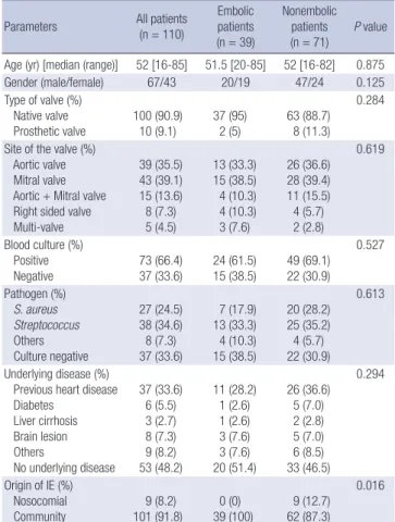
Embolic events occur in 39 of 110 patients (35.5%). The brain (n = 18, 38.5%) was main site of embolic infarction. Other sites included the spleen (n = 12, 30.8%), the lung (n = 11, 28.2%), the kidney (n = 9, 23.1%), the liver (n = 1, 2.6%) and main ves- sels (n = 1, 2.6%). Multi-site embolic infarctions were observed in 19 patients (48.7%). Embolic events were recognized at the time of hospital admission in 30 of 39 patients with embolic events (76.9%) and other 9 patients (23%) were recognized dur- ing hospital management due to new clinical symptom. There were no significant differences in the demographic characteris- tics (age, gender, underlying disease, the involved valve site, causative organism, the type of infected valve and the positivity of blood culture) between the patients with and without embo- lism. But nosocomial IE was observed only in the patients with- out embolism (0% vs. 12.7%, respectively, P = 0.016).
For the clinical outcome, the group of patients with embo- lism showed higher in-hospital mortality (46.2% vs. 8.5%, re- spectively, P = 0.03), more frequent ICU admission (53.8% vs.
35.2%, respectively, P = 0.045) and more accompanying other
cardiac complication (43.6% vs. 21.1%, respectively, P = 0.017).
Table 2 shows clinical influence of embolic infarction in the pa- tients with infective endocarditis.
In-hospital mortality
The in-hospital mortality rate was 18.2%. The leading causes of death were sepsis (n = 8, 33.3%) and heart failure (n = 5, 20.8%) and these were followed by brain complications (n = 4, 16.7%), multi-organ failure (n = 3, 12.5%), sudden death (n = 3, 12.5%) and hospital acquired pneumonia (n = 1, 4.2%). The causes of death in group of patients with embolism were surgery related
infection and sepsis (n = 6, 33.3%) and heart failure (n = 4, 22.2%) and followed by brain complications (n = 3, 16.7%: 2 brain he- morrhage: 1 massive brain infarction), multi-organ failure (n = 2, 11.1%), sudden death (n = 2, 11.1%) and hospital acquired pneu- monia (n = 1, 7.6%). In embolic group, there were no significant differences in in-hospital mortality between the patients with embolic event during management and the patients with em- bolism at the time of admission (55.5% vs. 43.4%, respectively, P = 0.23). On the multiple logistic regression analysis of the predictors of in hospital mortality, age (RR, 1.079; 95% CI, 1.036- 1.123, P = 0.001), embolic event (RR, 3.510; 95% CI, 1.271-9.69, P = 0.015) and staphylococcal infection (RR, 5.098; 95% CI, 1.308- 18.508, P = 0.023) were independently associated with in-hos- pital mortality (Tables 3, 4).
DISCUSSION
In this study, embolic events occurred in 35.5% of the patients with IE, and this was similar to that of the previous studies (30%- 44%) (6, 7, 15-17). Contrary to several other studies (2, 9-13), there was no significant difference of the demographic characteris- tics (age, gender, underlying disease, the involved valve site, the causative organism, the type of infected valve and positivity of blood culture) between the embolic and non-embolic groups in this study.
Staphylococcus infection shows no difference between the embolic and non-embolic groups, but there was an indepen- dent association of Staphylococcus infection with in-hospital mortality in our study. In some earlier studies, Staphylococcus infection has been associated with both higher rates of compli- Table 1. The demographic, clinical and microbiological characteristics of the patients
with and without embolism and who had infective endocarditis (IE)
Parameters All patients
(n = 110)
Embolic patients (n = 39)
Nonembolic patients
(n = 71) P value Age (yr) [median (range)] 52 [16-85] 51.5 [20-85] 52 [16-82] 0.875
Gender (male/female) 67/43 20/19 47/24 0.125
Type of valve (%) Native valve
Prosthetic valve 100 (90.9)
10 (9.1) 37 (95)
2 (5) 63 (88.7) 8 (11.3)
0.284
Site of the valve (%) Aortic valve Mitral valve Aortic + Mitral valve Right sided valve Multi-valve
39 (35.5) 43 (39.1) 15 (13.6) 8 (7.3) 5 (4.5)
13 (33.3) 15 (38.5) 4 (10.3) 4 (10.3) 3 (7.6)
26 (36.6) 28 (39.4) 11 (15.5) 4 (5.7) 2 (2.8)
0.619
Blood culture (%) Positive Negative
73 (66.4) 37 (33.6)
24 (61.5) 15 (38.5)
49 (69.1) 22 (30.9)
0.527
Pathogen (%) S. aureus Streptococcus Others Culture negative
27 (24.5) 38 (34.6) 8 (7.3) 37 (33.6)
7 (17.9) 13 (33.3) 4 (10.3) 15 (38.5)
20 (28.2) 25 (35.2) 4 (5.7) 22 (30.9)
0.613
Underlying disease (%) Previous heart disease Diabetes
Liver cirrhosis Brain lesion Others
No underlying disease
37 (33.6) 6 (5.5) 3 (2.7) 8 (7.3) 9 (8.2) 53 (48.2)
11 (28.2) 1 (2.6) 1 (2.6) 3 (7.6) 3 (7.6) 20 (51.4)
26 (36.6) 5 (7.0) 2 (2.8) 5 (7.0) 6 (8.5) 33 (46.5)
0.294
Origin of IE (%) Nosocomial
Community 9 (8.2)
101 (91.8) 0 (0)
39 (100) 9 (12.7) 62 (87.3)
0.016
Table 2. Clinical influence of embolic infarction in the patients with infective endocarditis Variables
All patients (n = 110)
No. (%)
Embolic patients (n = 39)
No. (%)
Nonembolic patients (n = 71) No. (%)
P value
Admission days (days)
[mean (range)] 36.1 (3-125) 34 (3-125) 37.3 (5-90) 0.488 In hospital mortality 24 (18.2) 18 (46.2) 6 (8.5) 0.030
Surgery 41 (37.3) 17 (43.6) 24 (33.8) 0.410
ICU admission 46 (41.8) 21 (53.8) 25 (35.2) 0.045 Accompanied with other
cardiac complications
32 (29.1) 17 (43.6) 15 (21.1) 0.017 ICU, intensive care unit.
Table 3. Simple logistic regression analysis of the potential independent risk factors for in hospital mortality
Variables RR 95% CI P value
Age 1.061 1.027-1.097 0.004
Total embolic events 3.000 1.086-8.287 0.034
CNS embolism 7.000 1.694-28.917 0.072
Multiple embolism 1.450 0.456-4.614 0.529
Surgery 0.366 0.125-1.070 0.066
S. aureus 4.068 2.205-22.508 0.005
Community origin 1.026 0.199-5.295 0.975
Cardiac complications 3.211 0.672-7.221 0.241
RR, relative risk; CI, confidence interval; CNS, central nervous system.
Table 4. Multiple logistic regression analysis of the potential independent risk factors for in hospital mortality
Variables RR 95% CI P value
Age 1.079 1.036-1.123 0.001
Total embolic events 3.510 1.271-9.690 0.015
S. aureus 5.098 1.308-18.508 0.023
Community origin 0.663 0.101-4.353 0.650
Cardiac complications 2.199 0.589-6.213 0.241
RR, relative risk; CI, confidence interval.
cations and mortality in patients with IE (4, 11, 18), whereas other reports did not find any association between embolism and the causative microorganism (7, 21). The high rate of cul- ture negative cases (33.6%) in this study could have affected in- terpreting the results about the causative organism. Previously receiving antibiotics is known to be the main cause of culture negative endocarditis (19, 20). In this study, out of 37 culture negative patients, 22 patients (59.5%) were referred from other hospitals under antibiotic treatment.
We evaluated the clinical impacts of embolism. The group of patients with embolism showed higher in-hospital mortality (46.2% vs. 8.5% respectively, P = 0.03), more frequent ICU ad- mission (53.8% vs. 35.2%, respectively, P = 0.045) and a higher tendency to be accompanied with other cardiac complications (43.6% vs. 21.1%, respectively, P = 0.017) as compared to the patients without embolism. Chu et al. (4) reported embolic events were closely related with surgery and mortality. Even though statistically significant differences were not observed, more pa- tients of the embolic group received surgery (43.6% vs. 33.8%, respectively, P = 0.410) in this study.
The in-hospital mortality rate was 18.2%, which was similar to those of other studies (3-5, 9, 18). Age, embolic events and Staphylococcus infection showed independent associations with in-hospital mortality in this study (Table 4). As for the predic- tors of mortality, several clinical variables have been investigat- ed in previous studies, including age (22), Staphylococcus infec- tion (4, 11, 18), prosthetic valve IE (2) and embolic events (2, 4, 9, 12, 13). Other studies have shown different results: Chu et al.
(4) reported male gender, diabetes mellitus and the APACHE II score were independent predictors of mortality, and Hasbun et al. (5) reported comorbidity, an abnormal mental status, mod- erate to severe congestive heart failure and medical therapy with- out an operation were also independent predictors of mortality.
Thuny et al. (9) showed a vegetation length > 15 mm as a pre- dictor of mortality. We could not find any correlation between the underlying diseases, including heart failure, diabetes or sur- gery, and the in-hospital mortality. Higher in-hospital mortality in the patients with a cardiac complications were observed, but there was no statistically significance. In the Asian population, Nomura et al. (23) reported “at least one complication” was a risk factor of mid–term mortality in 62 patients of a single cen- ter retrospective cohort. However there have been few studies on the predictors of mortality and the impact of embolism. Rec- ognition of these predictors of in-hospital mortality and the im- pact of embolism can be expected to improve the risk stratifica- tion and allow for more intensive treatment of patient with IE.
This study has several limitations. First, the study was perform- ed in two referral, large teaching hospitals. This is might have caused a selection bias, including a high culture negative rate and relatively faster operations, and it could have affected the evaluation of the predictors of in-hospital mortality. Second,
the data on the vegetation size was not prospectively collected, so we could not evaluate the correlation between the vegeta- tion size and the in-hospital mortality or embolism. Third, the study evaluated embolism on the basis of radiologic and clini- cal evidence. So clinically silent embolism may be not included in this study. Finally, this study may be affected by all of the limi- tations of the study’s retrospective design. Therefore, further pro- spective large multi-centered studies are required to provide more accurate results in Asian populations.
DISCLOSURES
The authors disclose no conflicts of interest.
ORCID
Su Jin Lee http://orcid.org/0000-0003-1907-9102 Doosoo Jeon http://orcid.org/0000-0002-8206-9487 Woo Hyun Cho http://orcid.org/0000-0002-8299-8008 Yun Seong Kim http://orcid.org/0000-0003-4328-0818
REFERENCES
1. Durack DT, Lukes AS, Bright DK. New criteria for diagnosis of infective endocarditis: utilization of specific echocardiographic findings. Duke Endocarditis Service. Am J Med 1994; 96: 200-9.
2. Bayer AS, Bolger AF, Taubert KA, Wilson W, Steckelberg J, Karchmer AW, Levison M, Chambers HF, Dajani AS, Gewitz MH, et al. Diagnosis and management of infective endocarditis and its complications. Circu- lation 1998; 98: 2936-48.
3. Wallace SM, Walton BI, Kharbanda RK, Hardy R, Wilson AP, Swanton RH. Mortality from infective endocarditis: clinical predictors of outcome.
Heart 2002; 88: 53-60.
4. Chu VH, Cabell CH, Benjamin DK Jr, Kuniholm EF, Fowler VG Jr, Enge- mann J, Sexton DJ, Corey GR, Wang A. Early predictors of in-hospital death in infective endocarditis. Circulation 2004; 109: 1745-9.
5. Hasbun R, Vikram HR, Barakat LA, Buenconsejo J, Quagliarello VJ. Com- plicated left-sided native valve endocarditis in adults: risk classification for mortality. JAMA 2003; 289: 1933-40.
6. Steckelberg JM, Murphy JG, Ballard D, Bailey K, Tajik AJ, Taliercio CP, Giuliani ER, Wilson WR. Emboli in infective endocarditis: the prognostic value of echocardiography. Ann Intern Med 1991; 114: 635-40.
7. De Castro S, Magni G, Beni S, Cartoni D, Fiorelli M, Venditti M, Schwartz SL, Fedele F, Pandian NG. Role of transthoracic and transesophageal echocardiography in predicting embolic events in patients with active infective endocarditis involving native cardiac valves. Am J Cardiol 1997;
80: 1030-4.
8. Adam O, Kramm T, Klein HH, Schäfers HJ. Intraaortic vegetations as a manifestation of infective endocarditis. N Engl J Med 2007; 356: 874-5.
9. Thuny F, Di Salvo G, Belliard O, Avierinos JF, Pergola V, Rosenberg V, Casalta JP, Gouvernet J, Derumeaux G, Iarussi D, et al. Risk of embolism and death in infective endocarditis: prognostic value of echocardiogra- phy: a prospective multicenter study. Circulation 2005; 112: 69-75.
10. Rohmann S, Erbel R, Görge G, Makowski T, Mohr-Kahaly S, Nixdorff U, Drexler M, Meyer J. Clinical relevance of vegetation localization by tran- soesophageal echocardiography in infective endocarditis. Eur Heart J 1992; 13: 446-52.
11. Fowler VG Jr, Sanders LL, Kong LK, McClelland RS, Gottlieb GS, Li J, Ryan T, Sexton DJ, Roussakis G, Harrell LJ, et al. Infective endocarditis due to Staphylococcus aureus: 59 prospectively identified cases with fol- low-up. Clin Infect Dis 1999; 28: 106-14.
12. Sanfilippo AJ, Picard MH, Newell JB, Rosas E, Davidoff R, Thomas JD, Weyman AE. Echocardiographic assessment of patients with infectious endocarditis: prediction of risk for complications. J Am Coll Cardiol 1991;
18: 1191-9.
13. Kupferwasser LI, Hafner G, Mohr-Kahaly S, Erbel R, Meyer J, Darius H.
The presence of infection-related antiphospholipid antibodies in infec- tive endocarditis determines a major risk factor for embolic events. J Am Coll Cardiol 1999; 33: 1365-71.
14. Li JS, Sexton DJ, Mick N, Nettles R, Fowler VG Jr, Ryan T, Bashore T, Co- rey GR. Proposed modifications to the Duke criteria for the diagnosis of infective endocarditis. Clin Infect Dis 2000; 30: 633-8.
15. Ben-Ami R, Giladi M, Carmeli Y, Orni-Wasserlauf R, Siegman-Igra Y.
Hospital-acquired infective endocarditis: should the definition be broad- ened? Clin Infect Dis 2004; 38: 843-50.
16. Schünemann S, Werner GS, Schulz R, Bitsch A, Prange HW, Kreuzer H.
[Embolic complications in bacterial endocarditis]. Z Kardiol 1997; 86:
1017-25.
17. Sandre RM, Shafran SD. Infective endocarditis: review of 135 cases over 9 years. Clin Infect Dis 1996; 22: 276-86.
18. Hill EE, Herijgers P, Claus P, Vanderschueren S, Peetermans WE, Her- regods MC. Clinical and echocardiographic risk factors for embolism and mortality in infective endocarditis. Eur J Clin Microbiol Infect Dis 2008; 27: 1159-64.
19. Hoen B, Selton-Suty C, Lacassin F, Etienne J, Briançon S, Leport C, Can- ton P. Infective endocarditis in patients with negative blood cultures: anal- ysis of 88 cases from a one-year nationwide survey in France. Clin Infect Dis 1995; 20: 501-6.
20. Werner M, Andersson R, Olaison L, Hogevik H. A clinical study of cul- ture-negative endocarditis. Medicine (Baltimore) 2003; 82: 263-73.
21. Deprèle C, Berthelot P, Lemetayer F, Comtet C, Fresard A, Cazorla C, Fascia P, Cathébras P, Chaumentin G, Convert G, et al. Risk factors for systemic emboli in infective endocarditis. Clin Microbiol Infect 2004; 10:
46-53.
22. Di Salvo G, Thuny F, Rosenberg V, Pergola V, Belliard O, Derumeaux G, Cohen A, Iarussi D, Giorgi R, Casalta JP, et al. Endocarditis in the elderly:
clinical, echocardiographic, and prognostic features. Eur Heart J 2003;
24: 1576-83.
23. Nomura A, Omata F, Furukawa K. Risk factors of mid-term mortality of patients with infective endocarditis. Eur J Clin Microbiol Infect Dis 2010;
29: 1355-60.