Submillisievert Computed Tomography of the Chest in Contact Investigation for Drug-Resistant Tuberculosis
Close contacts with infectious tuberculosis (TB) are persons at high risk for developing active disease. We preliminarily introduced submillisievert chest computed tomography (CT) scan (effective dose, 0.19–0.25 millisievert) in a contact investigation of multi-drug resistant (MDR)-TB. Baseline CT scan showed minimal nodules or branching opacities in two of six contacts. A two-month follow-up examination revealed a radiologic progression in contact 1, subsequently having the microbiologic diagnosis of MDR-TB at an
asymptomatic early stage, whereas nodules transiently increased after 3 months in contact 2, followed by a decrease after one year. Contact 1 was cured after 1.5-year of anti-MDR- TB treatment. In conclusion, early identification of secondary MDR-TB is feasible with submillisievert chest CT scans in contact investigations of MDR-TB, minimizing of MDR-TB transmission and offering a favorable treatment outcome. This was a clinical trial study and was registered at www.ClinicalTrials.gov (Identifier: NCT02454738).
Keywords: Tuberculosis; Drug-resistance; Computed Tomography Seung Chul Lee,1* Soon Ho Yoon,2,3*
Jin Mo Goo,2,3 Jae-Joon Yim,4 and Chang-Ki Kim5
1Division of HIV and Tuberculosis Control, Korea Centers for Disease Control and Prevention, Seoul, Korea; 2Department of Radiology, Seoul National University College of Medicine, Seoul, Korea;
3Institute of Radiation Medicine, Seoul National University Medical Research Center, Seoul, Korea;
4Division of Pulmonary and Critical Care Medicine, Department of Internal Medicine, Seoul National University College of Medicine, Seoul, Korea;
5Korean Institute of Tuberculosis, Seoul, Korea
* Seung Chul Lee and Soon Ho Yoon contributed equally to this work.
Received: 2 July 2017 Accepted: 12 August 2017 Address for Correspondence:
Jin Mo Goo, MD, PhD
Department of Radiology, Seoul National University College of Medicine, 103 Daehak-ro, Jongno-gu, Seoul 03080, Korea E-mail: [email protected]
Funding: This study was supported by the research fund of the Radiological Research Foundation of Korea (2013-01).
https://doi.org/10.3346/jkms.2017.32.11.1779 • J Korean Med Sci 2017; 32: 1779-1783
INTRODUCTION
Tuberculosis (TB) remains a major global health concern, with an estimated 9.6 million new cases and 1.2 million deaths in 2014 (1). Emerging drug resistance makes the control of TB es- pecially difficult. Currently, one-quarter of estimated multi-drug resistant tuberculosis (MDR-TB) patients are detected, and only a half of them are successfully treated whereas 63% of drug-sen- sitive TB are detected and 85% of them are successfully treated (1). In Korea, approximately 1,000 patients with newly diagnosed MDR-TB have been reported annually (2).
The transmission of the drug-resistant TB strain is primarily responsible for MDR-TB cases (3). Close contacts with MDR-TB patients are persons at high risk for latent TB infection (LTBI) and active MDR-TB. Contact investigation in households of MDR-TB patients showed 47.2% of LTBI and 7.8% of secondary active TB (4). Secondary active TB appeared within 1 year of di- agnosis of index MDR-TB in over half of patients, and had multi- drug resistance concordant with index MDR-TB in up to 90% (5).
LTBI treatment is important for contact with drug-sensitive TB in preventing the development of secondary active TB. Nev-
ertheless, there is no established effective treatment for contact with LTBI to MDR-TB (6), with frequent side effects and at least a 6-month period of treatment, which cause treatment discon- tinuation in 58%–100% of contacts (7). Management of contacts with MDR-TB largely depends on close observation (8). Thus, early identification of secondary active MDR-TB in contacts is crucial (3,5). However, close observation with chest radiograph- ic and symptom screening may be ineffective in detecting sec- ondary MDR-TB at an early stage, due to lack of symptoms, scar- city of sputum (9), and vagueness of chest radiograph (10). Im- munologic studies including tuberculin skin test and interferon gamma release assay (IGRA) had limitations in predicting the development of active TB from LTBI (11). Contacts are known to have low adherence to LTBI management, and are at risk of being lost to follow-up (12).
Several new biomarkers have been suggested for predicting progression from LTBI to active TB, but are not yet applicable and take time for clinical implementation (11). Computed to- mography (CT) is one of the most widely used imaging modali- ties, offering high-resolution anatomic evaluation, though radi- ation is of concern (13). We preliminarily introduced submil- ORIGINAL ARTICLE
Respiratory Diseases
1 / 1 CROSSMARK_logo_3_Test
2017-03-16 https://crossmark-cdn.crossref.org/widget/v2.0/logos/CROSSMARK_Color_square.svg
Lee SC, et al. • Submillisievert Chest CT for Contact Investigation of MDR-TB
lisievert chest CT scans to investigate early identification of ac- tive secondary MDR-TB in close contacts.
MATERIALS AND METHODS Contact investigation
The index patient was a 35-year-old man with a 3-month histo- ry of a productive cough and a poorly controlled diabetes. The acid-fast bacilli (AFB) smear of his sputum was positive with a 3+ grade. The Xpert/RIF assay was positive for Mycobacterium tuberculosis and rifampicin resistance, followed by culture posi- tivity. The drug susceptibility test confirmed resistances to iso- niazid (Carolina Medical Products Co., Farmville, NC, USA), ri- fampin (sanofi-aventis U.S. LLC, Bridgewater, NJ, USA), and ri- fabutin (Pfizer Inc., New York, NY, USA).
Investigation of the present study was initiated for eligible close contacts as follows: individuals older than 20 years old, with house- hold contact or working in the same room for more than 8 hours per day (ClinicalTrials.gov Identifier: NCT02454738). Close con- tacts underwent baseline, 3-month, 1-year follow-up submil- lisievert chest CT scans, along with conventional contact inves- tigation. For full technical details of contact investigation, bac- teriology and genotyping, see the Supplementary 1.
Submillisievert chest CT scanning
Unenhanced chest CT scans were obtained using a 64-channel multidetector CT scanner (Discovery CT750 HD; GE Health- care, Waukesha, WI, USA) at a peak kilo-voltage of 120 kV and a tube current of 5 mAs. Patients were scanned craniocaudally from the lung apex to the costophrenic angle in the supine po- sition at full inspiration during a single breath-hold. CT param- eters were collimation of 0.625 mm × 64, a pitch of 0.984, and a gantry rotation time of 0.5 seconds, with images reconstructed by model-based iterative reconstruction (Veo; GE Healthcare), and 1.25-mm slice thickness in the transverse plane and 2.5- mm slice thickness in the coronal plane. Effective doses of chest CT scans were estimated by multiplying dose-length product by a chest-conversion factor of 0.0145 (14).
Ethics statement
The study protocol was approved by our Institutional Review Board of Seoul National University Hospital (IRB No. 1501-113- 644). Written informed consents were obtained by all partici- pants.
RESULTS
Baseline contact investigation and CT scanning
Seven close contacts who worked with the index patient were identified. There was no family living together. One contact was reluctant to contact investigation and study enrollment, so con- tact investigation was performed in six contacts (contact 1 to 6) (mean age, 35 years; age range, 30–37; male:female, 5:1) (Table 1). All six contacts denied any symptoms or signs, and had nor- mal chest radiographic findings on baseline examination. Half of six contacts (contact 1, 3, and 6) turned out to have LTBI.
Baseline CT scans identified incidental parenchymal abnor- malities in two of six contacts. In contact 1, a few ill-defined nod- ules and branching opacities were localized in the superior seg- ment of the right lower lobe (Fig. 1). In contact 2, two sub-centi- meter nodules were found in the apical segment of the right up- per lobe (Fig. 2).
Follow-up contact investigation and CT scanning
Due to concern about branching opacities and nodules for base- line CT scan in contact 1, follow-up was advanced by one month.
Contact 1 denied any symptoms or signs at a 2-month follow- up. Sputum AFB smear, Xpert/RIF test, and mycobacterial cul- ture were negative. CT scan depicted increase of pre-existing nodular and branching opacities in the same lobe, while chest radiograph showed subtle opacities in the right upper lung zone (Fig. 1). The pulmonologist decided to perform a bronchoalve- olar lavage. Pre-procedural chest radiograph for bronchoscopy was taken 3 weeks later, showing overt progression of radiologic abnormalities. The bronchoalveolar lavage fluid was positive for M. tuberculosis and rifampin resistance on same-day Xpert/
RIF test, leading to immediate isolation. The mycobacterial cul-
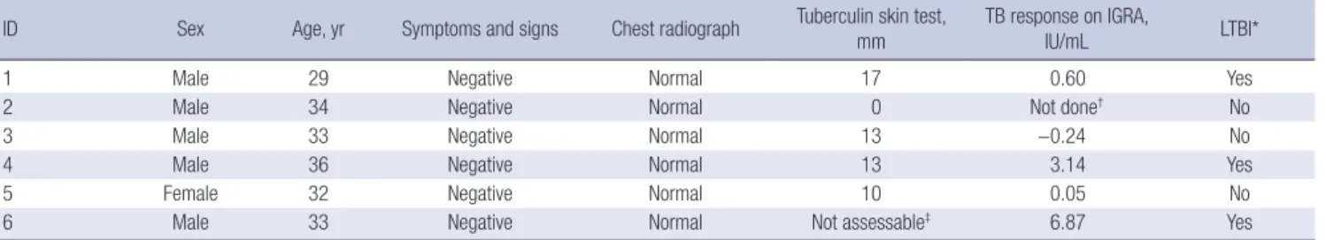
Table 1. Summary of contact investigation in close contacts to MDR-TB
ID Sex Age, yr Symptoms and signs Chest radiograph Tuberculin skin test,
mm TB response on IGRA,
IU/mL LTBI*
1 Male 29 Negative Normal 17 0.60 Yes
2 Male 34 Negative Normal 0 Not done† No
3 Male 33 Negative Normal 13 −0.24 No
4 Male 36 Negative Normal 13 3.14 Yes
5 Female 32 Negative Normal 10 0.05 No
6 Male 33 Negative Normal Not assessable‡ 6.87 Yes
The Korean national guideline for contact investigation for TB adopts a two-step strategy for LTBI, sequentially performing tuberculin skin test and IGRA.
MDR-TB = multi-drug resistant tuberculosis, TB = tuberculosis, IGRA = interferon gamma release assay, LTBI = latent tuberculosis infection.
*LTBI was assessed to be present when both tuberculin skin test and IGRA were positive. Cutoffs for positivity of tuberculin skin test and IGRA (corrected for Nil response) were 10 mm and 0.35 IU/mL, respectively. †Contact 2 did not have a positive conversion in the secondary tuberculin skin test which was performed 3 months later. ‡Contact 6 had a history of positive tuberculin skin test before, so tuberculin test was omitted.
Lee SC, et al. • Submillisievert Chest CT for Contact Investigation of MDR-TB
ture of bronchoalveolar lavage fluid was positive after 13 days in liquid media and 8 weeks in solid media. The result of the drug susceptibility test in contact 1 was identical with that of the index case. The pattern of spoligotyping and 24-loci myco-
bacterial interspersed repetitive units–variable-number tan- dem repeats (MIRU-VNTR) between index patient and contact 1 was matched identically (M. tuberculosis strain, Beijing; 24- loci MIRU-VNTR, 244233352544425173353824). Contact 1 was Fig. 1. Chest radiograph and CT of a secondary case of MDR-TB in contact 1. (A) Chest radiograph at baseline contact investigation is normal. (B) Baseline submillisievert com- puted tomographic scans of the chest shows tuberculous granuloma and focal bronchiolitis (arrows) in the superior segment of the right lower lobe on baseline scan. (C) Chest radiograph 2 months later shows subtle opacities (arrow) in the right upper lung zone. (D) Submillisievert computed tomographic scans of the chest 2 months later shows the progression of pre-existing lesions into ill-defined lobular consolidation (arrows) in the superior segment of the right lower lobe. (E) Results of spoligotyping 24-loci MIRU-VNTR analysis are identical between index and secondary MDR-TB. (F) Chest radiograph taken after completion of 1.5-year of anti-MDR-TB treatment shows small residual opacity (arrows) in the right upper lung zone.
CT = computed tomography, MDR-TB = multi-drug resistant tuberculosis, MIRU-VNTR = mycobacterial interspersed repetitive units-variable-number tandem repeats, TB = tu- berculosis.
Figure 1. E
Figure 1. F Figure 1. A
Figure 1. C
Figure 1. B
Figure 1. DA B
C D
F E
Baseline scan
2 months later
Lee SC, et al. • Submillisievert Chest CT for Contact Investigation of MDR-TB
treated with kanamycin, pyrazinamide, moxifloxacin, prothion- amide, and cycloserine. During the treatment, serial expecto- rated sputa were negative for smear and culture. He was cured after 1.5-year of treatment.
Other contacts did not have symptoms at 3-month and 1-year follow-up. In contact 2, a 3-month follow-up CT scan showed a slight increase in nodule size. One of two nodules decreased on follow-up CT scan 1 year later (Fig. 2). There were no abnormal- ities in follow-up CT scans in four of six contacts, and none with negative initial LTBI test had a positive conversion in the second- ary LTBI test.
Radiation dose of submillisievert chest CT scanning Median dose-length product and effective dose per CT scan were 14.91 milligray-cm (range, 13.08–16.90) and 0.21 millisievert (range, 0.19–0.25), respectively, with 1-year cumulative dose- length product of 44.52 milligray-cm (range, 43.28–49.18) and 1-year cumulative effective dose of 0.65 millisievert (range, 0.63–
0.71).
DISCUSSION
Radiation exposure ought to be the main concern for the use of chest CT scans in contact investigation of TB (13). Low dose ion- izing radiation of medical imaging below 100 millisievert may increase cancer risk in children, who are more radiosensitive than adults (15). However, the estimated effective dose of our submillisievert chest CT scans (average, 0.21 millisievert; range, 0.19–0.25) is much lower than that of conventional chest CT scans (average, 7.0 millisievert; range, 4.0–18.0), two times higher than
Fig. 2. Chest radiograph and CT in contact 2, having sub-centimeter-sized nodules. (A) Chest radiograph at baseline contact investigation is normal. (B) Baseline, 3-month, and 1-year follow-up submillisievert computed tomographic scans of the chest shows two suspected tuberculous granulomas (arrows). Lesions increased slightly 3 months later, and decreased after 1 year of observation.
CT = computed tomography.
Figure 2. A Figure 2. B
Baseline scan 3 months later 1 year later Figure 2. B
Baseline scan 3 months later 1 year later Figure 2. B
Baseline scanBaseline scan 3 months later3 months later 1 year later1 year later
A B
posteroanterior and lateral chest examinations (average, 0.10 millisievert; range, 0.05–0.24), and ten times higher than pos- teroanterior chest radiographs (average, 0.02 millisievert; range, 0.01–0.05) (16). Submillisievert radiation is negligible for cancer risk when compared with radiation from natural source of 3.1 millisievert per year in adults.
TB granuloma is the earliest pulmonary parenchymal abnor- mality (17). Baseline CT scan depicted sub-centimeter-sized nodules suspected of TB granuloma in contact 1 and 2, which were not shown on chest radiograph. However, the presence of parenchymal CT abnormalities in contacts are not indicative of active disease without overt CT findings of active TB, such as extensive tree-in-bud appearance and cavities (18). The hall- mark of active TB is the progression of parenchymal abnormali- ties on follow-up. Thus, we introduced repeated submillisievert CT scans for determining active disease. Indeed, repeated CT examinations revealed that TB granuloma gradually progressed in contact 1, while sub-centimeter-sized nodules was stabilized in contact 2, potentially in line with the inter-individual and with- in-host variability of TB (17).
Based on the radiologic progression of repeated submillisievert CT scans, contact 1 was further evaluated and diagnosed with secondary MDR-TB in asymptomatic periods. This led to a fa- vorable treatment outcome with minimal residue, and early iso- lation of MDR-TB at the lowest infectivity, consequently mini- mizing transmission of drug-resistant strain. In a risk-benefit perspective, submillisievert chest CT may be considered in adult contacts at risk for MDR-TB in high-resource setting, such as health care workers (19). Given that active TB most commonly develops in the first 2 years of TB exposure, annual follow-up
Lee SC, et al. • Submillisievert Chest CT for Contact Investigation of MDR-TB
chest CT may be performed up to the first 2 years. In addition, submillisievert chest CT may not be recommended in contacts with negative LTBI but it needs to be considered that false-neg- ative rate of LTBI test is 8%–19% and increased in immunocom- promised contacts (20). Further investigation is required for these issues.
In conclusion, early identification of secondary MDR-TB is feasible with submillisievert chest CT scans in contact investi- gations of MDR-TB, minimizing of MDR-TB transmission and offering a favorable treatment outcome.
DISCLOSURE
The authors have no potential conflicts of interest to disclose.
AUTHOR CONTRIBUTION
Conceptualization: Lee SC, Yoon SH, Goo JM, Yim JJ. Data cu- ration: Lee SC, Yoon SH, Goo JM, Yim JJ, Kim CK. Formal analy- sis: Lee SC, Yoon SH, Goo JM, Yim JJ, Kim CK. Investigation: Lee SC, Yoon SH, Goo JM, Yim JJ, Kim CK. Writing - original draft:
Yoon SH. Writing - review & editing: Lee SC, Yoon SH, Goo JM, Yim JJ, Kim CK.
ORCID
Seung Chul Lee https://orcid.org/0000-0003-1221-958X Soon Ho Yoon https://orcid.org/0000-0002-3700-0165 Jin Mo Goo https://orcid.org/0000-0003-1791-7942 Jae-Joon Yim https://orcid.org/0000-0002-9605-0074 Chang-Ki Kim https://orcid.org/0000-0003-4729-3114 REFERENCES
1. World Health Organization. Global tuberculosis report 2015 [Internet].
Available at http://apps.who.int/iris/bitstream/10665/191102/1/97892 41565059_eng.pdf [accessed on 29 August 2017].
2. Kim HJ, Yoon HH, Eun BW, Ahn Y, Ryoo S, Kim HJ. The rate of drug-resis- tant tuberculosis in Korean children and adolescents since 2007. J Kore- an Med Sci 2017; 32: 954-60.
3. Gandhi NR, Nunn P, Dheda K, Schaaf HS, Zignol M, van Soolingen D, Jen- sen P, Bayona J. Multidrug-resistant and extensively drug-resistant tuber- culosis: a threat to global control of tuberculosis. Lancet 2010; 375: 1830- 43.
4. Shah NS, Yuen CM, Heo M, Tolman AW, Becerra MC. Yield of contact in- vestigations in households of patients with drug-resistant tuberculosis:
systematic review and meta-analysis. Clin Infect Dis 2014; 58: 381-91.
5. Becerra MC, Appleton SC, Franke MF, Chalco K, Arteaga F, Bayona J, Mur- ray M, Atwood SS, Mitnick CD. Tuberculosis burden in households of pa-
tients with multidrug-resistant and extensively drug-resistant tuberculo- sis: a retrospective cohort study. Lancet 2011; 377: 147-52.
6. van der Werf MJ, Langendam MW, Sandgren A, Manissero D. Lack of evi- dence to support policy development for management of contacts of mul- tidrug-resistant tuberculosis patients: two systematic reviews. Int J Tuberc Lung Dis 2012; 16: 288-96.
7. Langendam MW, Tiemersma EW, van der Werf MJ, Sandgren A. Adverse events in healthy individuals and MDR-TB contacts treated with anti-tu- berculosis drugs potentially effective for preventing development of MDR- TB: a systematic review. PLoS One 2013; 8: e53599.
8. Falzon D, Jaramillo E, Schünemann HJ, Arentz M, Bauer M, Bayona J, Blanc L, Caminero JA, Daley CL, Duncombe C, et al. WHO guidelines for the programmatic management of drug-resistant tuberculosis: 2011 update.
Eur Respir J 2011; 38: 516-28.
9. Barry CE 3rd, Boshoff HI, Dartois V, Dick T, Ehrt S, Flynn J, Schnappinger D, Wilkinson RJ, Young D. The spectrum of latent tuberculosis: rethinking the biology and intervention strategies. Nat Rev Microbiol 2009; 7: 845-55.
10. Lee SW, Jang YS, Park CM, Kang HY, Koh WJ, Yim JJ, Jeon K. The role of chest CT scanning in TB outbreak investigation. Chest 2010; 137: 1057- 64.
11. Petruccioli E, Scriba TJ, Petrone L, Hatherill M, Cirillo DM, Joosten SA, Ottenhoff TH, Denkinger CM, Goletti D. Correlates of tuberculosis risk:
predictive biomarkers for progression to active tuberculosis. Eur Respir J 2016; 48: 1751-63.
12. Alsdurf H, Hill PC, Matteelli A, Getahun H, Menzies D. The cascade of care in diagnosis and treatment of latent tuberculosis infection: a systematic review and meta-analysis. Lancet Infect Dis 2016; 16: 1269-78.
13. Brenner DJ, Hall EJ. Computed tomography--an increasing source of ra- diation exposure. N Engl J Med 2007; 357: 2277-84.
14. Deak PD, Smal Y, Kalender WA. Multisection CT protocols: sex- and age- specific conversion factors used to determine effective dose from dose- length product. Radiology 2010; 257: 158-66.
15. Pearce MS, Salotti JA, Little MP, McHugh K, Lee C, Kim KP, Howe NL, Ron- ckers CM, Rajaraman P, Sir Craft AW, et al. Radiation exposure from CT scans in childhood and subsequent risk of leukaemia and brain tumours:
a retrospective cohort study. Lancet 2012; 380: 499-505.
16. Mettler FA Jr, Huda W, Yoshizumi TT, Mahesh M. Effective doses in radi- ology and diagnostic nuclear medicine: a catalog. Radiology 2008; 248:
254-63.
17. Lenaerts A, Barry CE 3rd, Dartois V. Heterogeneity in tuberculosis pathol- ogy, microenvironments and therapeutic responses. Immunol Rev 2015;
264: 288-307.
18. Im JG, Itoh H, Shim YS, Lee JH, Ahn J, Han MC, Noma S. Pulmonary tu- berculosis: CT findings--early active disease and sequential change with antituberculous therapy. Radiology 1993; 186: 653-60.
19. Yoon CG, Oh SY, Lee JB, Kim MH, Seo Y, Yang J, Bae KJ, Hong S, Yang ES, Kim HJ. Occupational risk of latent tuberculosis infection in health work- ers of 14 military hospitals. J Korean Med Sci 2017; 32: 1251-7.
20. de Visser V, Sotgiu G, Lange C, Aabye MG, Bakker M, Bartalesi F, Brat K, Chee CB, Dheda K, Dominguez J, et al. False-negative interferon-γ release assay results in active tuberculosis: a TBNET study. Eur Respir J 2015; 45:
279-83.
Lee SC, et al. • Submillisievert Chest CT for Contact Investigation of MDR-TB SUPPLEMENTARY MATERIAL
Supplementary 1. Full technical details of contact investigation, bacteriology and genotyping Contact investigation
To exclude active tuberculosis (TB), contact investigation was done with the sputum test and chest radiograph. For contacts exclud- ed from active TB, the latent TB infection (LTBI) test was first performed with a tuberculin skin test, followed by an interferon gam- ma release assay (IGRA) for contacts having a positive tuberculin skin test. This was the national guideline for diagnosis of LTBI, considering that Bacillus Calmette-Guérin vaccination is mandatory in Korea. A follow-up investigation was planned 3 months and 1 year after baseline examination. At the 3-month follow-up, a second tuberculin skin test and IGRA test were performed in those who were negative on initial LTBI tests.
Tuberculin skin tests were performed on the volar side of the forearm by administration of two units of tuberculin purified pro- tein derivative-RT23, with any induration measured in millimeters between 48 and 72 hours, using the ballpoint method. A Quan- tiFERON-TB Gold In-Tube test (Qiagen, Hilden, Germany) was done per manufacturer instructions. Cutoffs for positivity on tuber- culin skin test and IGRA (corrected for nil response) were 10 mm and 0.35 IU/mL, respectively.
Bacteriology and genotyping
Close contacts submitted sputum, and specimens were microscopically examined for acid-fast bacillus with auramine-rhodamine fluorescent staining and confirmed by Ziehl-Neelsen staining. The Xpert/RIF test (Cepheid, Sunnyvale, CA, USA) was performed according to manufacturer instructions within 24 hours of sputum submission. Sputum culture was done with the national refer- ence standard quality with solid culture on 3% Ogawa media for 8 weeks and liquid culture in BACTECTM MGITTM (Becton, Dickin- son and Company, Franklin Lakes, NJ, USA) for 6 weeks. Confirmed diagnosis of TB was determined if Mycobacterium tuberculosis was isolated in at least one solid or liquid culture.
Drug susceptibility test and genotyping were performed at the supranational reference laboratory for TB, the Korean Institute of Tuberculosis. The resistance to each anti-TB drug was defined as greater than 1% bacterial growth in Lowenstein-Jensen medium (Thermo Scientific, Waltham, MA, USA) using the absolute concentration method with following critical concentrations: isoniazid 0.2 µg/mL; streptomycin 10.0 µg/mL; ethambutol 2.0 µg/mL; rifampicin 40.0 µg/mL; paraaminosalicylic acid 1.0 µg/mL; prothio- namide 40.0 µg/mL; cycloserine 30.0 µg/mL; kanamycin 30.0 µg/mL; capreomycin 40.0 µg/mL; ofloxacin 4.0 µg/mL; levofloxacin 2.0 µg/mL; moxifloxacin 2.0 µg/mL; and rifabutin 20 µg/mL. Pyrazinamide susceptibility was assessed through a pyrazinamidase test. TB isolates were genotyped from index case and contacts with culture-positive TB, using spacer oligonucleotide typing spoli- gotyping) and 24-loci mycobacterial interspersed repetitive unit–variable-number tandem repeat analysis (1).
REFERENCE
1. Supply P, Allix C, Lesjean S, Cardoso-Oelemann M, Rüsch-Gerdes S, Willery E, Savine E, de Haas P, van Deutekom H, Roring S, et al. Proposal for standard- ization of optimized mycobacterial interspersed repetitive unit-variable-number tandem repeat typing of Mycobacterium tuberculosis. J Clin Microbiol 2006; 44: 4498-510.