Introduction
Panoramic radiography has been widely used as a diag- nostic tool because it is easy to provide a radiographic overview of the teeth and surrounding anatomical struc- tures.1,2 Recently, a number of dental practitioners have come to prefer digital radiography over conventional film radiography.3-6One of the main advantages of digital radio- graphy compared with conventional film radiography is the possibility of reducing the radiation dose to patients.7 Diagnostic benefits and possible dose hazard trade-offs
are important considerations in the choice of radiographic procedures. Since X-ray risks are cumulative, the choice of radiographic unit should be considered in examining all patients for dose reduction.8Digital panoramic equip- ment requires the evaluation of the radiation dose to pati- ents according to the machines, and in maxillofacial pano- ramic imaging, the radiation dose of the machines should follow the principle of keeping the dose as low as reason- ably achievable (ALARA).
The effective dose (E) is a concept and approach recom- mended by the International Commission on Radiological Protection (ICRP) to estimate the damage from radiation to the exposed population.9E is a widely used calculation that permits comparison of the detriment of different expo- sures, including specific area exposure,10to ionizing radi- ation to an equivalent detriment produced by a full body dose of radiation.8White11used this approach in 1992 in
Effective dose from direct and indirect digital panoramic units
Gun-Sun Lee, Jin-Soo Kim, Yo-Seob Seo, Jae-Duk Kim
Department of Oral and Maxillofacial Radiology, School of Dentistry, Oral Biology Research Institute, Chosun University, Gwangju, Korea
ABSTRACT
Purpose: This study aimed to provide comparative measurements of the effective dose from direct and indirect digital panoramic units according to phantoms and exposure parameters.
Materials and Methods: Dose measurements were carried out using a head phantom representing an average man (175 cm tall, 73.5 kg male) and a limbless whole body phantom representing an average woman (155 cm tall, 50 kg female). Lithium fluoride thermoluminescent dosimeter (TLD) chips were used for the dosimeter. Two direct and 2 indirect digital panoramic units were evaluated in this study. Effective doses were derived using 2007 International Commission on Radiological Protection (ICRP) recommendations.
Results: The effective doses of the 4 digital panoramic units ranged between 8.9μSv and 37.8 μSv. By using the head phantom, the effective doses from the direct digital panoramic units (37.8μSv, 27.6 μSv) were higher than those from the indirect units (8.9μSv, 15.9 μSv). The same panoramic unit showed the difference in effective doses according to the gender of the phantom, numbers and locations of TLDs, and kVp.
Conclusion: To reasonably assess the radiation risk from various dental radiographic units, the effective doses should be obtained with the same numbers and locations of TLDs, and with standard hospital exposure. After that, it is necessary to survey the effective doses from various dental radiographic units according to the gender with the corresponding phantom. (Imaging Sci Dent 2013; 43: 77-84)
KEY WORDS: Radiography, Dental, Digital; Radiography, Panoramic; Radiation Dosage
*This study was supported by the research fund from Chosun University, 2012.
Received September 29, 2012; Revised October 30, 2012; Accepted November 7, 2012 Correspondence to : Prof. Jae-Duk Kim
Department of Oral and Maxillofacial Radiology, School of Dentistry, Chosun Uni- versity, 309 Pilmun-daero, Dong-gu, Gwangju 501-759, Korea
Tel) 82-62-220-3880, Fax) 82-62-227-0270, E-mail) [email protected]
Copyright ⓒ 2013 by Korean Academy of Oral and Maxillofacial Radiology
This is an Open Access article distributed under the terms of the Creative Commons Attribution Non-Commercial License (http://creativecommons.org/licenses/by-nc/3.0) which permits unrestricted non-commercial use, distribution, and reproduction in any medium, provided the original work is properly cited.
Imaging Science in Dentistry∙pISSN 2233-7822 eISSN 2233-7830
assessing the radiation risk from various dental radio- graphic examinations. The unit of the effective dose is the sievert (Sv) and it is calculated by the equation: E==»HT
×WT, where E is the product of the tissue weighting fac- tor (WT), which represents the relative contribution of each organ or tissue to the overall risk, and the equivalent dose (HT).12The whole-body risk is acquired by the summation of the weighted equivalent doses to all exposed tissues or organs. The earlier 1990 ICRP tissue-weighting factors,13 2005 weighting factors, and the new 2007 weighting fac- tors12were used to calculate the effective dose.7,8,14Newly adopted recommendations from the ICRP provide revi- sion of tissue-weighting factors and inclusion of salivary glands as a weighted tissue. These changes resulted in an upward reassessment of the effective dose from oral and maxillofacial radiographic examinations.12
The studies of the absorbed dose or effective dose from each exposure program of the film-based panoramic machine with a limbless whole body phantom and a head phantom were reported by Lecomber et al in 200015and Choi et al in 2001,16respectively. The tissue-absorbed dose and the whole-body effective dose (E) for a new genera- tion film-based panoramic machine (Planmeca PM 2002 CC Proline, Planmeca, Helsinki, Finland), operating in the panoramic examination mode with a head phantom, was studied by Danforth et al in 2000.14 Gijbels et al7 reported that the effective radiation doses ranged from 4.7 μSv to 14.9 μSv for various digital panoramic units with a head phantom using the 2005 recommendations of the ICRP.
Few studies have reported the difference in radiation dose between direct and indirect digital panoramic units.
Also, to our knowledge, no studies have been published addressing the difference in the assessment of the effec- tive dose using a head versus a whole body phantom.
Ludlow and Ivanovic8reported that there was large dif- ference in the radiation dose between the “standard” and
“ultra” exposure in the Iluma CBCT unit. They also men- tioned that a substantial difference in the effective dose was found according to variations in technique even when they evaluated the same CBCT unit. Therefore, it is neces-
sary to prepare standardized conditions for calculation of the effective dose to compare the radiation risk from vari- ous dental radiographic examinations.
The aim of the present study is to provide the compara- tive measurements of the effective dose from direct and indirect digital panoramic units according to phantoms and exposure parameters. The average tissue-absorbed dose, weighted (equivalent) radiation dose, and effective dose were calculated for the anatomy of the head and neck area and the whole body with the corresponding phan- tom, respectively.
Materials and Methods
Exposures
The digital panoramic units used for this study were the ProMax (Planmeca, Helsinki, Finland), Orthopantomo- graph OP100 (Imaging Instrumentarium, Tusula, Finland), and 2 ProlineXC units (Planmeca, Helsinki, Finland).
Table 1 shows the technical and phantom parameters for the direct and indirect panoramic units used. Both the radiation exposure dose and the latitude of the TLD are too small to be measured with reliability; therefore, the TLD loaded phantom was exposed to radiation by the dosimeters ten times. Then, the obtained values were divided by ten to obtain one individual value for each region, subtracting each background dose. The phantom was positioned in accordance with the manufacturer’s specifications for each machine, following the reference lines and head rests. The standard examination was carried out for each unit and the dosimetry was performed for each technique in order to ensure reliability.
Dosimetry
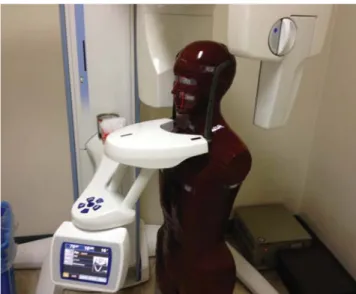
Dose measurements were carried out using two anthro- pomorphic phantoms. The head phantom (Rando ART- 210, Alderson Research Laboratories, Long Beach, CA, USA) representing an average man (175 cm tall, 73.5 kg male) (Fig. 1) and the limbless whole body phantom (Rando ART-300, Alderson Research Laboratories, Long
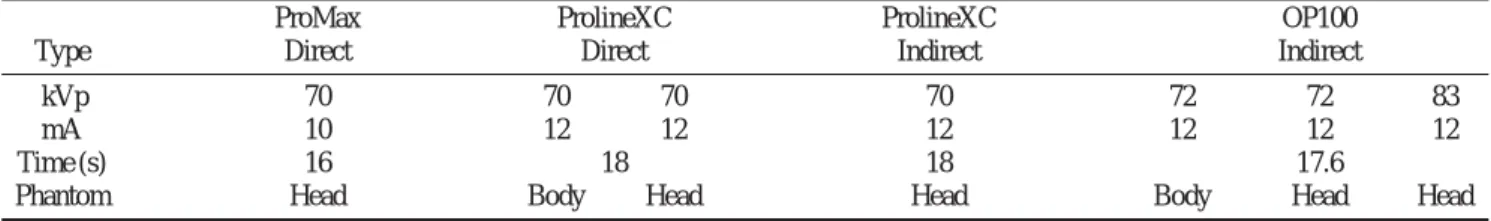
Table 1.Exposure and phantom parameters for the panoramic units
ProMax ProlineXC ProlineXC OP100 Type Direct Direct Indirect Indirect
kVp 70 70 70 70 72 72 83
mA 10 12 12 12 12 12 12
Time (s) 16 18 18 17.6
Phantom Head Body Head Head Body Head Head
Beach, CA, USA) representing an average woman (155 cm tall, 50 kg female) (Fig. 2) consisted of 9 and 31 trans- verse sections, respectively. The reservoirs for the place- ment of radiation dosimetry measuring devices were pre- pared to correspond to the anatomic sites of interest. Lithi- um fluoride thermoluminescent dosimeter (TLD) chips (Harshaw TLD-100, Thermo Electron Co., Oakwood Vil- lage, OH, USA) were used as dosimeters. The absorbed doses at the selected locations, corresponding to the radio-
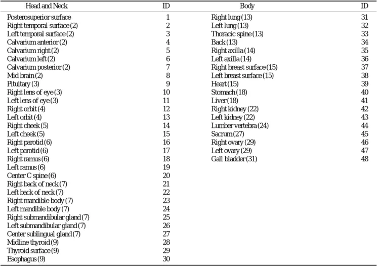
sensitive organs of interest, were measured using two sets of 60 and 96 TLDs. The 30 and 48 phantom sites both inside and on the surface of each phantom can be seen in Table 2. Two dosimeters were placed in each anatomical site to calculate the mean value of each location, while retaining the same dosimeters in the same positions for each exposure. A set of dosimeters were kept separately to record the background radiation. Before the study, all of the dosimeters were calibrated using the same type and range of radiation that would be used during the experi- ments. Prior to every exposure, the dosimeters were anneal- ed at 100�C for 1 hour, 400�C for 2 hours, and then cooled to 35�C. All of the TLDs were read in accordance with the manufacturer’s directions after each exposure using a Harshaw 3500 TLD Reader (Harshaw/Bicron, Solon, OH, USA).
Dose calculations
After reading, an individual sensitivity value was appli- ed for each TLD. Exposure doses were recorded in nano- coulombs (nC) and, after the application of energy calibra- tion factors (RCF, reader calibration factor; and ECC, ele- ment correction coefficient), the dosimetry data were con- verted into micrograys (μGy) and subsequently recorded.
The standard deviation of the readings from the TLDs was less than 30%. Doses from two TLDs located at different points in the same tissue or organ were averaged, result- ing in the average organ absorbed dose. The weighted dose for bone marrow of the whole body phantom was calculat- ed using the sum of the radiation from the calvarium, mandibular body and ramus, cervical, thoracic, and lumbar vertebrae, and sacrum. The weighted dose for the bone marrow of the head phantom was calculated using the sum of the radiation from the calvarium, mandibular body and ramus, and cervical vertebra. Sublingual, submandibular, and parotid salivary gland doses were used for calculating the weighted dose for the salivary glands. The thyroid gland dose was individually calculated taking its specific weighted factor into consideration. For the skin surface area, fifteen points were measured: the posterosuperior surface of the head, thyroid surface, back of the thorax, and both sides of the right and left of the temporal region, lens of the eye, cheek, back of the neck, axilla, and breast (Table 2).
The products of these average organ absorbed doses and the percentage of a tissue or organ irradiated (Tables 3 and 4) in a radiographic examination were used to cal- culate the equivalent dose (HT) in microsieverts (μSv).12
Fig. 1.The limbless female whole body phantom.
Fig. 2.The head phantom representing a man.
The effective dose was calculated by multiplying actual organ doses (equivalent dose; HT) by ‘risk weighting fac- tors’ as follows: E==»HT×WT. The tissue weighting factor (WT) represents the contribution that each specific tissue or organ makes to the overall risk. The whole-body risk was found by the summation of the tissue weighted equivalent doses to all tissues or organs exposed. Effec- tive doses were derived using 2007 ICRP recommenda- tions12(Table 5). This dose was expressed in microsieverts (μSv). The obtained effective dose from each panoramic unit was compared with the dosimetric values calculated from the 24 anatomical sites of the phantoms used by Lud- low and Ivanovic8.
Results
Table 6 shows the dosimetric results. The equivalent doses and the effective doses were expressed in μSv. The greatest individual organ doses for all examinations were measured in the salivary tissue.
The effective radiation doses ranged between 8.9μSv
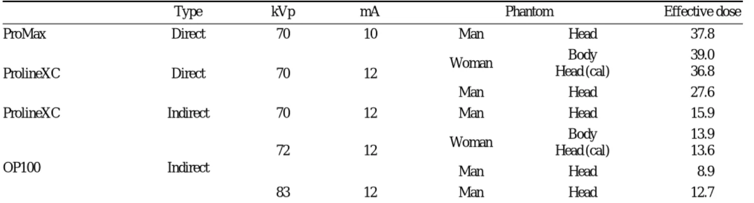
and 37.8μSv for the 4 digital panoramic units. By using a head phantom, the effective doses from the ProMax (37.8 μSv) and ProlineXC (27.6 μSv) direct digital panoramic units were higher than those from the Orthopantomograph OP100 (8.9μSv) and ProlineXC (15.9 μSv) indirect units.
The effective dose from the same panoramic unit (Ortho- pantomograph OP100) using the same head phantom was higher at 83 kVp than at 72 kVp.
Comparison of effective doses with parameters; the exposure, phantom (gender), and calculation method can be found in Table 7.
The effective dose from the ProlineXC (direct) with an exposure of 70 kVp and 12 mA was 36.8μSv by the cal- culation for the head region of the whole body phantom and 27.6μSv by the head phantom. The effective dose from the Orthopantomograph OP100 (indirect) with an exposure of 72 kVp and 12 mA was 13.6μSv by the cal- culation for the head region of the whole body phantom and 8.9μSv by the head phantom. The effective doses from the direct and indirect digital units were both higher with the female phantom than with the male one.
Table 2.Locations of TLD chips in phantoms
Head and Neck ID Body ID
Posterosuperior surface 1 Right lung (13) 31
Right temporal surface (2) 2 Left lung (13) 32
Left temporal surface (2) 3 Thoracic spine (13) 33
Calvarium anterior (2) 4 Back (13) 34
Calvarium right (2) 5 Right axilla (14) 35
Calvarium left (2) 6 Left axilla (14) 36
Calvarium posterior (2) 7 Right breast surface (15) 37
Mid brain (2) 8 Left breast surface (15) 38
Pituitary (3) 9 Heart (15) 39
Right lens of eye (3) 10 Stomach (18) 40
Left lens of eye (3) 11 Liver (18) 41
Right orbit (4) 12 Right kidney (22) 42
Left orbit (4) 13 Left kidney (22) 43
Right cheek (5) 14 Lumber vertebra (24) 44
Left cheek (5) 15 Sacrum (27) 45
Right parotid (6) 16 Right ovary (29) 46
Left parotid (6) 17 Left ovary (29) 47
Right ramus (6) 18 Gall bladder (31) 48
Left ramus (6) 19
Center C spine (6) 20
Right back of neck (7) 21
Left back of neck (7) 22
Right mandible body (7) 23
Left mandible body (7) 24
Right submandibular gland (7) 25
Left submandibular gland (7) 26
Center sublingual gland (7) 27
Midline thyroid (9) 28
Thyroid surface (9) 29
Esophagus (9) 30
The effective doses calculated by the method of Lud- low and Ivanovic were lower than those calculated by the authors from each panoramic examination.
Discussion
The equivalent dose is used to compare the effects of different types of radiation on tissues or organs, presented in sieverts (Sv). The equivalent dose (HT) was calculated according to the equation: HT==»WR×DT, where the equivalent dose (HT) for a tissue or organ is the product of the radiation weighting factor (WR) and the average absorb- ed dose (DT) measured for that specific organ.13The pro- ducts of these values and the percentage of a tissue or organ irradiated (Table 3) in a radiographic examination were used to calculate the equivalent dose (HT) in micro- sieverts (μSv).12
The equivalent doses from the indirect and direct digi- tal panoramic units were highest in the remainders and salivary glands (Table 6). The next highest equivalent doses to other organs were the following, in order: bone surface, thyroid gland, and bone marrow. Several studies on panoramic units have pointed out that the salivary gland tissue is one of the organs receiving the highest individual organ doses during maxillofacial imaging7,15,17 and our
results confirmed these findings. Lecomber et al15indicat- ed that the influence on the calculated effective dose of treating the salivary tissue as a remainder organ deserved discussion in 2000. Ludlow and Ivanovic8explained this by the fact that the rotational centers of panoramic units for the jaws coincide with the location of the salivary glands.
Because anatomy at the rotational center is continuously
Table 3. Estimated percentage of tissue irradiated with the head phantom
Head and Neck ID %
Bone marrow 16.5
Mandible 18, 19, 23, 24 1.3
Calvaria 1-7 11.8
Cervical spine 20 3.4
Thyroid 28, 29 100.0
Esophagus 30 10.0
Skin 1-3 ,10, 11, 14, 15, 21, 22, 29 9.0
Bone surface* 16.5
Mandible 18, 19, 23, 24 1.3
Calvaria 1-7 11.8
Cervical spine 20 3.4
Salivary glands 100.0
Parotid 16, 17 100.0
Submandibular 25, 26 100.0
Sublingual 27 100.0
Brain 8, 9 100.0
Remainders
Lymphatic nodes 16-20, 23-28, 30 10.0
Muscle 16-20, 23-28, 30 5.0
Extrathoracic airway 12, 13, 16-20, 23-28, 30 100.0
Oral mucosa 16-19, 23-27 100.0
*Bone surface dose==bone marrow dose×bone/muscle mass energy absorp- tion, coefficient ratio==-0.0618×2/3 kV peak++6.9406 using data.8
Table 4. Estimated percentage of tissue irradiated with the whole body phantom
Whole body ID %
Bone marrow
Mandible 18, 19, 23, 24 1.3
Calvaria 1-7 11.8
Cervical spine 20 3.4
Thoracic spine 33 14.1
Lumbar vertebra 44 10.9
Sacrum 45 13.9
Colon 41-43, 48 100.0
Lung 31, 32, 39 100.0
Stomach 39, 40 100.0
Breast 37, 38 100.0
Gonads 46, 47 100.0
Bladder 48 100.0
Liver 41 100.0
Thyroid 28, 29 100.0
Esophagus 30, 39 100.0
Skin 1-3,10, 11, 14, 15, 21, 22,
29, 34-38 36.0
Bone surface* 16.5
Mandible 18, 19, 23, 24 1.3
Calvaria 1-7 11.8
Cervical spine 20 3.4
Thoracic spine 33 14.1
Lumbar vertebra 44 10.9
Sacrum 45 13.9
Salivary glands 100.0
Parotid 16, 17 100.0
Submandibular 25, 26 100.0
Sublingual 27 100.0
Brain 8, 9 100.0
Remainders
Lymphatic nodes 16-20, 23-28, 30, 35, 36, 39 40.0 Muscle 16-20, 23-28, 30, 33, 44, 45 43.9 Extrathoracic airway 12, 13, 16-20, 23-28, 30 100.0
Oral mucosa 16-19, 23-27 100.0
Adrenals 40 100.0
Gall bladder 41 100.0
Heart 39 100.0
Kidneys 42, 43 100.0
Pancreas 40 100.0
Small intestine 42, 43 100.0
Spleen 40 100.0
Thymus 39 100.0
Uterus/cervix 48 100.0
*Bone surface dose==bone marrow dose×bone/muscle mass energy absorp- tion coefficient ratio==-0.0618×2/3 kV peak++6.9406 using data.8
exposed, effective doses from dental panoramic imaging will be larger than imaging procedures that produce a more uniform distribution of absorbed energy within the scanned region. It seems reasonable to include salivary gland expo- sures in calculations of effective dose until these exposures could be shown not to be significant.18The effective doses in this study were calculated with inclusion of the salivary gland tissue using 0.01 according to the 2007 ICRP tissue
weights.12 Dental radiographic examinations resulted in negligible doses to the gonads, and hereditary doses were not effective for the calculation of detriment in this study.
The use of a head phantom or only the head region calcu- lation of the whole body phantom is thought to be reason- able for the calculation of detriment from dental radio- graphic examinations.
The effective dose is the product of the ICRP’s tissue weighting factor (WT) for the type of tissue or body and the human-equivalent dose for tissue (HT). The effective dose is calculated by multiplying actual organ doses by
‘risk weighting factors’ (associated with individual organ sensitivities) and represents the dose that the total body could receive and that would provide the same cancer risk as the application of different doses to various organs.13 One sievert of effective dose carries with it a 4.1% chance of developing fatal cancer in an adult worker and a 5.5%
chance in a whole population, and a 0.8% chance of hered- itary defect in future offspring.12 The ICRP recommends limiting artificial irradiation of the public to an average effective dose of 1 mSv per year, not including medical and occupational exposures.12
The effective doses from the ProMax (37.8μSv) and ProlineXC (27.6μSv) direct digital panoramic units were higher than those from the Orthopantomograph OP100 (8.9μSv) and ProlineXC (15.9 μSv) indirect units by using the head phantom (Table 7). Gijbels et al7reported that comparable results (9.35μSv for CCD, 8.1 μSv for storage phosphor) were found for the various digital panoramic
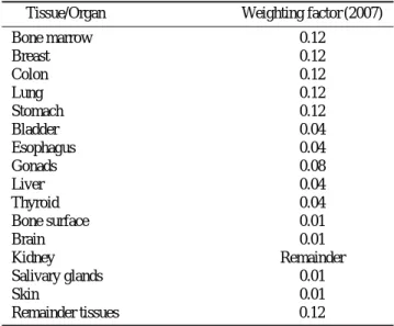
Table 5. Tissue weighting factors for calculation of effective dose 2007 recommendations12
Tissue/Organ Weighting factor (2007)
Bone marrow 0.12
Breast 0.12
Colon 0.12
Lung 0.12
Stomach 0.12
Bladder 0.04
Esophagus 0.04
Gonads 0.08
Liver 0.04
Thyroid 0.04
Bone surface 0.01
Brain 0.01
Kidney Remainder
Salivary glands 0.01
Skin 0.01
Remainder tissues 0.12†
†: Adrenals, extrathoracic region, gall bladder, heart, kidneys, lymphatic nodes, muscle, oral mucosa, pancreas, prostate, small intestine, spleen, thymus, and uterus/cervix. Italicized text represents remainder tissues used for calculation of maxillofacial dose.
Table 6.Equivalent dose (HT) to tissues/organs and effective doses from direct and indirect digital panoramic units (unit; μSv) ProMax ProlineXC ProlineXC OP100
Direct Direct Indirect Indirect
kVp (mA) 70 (10) 70 (12) 70 (12) 72 (12) 83 (12) Phantom Head Head Body Head Head Body Head Bone marrow 33.4 18.5 24.6 14.2 8.2 8.9 8.1
Colon 1.0 0.0
Lung 3.4 0.7
Stomach 2.0 0.0
Breast 0.8 0.0
Gonads 2.4 0.0
Bladder 2.3 0.0
Liver 0.2 0.0
Thyroid 73.7 37.5 41.8 26.9 28.8 14.9 25.0
Esophagus 10.6 4.4 22.3 4.6 4.4 7.0 3.5
Skin 10.2 7.0 10.2 5.7 3.4 3.8 3.7
Bone surface* 135.6 75.0 99.8 57.7 32.7 35.3 28.6 Salivary glands 1161.1 880.9 1214.6 522.5 203.0 447.1 414.0
Brain 29.2 14.9 25.0 1.6 5.2 9.2 6.5
Remainders 1853.2 1510.9 2036.2 761.8 451.3 754.2 652.9 Effective dose 37.8 27.6 39.0 15.9 8.9 13.9 12.7
units when the effective dose data of the direct panoramic units were averaged and compared with the indirect units.
The effective doses from both types of digital panoramic unit evaluated in this study were larger than the results of Gijbels et al.
Given that comparison of the effective doses from the different panoramic units was difficult because of the dif- ferent exposure settings, Gijbels et al7 reported that by considering per unit of exposure (mAs), the Orthoralix yielded the lowest dose per mAs (0.10μSv mAs-1), but the Veraviewepocs the highest (0.17μSv mAs-1), and that the influence of the tube potential was less clear. Actually, for the comparison of the effective doses from the differ- ent units having different exposure settings recommended for the preservation of diagnostic image quality, the doses per unit of exposure are thought not to be significant. In the present study design, the X-ray parameters used were those for an adult as usual in the hospital. The results obtained are thought to be reasonable for the comparison of effective doses from several digital panoramic units considering the respective technical aspects of each radia- tion unit for good image quality, such as the size of the radiation field and shape of the focal trough.
Considering the phantom parameters, the effective dose from the indirect digital panoramic units by the calcula- tion for the head region of the whole body phantom (13.6 μSv) was higher than that of the head phantom (8.9 μSv).
The effective dose from the direct units by the calculation for the head region of the whole body phantom (36.8μSv) was also higher than that of the head phantom (27.6μSv) (Table 7). In addition, the effective dose was higher with the female phantom than with the male phantom in both types of digital units. This is because the absorbed dose at each TLD dose in the phantom depends on the skull size19 and soft tissue morphology of the phantom, which simu-
late an actual man and woman.
The effective dose calculated from the 24 anatomical sites of the phantom used by Ludlow and Ivanovic8 was lower than the obtained effective dose from each panoram- ic unit in this study. The numbers of anatomical sites of the phantoms for their calculation of the effective dose were less than the numbers of anatomical sites in this study.
Furthermore, the difference in the location (left, center, right) and numbers of sites for the TLDs at the same select- ed organs can account for the small difference in effective dose between the calculations of the present study and their study.
Besides, small variations in collimator adjustment or phantom position within the unit may account for the very nearly 23% difference seen between the dosimeter values for the 2 modes of one unit.8The slight discrepancy in the location of the TLD or phantom position are magnified as the TLD is positioned inside or outside of the field of direct radiation.18
The effective doses from direct digital panoramic units were higher than from the indirect units. Even the same panoramic unit showed a difference in effective doses according to the gender of the phantom, number and loca- tions of TLDs, and kVp. To reasonably assess the radiation risk from various dental radiographic units, the effective doses should be obtained with the same number and loca- tions of TLDs, and with usual hospital exposure. After that, a survey on the effective doses from various dental radiographic units must be performed according to the gender with corresponding phantoms.
References
1. Rushton VE, Horner K, Worthington HV. Aspects of panoram- ic radiography in general dental practice. Br Dent J 1999; 186:
342-4.
Table 7.Comparison of effective doses with parameters (unit: μSv)
Type kVp mA Phantom Effective dose
ProMax Direct 70 10 Man Head 37.8
Body 39.0
ProlineXC Direct 70 12 Woman
Head (cal) 36.8
Man Head 27.6
ProlineXC Indirect 70 12 Man Head 15.9
Woman Body 13.9
OP100 Indirect
72 12 Head (cal) 13.6
Man Head 8.9
83 12 Man Head 12.7
Head (cal): calculation of only head portion of whole body phantom.
2. Tugnait A, Clerehugh V, Hirschmann PN. Radiographic equip- ment and techniques used in general dental practice: a survey of general dental practitioners in England and Wales. J Dent 2003; 31: 197-203.
3. Hildebolt CF, Couture RA, Whiting BR. Dental photostimu- lable phosphor radiography. Dent Clin North Am 2000; 44:
273-97.
4. Macdonald R. Digital imaging for dentists. Aust Dent J 2001;
46: 301-5.
5. Brennan J. An introduction to digital radiography in dentistry.
J Orthod 2002; 29: 66-9.
6. Parks ET, Williamson GF. Digital radiography: an overview.
J Contemp Dent Pract 2002; 3: 23-39.
7. Gijbels F, Jacobs R, Bogaerts R, Debaveye D, Verlinden S, Sanderink G. Dosimetry of digital panoramic imaging. Part I:
Patient exposure. Dentomaxillofac Radiol 2005; 34: 145-9.
8. Ludlow JB, Ivanovic M. Comparative dosimetry of dental CBCT devices and 64-slice CT for oral and maxillofacial radio- logy. Oral Surg Oral Med Oral Pathol Oral Radiol Endod 2008;
106: 106-14.
9. Radiation protection. Protection from potential exposures:
application to selected radiation sources. A report of a task group of the International Commission on Radiation Protec- tion. Ann ICRP 1997; 27: 1-61.
10. Cho JY, Han WJ, Kim EK. Absorbed and effective dose from periapical radiography by portable intraoral X-ray machine.
Korean J Oral Maxillofac Radiol 2007; 37: 149-56.
11. White SC. 1992 assessment of radiation risk from dental radio-
graphy. Dentomaxillofac Radiol 1992; 21: 118-26.
12. The 2007 Recommendations of the International Commission on Radiological Protection. ICRP Publication 103. Ann ICRP 2007; 37: 1-332.
13. 1990 Recommendations of the International Commission on Radiological Protection. Ann ICRP 1991; 21: 1-201.
14. Danforth RA, Clark DE. Effective dose from radiation absorb- ed during a panoramic examination with a new generation machine. Oral Surg Oral Med Oral Pathol Oral Radiol Endod 2000; 89: 236-43.
15. Lecomber AR, Downes SL, Mokhtari M, Faulkner K. Optimi- sation of patient doses in programmable dental panoramic radiography. Dentomaxillofac Radiol 2000; 29: 107-12.
16. Choi SC, Lee SM. The absorbed dose from each exposure program of the Orthopos®panoramic machine. Korean J Oral Maxillofac Radiol 2001; 31: 215-9.
17. Lecomber AR, Yoneyama Y, Lovelock DJ, Hosoi T, Adams AM. Comparison of patient dose from imaging protocols for dental implant planning using conventional radiography and computed tomography. Dentomaxillofac Radiol 2001; 30:
255-9.
18. Ludlow JB, Davies-Ludlow LE, Brooks SL. Dosimetry of two extraoral direct digital imaging devices: NewTom cone beam CT and Orthophos Plus DS panoramic unit. Dentomaxil- lofac Radiol 2003; 32: 229-34.
19. Lee JN, Han WJ, Kim EK. Absorbed and effective dose from newly developed cone beam computed tomography in Korea.
Korean J Oral Maxillofac Radiol 2007; 37: 93-102.