Received on August 4, 2008. Revised on August 14, 2008. Accepted on August 21, 2008.
*Corresponding Author. Tel: 82-33-250-8862; Fax: 82-33-242-7571; E-mail: [email protected]
This work was supported by the Vascular System Research Center grant from the Korea Science and Engineering Foundation.
Keywords: platelet, HUVEC, proliferation, CD40, CD154, VEGF
Platelets Induce Proliferation of Human Umbilical Vein
Endothelial Cells via CD154-CD40 Pathway Independently of VEGF
Whajung Cho1,2, Eun-Mi Ko1,2, In Su Cheon1,2, Doo-Il Jeoung2, Young-Myeong Kim2 and Jongseon Choe1,2*
1Department of Microbiology and Immunology, College of Medicine, 2Vascular System Research Center, Kangwon National University, Chuncheon, Korea
Background: Platelets take part in repairing the lesions of endothelial damage. To understand the molecular mechanism of this process, we tested the hypothesis that CD154 ex- pressed on activated platelets stimulates proliferation of hu- man endothelial cells. Methods: The expression levels of CD154 and CD40 on platelets and endothelial cells, re- spectively, were measured by flow cytometry and confocal microscopy. Function-blocking monoclonal antibody against CD154 was developed after immunization with CD154- transfected L cells. Results: An anti-CD40 agonist antibody and soluble CD154 both induced significant proliferation of endothelial cells. In addition, a function-blocking anti-CD154 antibody inhibited the platelet-induced proliferation of endo- thelial cells, indicating that the CD154-CD40 pathway is in- volved in these cellular interactions. An anti-VEGF antibody failed to inhibit the proliferation. This, in addition to the fact that very small amounts of VEGF are released from platelets or endothelial cells, suggests that VEGF does not play an im- portant role in the platelet-stimulated proliferation of endo- thelial cells. Conclusion: Our results indicate that platelets induce proliferation of endothelial cells by CD154-CD40 in- teractions independently of VEGF.
[Immune Network 2008;8(3):75-81]
INTRODUCTION
In addition to their well-known role in hemostasis, platelets are thought to function in the homeostasis of the vascular
endothelium. Once the endothelium is damaged by mechanical and chemical insults, platelets that are activated at the sites of vascular injury may promote proliferation of endothelial cells and contribute to the repair of endothelial lesions (1). Clinical use of platelet-rich plasmas (PRPs) to accelerate the healing process supports the idea of the active role of platelets in endothelial repair (2-5). Several reports indicate that platelets induce pro- liferation of endothelial cells (5-8). However, the molecules that mediate platelet-induced endothelial proliferation have not been fully identified.
CD154 is a glycoprotein exhibiting various activities in a variety of cells by binding to the receptor CD40 (9). CD154 was origi- nally discovered as a protein expressed in T lymphocytes (10) and then shown to be one of the most potent proliferative stimuli for B lymphocytes (11). CD40 signaling elicits various outcomes in distinct cell types, ranging from proliferation, survival, and dif- ferentiation to growth suppression and apoptosis (12). In addi- tion, the molecular interactions between CD154 and CD40 have been shown to play important roles in the immune system as well as in the vascular system (13-15). The involvement of plate- lets in physiologic and pathologic vascular conditions via CD40-CD154 interactions was suggested by Henn et al. when they demonstrated that human platelets expressed CD154 and triggered an inflammatory reaction of endothelial cells by binding to CD40 (14). Furthermore, CD154 and an anti-CD40 antibody induced proliferation of endothelial cells (12,16,17). Based on these results, we tested the hypothesis that CD154 expressed on activated platelets stimulates the proliferation of endothelial cells.
MATERIALS AND METHODS Preparation of platelets
Human platelets were prepared as described previously (18).
Briefly, peripheral blood of healthy volunteers was drawn in- to a syringe containing EDTA after discarding the first 2 ml of blood. PRP was obtained by centrifuging the blood at 200×g for 15 min. PRP was washed with HEPES buffer con- taining 300 ng/ml of prostacyclin (Sigma) and then spun at 1,000×g for 20 min. The isolated platelets were washed one more time and resuspended in M199 media for cell counting.
Platelets were stimulated by incubation with 0.1 U/ml of thrombin (Sigma) for 5 min.
Human Umbilical Vein Endothelial Cells (HUVECs) proliferation assay
HUVECs were cultured as described previously (19). After culturing with platelets or in other conditions, the degree of cellular proliferation was measured by using cell-counting kit-8 reagent (20) or by directly counting the number of viable cells with a hemocytometer under the microscope. FGF-2 (3 ng/ml, Upstate Biotechnology) was included in the positive controls of HUVEC proliferation. VEGF (R&D) was used at 5 ng/ml after determining the optimal concentration by HUVEC proliferation. Goat anti-VEGF neutralizing antibody (R&D) was used at 5μg/ml. Anti-CD40 agonist antibody G28-5 was obtained from ATCC. sCD154 trimer was used as described previously (21). Human fibroblasts isolated from tonsil specimens (22) were used as a control in HUVEC proliferation.
Production of anti-CD154 neutralizing mAb
mAbs against CD154 were raised by immunizing Balb/c mice (Daehan Biolink) three times at 2-week intervals with CD154-transfected L cells (1×10⁷) (23). Fusion of splenocytes with myelomas was carried out as described elsewhere (18).
Hybridoma screening was performed by cell-based ELISA, flow cytometry, and a function-blocking assay. Hybridoma supernatants were selected for binding to CD154-trasfected L cells (CD154-L cells) but not to control L cells first with cell-based ELISA and then by a FACSCalibur (Becton Dickinson). The binding specificity of selected hybridomas was confirmed using another cell lines, CD154-positive and
?negative Jurkat cells, clones D1.1 and B2.7, respectively.
The Jurkat clones were generously provided by Dr. D.-H.
Yoo (Hanyang University College of Medicine, Seoul).
Positive supernatants were then added to the soluble CD154 (sCD154)-stimulated B cell proliferation experiments to select mAbs that exhibited the function-blocking activity. B cell pro- liferation was carried out in the presence of IL-2, IL-4 and IL-10 (21). The resultant 3D11 hybridoma (IgG1) was cloned by limiting dilution.
Flow cytometry and confocal scanning fluorescence microscopy
Flow cytometry and confocal fluorescence microscopy were carried out as described previously (24).
ELISA to measure VEGF concentrations
The concentrations of VEGF in the supernatants of activated platelets were determined by ELISA. In brief, ELISA plates (Nunc) were coated with 0.5μg/ml of goat anti-VEGF anti- body (R&D) in carbonate buffer overnight at 4oC, followed by blocking with PBS containing 1% BSA for 30 min at RT.
After washing with PBS containing 0.05% Tween-20, the plates were incubated with 1:10 dilution of samples or stand- ards for 2 h at RT. The plates were washed three times and then incubated with 0.5μg/ml of murine anti-VEGF mAb (R&D) for 2 h at RT, followed by washing and further in- cubation with 1:1,000 diluted HRP-conjugated goat an- ti-mouse Ig (Jackson ImmunoResearch) for 30 min at RT. The plates were washed 5 times before the addition of 0.4 mg/ml of OPD substrate solution. Color development was stopped with 0.05 ml of 4M H2SO4 solution, and the plates were read at 492 nm.
Statistical analysis
Statistical analysis and graphic presentation were carried out with GraphPad Prism 4.0 (GraphPad). Results are presented as the mean and standard error of the mean (SEM). Statistical significance of differences was determined by Student’s t test;
p<0.05 was considered significantly different.
RESULTS
In our previous report, we showed that platelets promote proliferation of endothelial cells by direct cell-to-cell-contact (18). Since the potent role of CD154 in B cell proliferation is well known (9), we hypothesized that CD154 on platelets may stimulate endothelial proliferation via the CD40 receptor.
We first measured the expression levels of CD154 on platelets before and after stimulation with thrombin. Freshly isolated
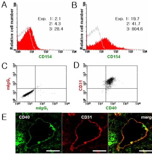
Figure 1. Platelets and endothelial cells express CD154 and CD40, respectively.
The expression levels of CD154 on freshly isolated platelets were measured with a flow cytometer before (A) and after (B) stimulation with thrombin. The numbers represent specific mean fluorescence intensity of platelets stained with FITC-labeled anti-CD154 antibody from three independent experiments.
The expression of CD40 on HUVECs was determined after dual staining with anti-CD40 and anti-CD31 (D) or control antibodies (C). The co-localization of CD40 and CD31 on tonsillar endothelial cells was determined by confocal microscopy (E). Scale bar, 50μm.
platelets expressed low levels of CD154, which increased more than 10-fold after thrombin treatment (Fig. 1A and B).
The weak expression of CD154 on freshly isolated platelets may have been induced during the isolation processes, in- dicating the vulnerability of platelets to activation (8,25). The expression of CD40 on endothelial cells was examined by du- al staining of HUVECs and frozen tonsil sections with an an- ti-CD40 antibody and an anti-CD31 antibody, an endothelial cell-specific marker. Flowcytometric and confocal analyses clearly indicate that endothelial cells express CD40 in vitro and in situ, respectively (Fig. 1C-E). These results confirm the data reported by other investigators (14,15). Since endothelial cells express CD40 and platelets express CD154, we next ex- amined whether an anti-CD40 antibody acting as a CD40 ago- nist would stimulate HUVEC proliferation. Unlike an iso- type-matched control antibody, G28-5 induced HUVEC pro- liferation in a dose-dependent manner (Fig. 2A and B).
sCD154 trimer also displayed the mitogenic effect on HUVECs
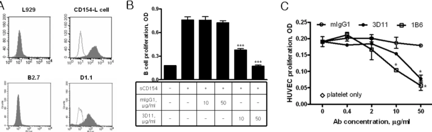
(Fig. 2C). This proliferation by sCD154 was specific to HUVECs because proliferation of fibroblast cells was not modulated by sCD154. To investigate whether CD154 on pla- telets could stimulate endothelial proliferation, we tested sev- eral commercially available reagents, such as an anti-CD154 mAb (Becton-Dickinson Pharmingen), CD40-Fc (Ancell), and CD40-COMP (Alexis). However, we failed to observe any modulating effects on sCD154- or platelet-induced HUVEC proliferation using these reagents. This failure may be due to the potent functional activity of sCD154 trimers expressed in animal cells as compared with CD154 monomers com- monly produced in bacterial system. We thus decided to de- velop a function-blocking mAb against CD154. The screening of mAbs was carried out in 4 steps. The first step was a cell-based ELISA that used CD154-L cells and L cells to meas- ure differential binding ability. The binding ability of positive hybridomas was confirmed in the second screening test using flow cytometry. The binding specificity of the selected mAbs
Figure 2. CD40-CD154 interactions contribute to the platelet-induced HUVEC proliferation. (A) A dose-response experiment was performed by culturing HUVECs with the indicated concentrations of control or anti-CD40 agonist antibodies. (B) The kinetics of anti-CD40-stimulated proliferation of HUVECs was carried out. HUVECs were cultured with control or anti-CD40 agonist antibodies (100μg/ml). (C) Human tonsillar fibroblasts or HUVECs were cultured in the presence of graded concentrations of sCD154. The degree of HUVEC proliferation was estimated by cell-counting kit-8 reagent (A, C) or viable cell counting (B) after 72 h culture. Asterisks indicate significant difference compared to controls (**p<0.01, ***p<0.001). These results were reproducible in at least three independent experiments.
Figure 3. Function-blocking anti-CD154 mAb inhibits the platelet-induced HUVEC proliferation. (A) The specificity of the anti-CD154 mAb, 3D11, was examined with indicated cell lines by flowcytometric analysis. Blank histograms represent controls stained with isotype-matched mAb. (B) The function-blocking activity of 3D11 was tested by adding to the culture of human B cells in the presence of sCD154 (1μg/ml). The degree of B cell proliferation was estimated by cell-counting kit-8 reagent after 72 h culture. (C) HUVECs (5 × 10³ cells/100μl/well) were co-cultured with platelets (2×10⁶ cells/well) in the presence of 3D11 or control antibody for 72 h. 1B6 mAb was used as a positive control for the inhibition of platelet-induced HUVEC proliferation (18). Asterisks indicate significant differences compared with controls (*p<0.05, ***p<0.001).
Representative of three reproducible experiments is shown.
was tested in the third step by using CD154-positive Jurkat clone D1.1 and CD154-negative Jurkat clone B2.7 cell lines (Fig. 3A). The selected mAbs were added to B cell cultures that were stimulated by sCD154. The new mAb 3D11 con- sistently and completely neutralized the potent activity of sCD154 (Fig. 3B). Furthermore, 3D11 significantly inhibited the platelet-induced HUVEC proliferation in a dose-dependent manner (Fig. 3C), indicating that platelet CD154 indeed con- tributes to the endothelial proliferation.
VEGF was reported to mediate the CD40-stimulated endo-
thelial proliferation (17). To investigate whether VEGF is re- sponsible for the platelet-induced HUVEC proliferation in our experimental system, we utilized a function-blocking an- ti-VEGF mAb. The mAb completely and specifically abrogated the proliferation-stimulating activity of VEGF (Fig. 4A).
However, HUVEC proliferation stimulated by either sCD154 or platelets was not affected by the presence of anti-VEGF mAb, suggesting that VEGF was not involved in these cultures. Since VEGF was added to a concentration of 5 ng/ml, it was possible that the amounts of VEGF produced
Figure 4. Platelet-induced HUVEC proliferation is independent of VEGF. (A) The effect of an anti-VEGF neutralizing mAb was examined in HUVEC cultures carried out under the indicated conditions.
HUVECs (5×10³ cells/100μl/well) were incubated with media, sCD154 (1μg/ml), platelets (2×10⁶ cells/well), or VEGF (5 ng/ml) in the presence or absence of control or anti-VEGF (5μg/ml) neutralizing antibodies. The degree of HUVEC proliferation was estimated using the cell-counting kit-8 reagent. A representative of three reproducible experiments is shown. Asterisks indicate significant differences between the indicated comparison (**p<0.01). (B) The concen- trations of VEGF released from platelets stimulated in the presence or absence of thrombin (0.2 U/ml) were measured by ELISA as described in Materials and Methods.
during the co-culture of platelets with HUVEC might have ex- ceeded the neutralizing capacity of anti-VEGF mAb. To exam- ine this possibility, the concentrations of VEGF that was re- leased from activated platelets and produced during the co-culture were measured. Less than 200 pg/ml of VEGF was released from 1×10⁸ platelets irrespective of the addition of thrombin (Fig. 4B). Considering the numbers of platelets used in the co-culture with HUVECs (8×10⁶), we propose that platelets are not releasing enough VEGF to induce HUVEC proliferation. Furthermore, the concentrations of VEGF in the conditioned media that were obtained daily in the course of platelet and HUVEC co-culture were below the
detection limit (data not shown), suggesting that plate- let-stimulated HUVECs are not producing significant amounts of VEGF. Based on these results, we conclude that platelet-in- duced HUVEC proliferation is independent of VEGF.
DISCUSSION
This study extends our recent report (18) and shows that pla- telets stimulate the proliferation of endothelial cells via direct cell-to-cell contacts and that the molecular interactions be- tween CD40 and CD154 play significant roles in the process.
The active role of platelets in stimulating endothelial pro- liferation was recognized previously (5-8). However, the mo- lecular mechanisms of this proliferation were poorly understood. Given the potent proliferative activity of CD154 on B lymphocytes, our results make sense. In support of our results, CD154 signaling has already been reported to induce proliferation of endothelial cells (12,16,17). By developing a mAb that neutralizes the activity of sCD154 trimers, we dem- onstrate that CD154 is involved in the platelet-induced pro- liferation of endothelial cells. Our observation suggests a physiological significance for the potential proangiogenic ac- tivity of platelets in vivo because the concentration of plate- lets used in our studies is 1×10⁷/ml, which is far below the physiological concentration.
Expression levels of CD154 on the platelet surface were re- markably enhanced after stimulation with thrombin. This re- sult suggests that preformed CD154 molecules stored inside of platelets are translocated to the surface in response to stim- uli such as thrombin or cultured HUVECs. In line with our results, other investigators reported the activation of resting platelets on co-culture with endothelial cells (8,25) and up-regulation of CD154 expression levels on the surface of platelets upon adhesion to endothelial cells or fibrinogen (26,27).
Although platelets produce several endothelial growth fac- tors including VEGF, it was not known whether the factors are produced in enough amounts to induce endothelial proliferation. Our data suggest that the concentrations of VEGF released during the interactions between platelets and endothelial cells are too low to exhibit the mitogenic activity.
This result is in agreement with our previous observation that direct cell-to-cell contacts are required for the platelet-stimu- lated HUVEC proliferation (18). However, we do not exclude the possibility of there being diffusible growth factors other than VEGF secreted from either cell type after the initial con-
tacts, including CD154-CD40 interactions. Our result suggest- ing that VEGF is not involved in platelet- or CD154-stimulated endothelial proliferation appears to contradict the results ob- tained by Melter et al. (17). It is difficult for us to explain the discrepancy because different reagents were used in the current study and theirs. For example, sCD154 used by Melter et al. was an undefined culture supernatant while purified sCD154 trimer was used in our experiments. In addition, neu- tralizing anti-VEGF antibodies were obtained from different companies. We are currently investigating whether plate- let-activating factor (PAF) is involved in this process. PAF is a potent phospholipid mediator of inflammation and inter- cellular communication. It has been reported to mediate CD40-dependent angiogenesis (28) and to promote pro- liferation of tumor cells and angiogenesis (29).
In conclusion, the current study provides a molecular mechanism for the platelet-induced proliferation of endothe- lial cells. Our results suggest that platelets play important roles in vascular endothelial regeneration and CD40 and CD154 may be therapeutic targets in the control of endothe- lial proliferation.
REFERENCES
1. Ware JA, Heistad DD: Platelet-endothelium interactions. N Engl J Med 328;628-635, 1993
2. Anitua E, Andia I, Sanchez M, Azofra J, del Mar Zalduendo M, de la Fuente M, Nurden P, Nurden AT: Autologous preparations rich in growth factors promote proliferation and induce VEGF and HGF production by human tendon cells in culture. J Orthop Res 23;281-286, 2005
3. Eppley BL, Woodell JE, Higgins J: Platelet quantification and growth factor analysis from platelet-rich plasma: im- plications for wound healing. Plast Reconstr Surg 114;1502- 1508, 1994
4. Frechette JP, Martineau I, Gagnon G: Platelet-rich plasmas:
growth factor content and roles in wound healing. J Dent Res 84;434-439, 2005
5. Martineau I, Lacoste E, Gagnon G: Effects of calcium and thrombin on growth factor release from platelet concen- trates: kinetics and regulation of endothelial cell proli- feration. Biomaterials 25;4489-4502, 2004
6. Falke P, Mattiasson I, Stavenow L: Effects of platelets from men with risk factors for atherosclerosis on endothelial cell proliferation and prostacyclin production in vitro. Scand.
J Clin Lab Invest 53;297-303, 1993
7. Kim HK, Song KS, Chung JH, Lee KR, Lee SN: Platelet mi- croparticles induce angiogenesis in vitro. Br J Haematol 124;376-384, 2004
8. Verheul HMW, Jorna AS, Hoekman K, Broxterman HJ, Gebbink MFBG, Pinedo HM: Vascular endothelial growth
factor-stimulated endothelial cells promote adhesion and activation of platelets. Blood 96;4216-4221, 2000 9. Banchereau J, Bazan F, Blanchard D, Briere F, Galizzi JP,
van Kooten C, Liu YJ, Rousset F, Saeland S: The CD40 anti- gen and its ligand. Annu Rev Immunol 12;881-922, 1994 10. Armitage RJ, Macduff BM, Spriggs MK, Fanslow WC:
Human B cell proliferation and Ig secretion induced by re- combinant CD40 ligand are modulated by soluble cyto- kines. J Immunol 150;3671-3680, 1993
11. Nishioka Y, Lipsky PE: The role of CD40-CD40 ligand inter- action in human T cell-B cell collaboration. J Immunol 153;1027-1036, 1994
12. Deregibus MC, Buttiglieri S, Russo S, Bussolati B, Camussi G: CD40-dependent activation of phosphatidylinositol 3-kinase/Akt pathway mediates endothelial cell survival and in vitro angiogenesis. J Biol Chem 278;18008-18014, 1997
13. Dechanet J, Grosset C, Taupin J-L, Merville P, Banchereau J, Ripoche J, Moreau JF: CD40 ligand stimulates proin- flammatory cytokine production by human endothelial cells. J Immunol 159;5640-5647, 1997
14. Henn V, Slupsky JR, Grafe M, Anagnostopoulos K, Forster R, Muller-Berghaus G, Kroczek RA: CD40 ligand on acti- vated platelets triggers an inflammatory reaction of endo- thelial cells. Nature 391;591-594, 1998
15. Karmann K, Hughes CCW, Schechner J, Fanslow WC, Pober JS: CD40 on human endothelial cells: Inducibility by cytokines and functional regulation of adhesion molecule expression. Proc Natl Acad Sci USA 92;4342-4346, 1995 16. Choudhury JA, Russell CL, Randhawa S, Young LS, Adams
DH, Afford SC: Differential induction of nuclear factor-kB and activator protein-1 activity after CD40 ligation is asso- ciated with primary human hepatocyte apoptosis or intra- hepatic endothelial cell proliferation. Mol Biol Cell 14;1334-1345, 2003
17. Melter M, Reinders MEJ, Sho M, Pal S, Geehan C, Denton MD, Mukhopadhyay D, Briscoe DM: Ligation of CD40 in- duces the expression of vascular endothelial growth factor by endothelial cells and monocytes and promotes angio- genesis in vivo. Blood 96;3801-3808, 2000
18. Ko E-M, Lee IY, Cheon IS, Kim J, Choi JS, Hwang JY, Cho JS, Lee DH, Kang D, Kim SH, Choe J: CD9 is involved in platelet-induced proliferation of human umbilical vein en- dothelial cells. Mol Cells 22;70-77, 2006
19. Lee IY, Kim J, Ko EM, Jeoung EJ, Kwon YG, Choe J:
Interleukin-4 inhibits the vascular endothelical growth fac- tor- and basic fibroblast growth factor-induced angio- genesis in vitro. Mol Cells 14;115-121, 2002
20. Lee IY, Ko EM, Kim SH, Jeoung DI, Choe J: Human fol- licular dendritic cells express prostacyclin synthase: a novel mechanism to control T cell numbers in the germinal center. J Immunol 175;1658-1664, 2005
21. Choe J, Li L, Zhang X, Gregory CD, Choi YS: Distinct role of follicular dendritic cells and T cells in the proliferation, differentiation, and apoptosis of a centroblast cell line, L3055. J Immunol 164;56-63, 2000
22. Kim HS, Zhang X, Choi YS: Activation and proliferation of follicular dendritic cell-like cells by activated T lympho-
cytes. J Immunol 153;2951-2961, 1994
23. Cho CS, Cho ML, Min SY, Kim WU, Min DJ, Lee SS, Park SH, Choe J, Kim HY: CD40 engagement on synovial fibro- blast up-regulates production of vascular endothelial growth factor. J Immunol 164;5055-5061, 2000
24. Lee IY, Choe J: Human follicular dendritic cells and fibro- blasts share the 3C8 antigen. Biochem Biophys Res Commun 304;701-707, 2003
25. Marcondes S, Lafay M, Brohard-Bohn B, de Nucci G, Rendu F: Platelets induce human umbilical vein endothelial cell proliferation through P-selectin. Life Sci 66;1817-1826, 2000 26. Danese S, Katz JA, Saibeni S, Papa A, Gasbarrini A, Vecchi
M, Fiocchi C: Activated platelets are the source of elevated levels of soluble CD40 ligand in the circulation of in- flammatory bowel disease patients. Gut 52;1435-1441, 2003
27. May AE, Kalsch T, Massberg S, Herouy Y, Schmidt R, Gawaz M: Engagement of glycoprotein IIb/IIIa (αIIbβ3) on platelets upregulates CD40L and triggers CD40L-dependent matrix degradation by endothelial cells. Circulation 106;
2111-2117, 2002
28. Russo S, Bussolati B, Deambrosis I, Mariano F, Camussi G:
Platelet-activating factor mediates CD40-dependent angio- genesis and endothelial-smooth muscle cell interaction. J Immunol 171;5489-5497, 2003
29. Bussolati B, Biancone L, Cassoni P, Russo S, Rola-Plesz- czynski M, Montrucchio G, Camussi G: PAF produced by human breast cancer cells promotes migration and pro- liferation of tumor cells and neo-angiogenesis. Am J Pathol 157;1713-1725, 2000