179
Immune Network
Effects of IL-12N220L-expressing Adenovirus by Co-delivery of DOTAP
Je-In Youn1, Hyun-Tak Jin2 and Young-Chul Sung1
1Division of Molecular and Life Science, Pohang University of Science and Technology, 2Research Institute, Genexine Co. Ltd., Pohang, Korea
ABSTRACT
Background: Adenovirus (Ad) vectors have been widely used for many gene therapy applications because of their high transduction ability and broad tropism. However, their utility for cancer gene therapy is limited by their poor transduction into cancer cells lacking the primary receptor, coxsackievirus and adenovirus receptor (CAR). Methods:
To achieve CAR-independent gene transfer via Ad, we pretreated Ad with 1,2-dioleoyl-3- trimethylammonium propane (DOTAP) and analyzed their transduction efficiency into cancer cells in vitro and in vivo comparing with the virus alone. Results: Treatment of DOTAP significantly increased adenoviral gene transfer in tumor cells in vitro. Moreover, DOTAP at an optimum dose (10 μg/ml) enhanced IL-12 transgene expression by fivefold in tumor, and twofold in serum after intratumoral injection of adenovirus expressing IL-12N220L (Ad/IL-12N220L). In addition, cotreatment of DOTAP decreased tumor growth rate in the Ad/IL-12N220L-transduced tumor model, finally leading to enhanced survival rate. Conclusion: Our results strongly suggest that DOTAP could be of great utility for improving adenovirus-mediated cancer gene therapy. (Immune Network 2007;7(4):179-185)
Key Words: Adenovirus, DOTAP, liposome, tumor
Correspondence to: Young-Chul Sung, Division of Molecular and Life Science, Pohang University of Science and Technology, San 31, Hyoja- dong, Pohang 790-784, Korea (Tel) 82-54-279-2294, (Fax) 82-54-279- 5544, (E-mail) [email protected]
This research was supported both by Generic Technology Develop- ment Program, Minister of Commerce, Industry and Energy (10020817) and by Technology Innovation Development Program, SMEs (Small and Medium enterprises) (S0703222-C1449600-10000011).
Introduction
Adenovirus vectors (Ads) are widely used in many gene therapy applications because of their several pro- minent advantages over other vectors. Ad can produce high levels of transgene expression compared with oth- er established gene transfer methods, including retro- viruses and cationic lipids. In addition, Ad is capable of efficiently delivering gene into various cell types (either dividing or non-dividing cells). Adenoviral DNA is not integrated into the host genome, thereby result- ing in a low risk of insertional mutagenesis. Further- more, Ad can accommodate the large-size transgene and
is easy to manipulate by classical recombinant DNA techniques. Finally, the production of high titers of Ad, which is necessary for clinical trials, is well estab- lished (1,2).
Ads have been commonly used to transfer tumor suppressor genes, suicide genes, anti-angiogenic fac- tors, prodrug activating genes and immunostimulatory genes for cancer gene therapy (3,4). However, the util- ity of Ad is limited by their low transduction efficiency in certain types of cancer cells due to the low ex- pression level of the primary adenovirus receptor, cox- sackievirus and adenovirus receptor (CAR) (5). Althou- gh CAR is expressed ubiquitously on most normal epi- thelial tissues, its lack or down-regulation has been re- ported in various tumor types (6-8).
There have been many studies designed to improve Ad-mediated gene therapy in cells expressing a low level of CAR. One of them is the modification of Ad through the attachment of ligand for cellular receptors and the incorporation of chimeric envelope glycoprotein.
Moreover, biochemical reagents such as polybrene, lip- ofectamine, protamine sulfate and DOTAP have been used to facilitate the entry of virus particles into the target cells. Complexing adenovirus with polycations has been shown to be effective in increasing Ad-medi- ated gene transfer (9-13). In vitro studies have demon- strated that polycations can efficiently increase Ad de- livery into various tumor cells (14-17). However, in vivo studies showing same dramatic effects as in vitro re- sults are lacking (14,15,18,19). Therefore, the low effi- ciency of polycation-mediated Ad delivery in vivo re- mains to be a major obstacle to achieving its successful application for cancer gene therapy.
DOTAP which is widely known as transfection lipid consists of a monocationic trimethylammonium head group and two unsaturated hydrocarbon chains. Previous studies revealed that DOTAP can efficiently transduce plasmid DNA to various cells in vitro and improve im- mune response and protective immunity by co-immu- nization with DNA or recombinant tumor-associated antigen (20-22). Additionally, it was reported that DOTAP can improve transduction efficiency of Ad in several tumor cells in vitro and enhance pulmonary gene transfer in vivo (15,23). However, its therapeutic application usage, especially in cancer gene therapy, has not been well studied.
In this study, the effect of DOTAP on adenoviral transduction in tumor cells is investigated using Ad expressing IL-12N220L (Ad/IL-12N220L) which was known to have potent antitumor effects (24). Our stu- dy’s results show that the combined use of DOTAP and Ad greatly improved transduction in mouse and hu- man melanoma which are resistant to adenoviral infec- tion. Furthermore, DOTAP improved the therapeutic antitumor effect of Ad/IL-12N220L in mouse melano- ma model. These results suggest that DOTAP can be a promising strategy for improving Ad-based cancer gene therapy.
Materials and Methods
Mice. Six- to eight-week old C57BL6 mice were pur- chased from Charles River Breeding Laboratories (Shi- zuoka, Japan). The mice were housed in a specific patho- gen-free environment in an internally approved viva- rium at the institute.
Cell lines. B16F10, the mouse melanoma cell line, A375, the human melanoma cell line were purchased from
American Type culture Collection (Manassas, VA).
These were maintained in DMEM supplemented with 10% fetal bovine serum (Hyclone, Logan, UT) and 1% w/v each of penicillin/streptomycin (Life Technolo- gies, Inc., Grand Island, NY) per 100 ml.
Cationic liposome. 1,2-dioleoyl-3-trimethylammonium pro- pane (DOTAP) was purchased from Roche (Mannheim, Germany).
Construction of replication-defective adenoviral vectors. Re- combinant replication-defective adenoviruses (Ads) were generated according to the AdEasyTM Vector System (Q Biogene) and Ad/IL-12N220L was constructed as described earlier (24). Briefly, the cDNAs of murine IL-12N220L and EGFP were subcloned into the ad- enoviral shuttle vector, pShuttleCMV. After recombi- nation with the adenoviral backbone vector, pAdEasy in Escherichia coli BJ5183, the recombinant adenoviruses were generated and expanded in 293 cells.
In vitro adenovirus transduction. Cells were seeded into 48-well plates at 3×10⁴ cells/well and were incubated overnight at 37oC. Adenovirus was diluted in DMEM without serum to achieve a 2× virus dilution. DOTAP was similarly diluted into DMEM without serum to yield 2× dilutions. Adenovirus and DOTAP mixtures were combined at a 1:1 ratio and were allowed to incubate for 30 min at room temperature. Cell mono- layers were washed with PBS and overlaid with 150 μl of virus/DOTAP mixture. After 2 h incubation in a CO2 incubator at 37oC, the cells were washed with PBS to remove the complexes, and 300 μl fresh se- rum-containing medium was added. The cells were then incubated for an additional 46 h before assessing GFP expression. Gene transduction efficiency was as- sessed by flow cytometry on a FACScalibur using Cell- Quest software (Becton Dickinson, Tokyo, Japan), ac- quiring 10,000 events by forward and side scatter gat- ing to exclude cell debris.
In vivo adenovirus transduction. A dose of 5×10⁵ B16F10 cells in 100 μl of PBS was subcutaneously injected into the right hind flank of C57BL/6 syngenic mice.
After palpable tumor formation (a mean diameter of 7 mm), the animals were intratumorally injected with 1×10⁸ PFU of Ad/IL-12N220L pre-incubated with 50 μl PBS or 5, 10, 20 μg/ml of DOTAP in equal vol- ume of PBS. The serum samples were taken 24 h and 48 h after viral injections. At 48 h after viral injection, the mice were sacrificed, and the tumors of sacrificed
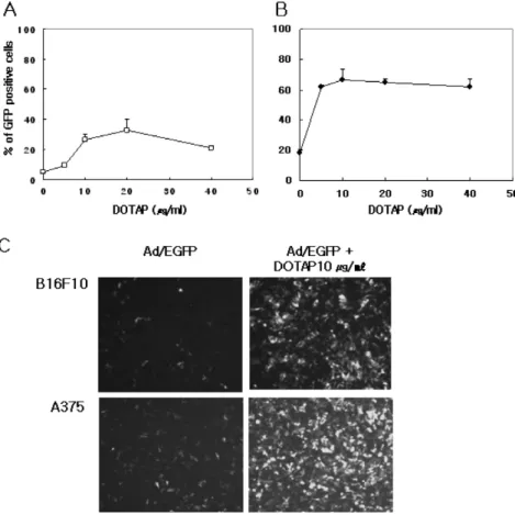
Figure 1. Enhanced adenovirus trans- duction of mouse (B16F10) and hu- man (A375) melanoma cells using DOTAP. Ad/EGFP was admixed with indicated amounts of DOTAP.
B16F10 (A) and A375 (B) were res- pectively transduced at an MOI of 200, 10 for 2 h, incubated for an additional 46 h, and the percentage of GFP expressing cells was deter- mined by FACS analysis. Represen- tative samples (C) were visualized microscopically for GFP under stan- dard excitation/emission parameters.
Each data point represents the average of triplicate wells±SEM.
animals were disaggregated in PBS with a tissue ho- mogenizer for 1 min on ice. After centrifugation, the supernatant was harvested. IL-12 concentration in the tumor and serum samples was detected by IL-12p70 ELISA kits (R&D System, Minneapolis, MN) accord- ing to the manufacturer’s instructions. The results are plotted as mean±SEM of three animals/data point.
Antitumor effects in the in vivo tumor model. B16F10 cells were seeded at an initial density of 2×10⁶ in a 100 mm dish and were incubated overnight at 37oC. The cells were infected with adenovirus at a multiplicity of in- fection (MOI) of 100 in 4 ml serum-free medium at 37°C for 2 h. The culture medium was replaced, and the cells were additionally incubated for 4 h. Infected cells were trypsinized, washed, and injected into C57BL/6 mice subcutaneously. At 24 h after tumor injection, the serum samples were taken and the expression level of IL-12 in the serum was measured by IL-12p70 ELISA kits. Tumor size was measured at least twice in a week with a digital caliper for two-dimensional longest axis (L in mm) and shortest axis (W in mm), and tumor volume was calculated using the following formula:
volume in mm3=(L×W2)/2. Mice bearing tumors that
exceeded 15 mm in two perpendicular diameters or 20 mm in one diameter were sacrificed for ethical rea- sons according to institutional guidelines.
Statistical analysis. We used student’s t-test to measure statistical difference between groups. For all cases, di- fferences were considered significant when the p values were <0.05.
Results
DOTAP increased the transduction efficiency of adenovirus into tumor cells. To investigate the effect of DOTAP on the efficiency of adenovirus-mediated gene ex- pression, mouse (B16F10) and human (A375) melano- ma cells were infected with Ad/EGFP pre-incubated with DOTAP at different concentrations. When the expression level of GFP was analyzed by flow cy- tometry at 48 hr after infection, DOTAP augmented GFP expression by facilitating the Ad transduction in a dose-dependent manner (Fig. 1). In particular, trans- gene expression was increased up to by 5 (B16F10), 3.5 (A375)-fold at 10 μg/ml of DOTAP compared to the virus alone. At optimal concentrations of DOTAP, we did not observe any cytotoxicity under the light
Figure 2. Effect of adenovirus dose on DOTAP-mediated adeno- virus transduction. Transductions were performed at various doses of Ad/EGFP (10, 50, 100, 250, 500 MOI) complexed with DOTAP (10 μg/ml) and compared with parallel infections with Ad/EGFP alone. At 48 h the percentage of GFP expressing cells was determined by FACS analysis. Each data point represents the average of triplicate wells±SEM.
Figure 3. In vivo IL-12 levels in serum (A) and tumor (B) fol- lowing intratumoral injection of Ad/IL-12N220L with DOTAP.
Recombinant adenovirus encoding IL-12N220L (3×10⁸ pfu) with or without DOTAP was injected into B16F10 established tumors.
The serum samples were taken 24 and 48 hr after viral injec- tions. Then 48 h after viral injection, the mice were sacrificed and the tumor samples were taken. The results are expressed relative to values with infection with adenovirus alone, and are presented as mean±SEM of three mice.
microscopy. Based on the optimal dose DOTAP (10 μg /ml), the transduction efficiency of Ad with DOTAP was assessed at various doses of Ad (MOI of 10-500).
As shown in Fig. 2, DOTAP allowed higher levels of transduction at all doses of Ad tested. These results indicate that the cotreatment of Ad with DOTAP can markedly enhance the transduction efficiency of Ad in- to melanoma without notable cytotoxicity.
DOTAP augmented adenovirus transduction into tumors fol- lowing intratumoral injection. The effect of DOTAP on in vivo gene transfer of Ad in tumors was evaluated using s.c. B16F10 tumors in C57BL/6 mice. Mice bea- ring tumors were intratumorally injected with 3×10⁸ pfu Ad/IL-12N220L pre-incubated with or without 5, 10, 20 μg/ml of DOTAP, respectively. When IL-12 levels in serum and tumor were determined by IL-12 p70 ELISA, DOTAP enhanced in vivo IL-12 expre- ssion in a dose-dependent manner (Fig. 3). Based on this in vitro result, the optimum dose of DOTAP was established at 10 μg/ml. At this concentration DOTAP improved IL-12 expression by twofold in serum at 24 hr after viral injection, and fivefold in tumor at 48 hr.
Collectively, these results demonstrated that DOTAP could augment the transduction efficacy of adenoviral vector in B16F10 melanoma both in vitro and in vivo.
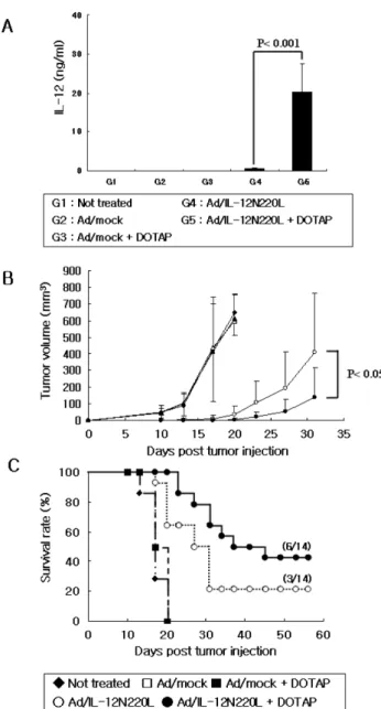
Enhanced antitumor effect of Ad/IL-12N220L by cotreatment of DOTAP in the mouse B16F10 tumor model. We pre- viously found that Ad encoding IL-12N220L (Ad/IL- 12N220L), which selectively decreases the secretion of p40 as a natural antagonist of IL-12, showed potent antitumor effects even when compared to that of enco- ding native IL-12 (24). To test the effect of DOTAP on Ad/IL-12N220L-mediated antitumor activity, B16F10 cells were infected by rAd/IL-12N220L in vitro with or without DOTAP and then were subcutaneously in- jected into syngenic C57BL/6 mice. When the level of IL-12 in serum was determined by ELISA at 24 hrs after tumor injection, the expression level of this transgene increased 40-fold by DOTAP, which is high- er than even that by adenovirus alone (Fig. 4A). In addition, mice bearing B16F10 transduced with Ad/IL- 12N220L + DOTAP showed the further retardation of tumor growth at a great degree (Fig. 4B, p<0.05) and significantly increased the survival rate compared to those with only Ad/IL-12N220L (42.8% versus 21.4%, Fig. 4C). Taken together, cotreatment with DOTAP increased the transduction efficiency of Ad in tumors
Figure 4. The enhanced antitumor effects by cotreatment of DOTAP. B16F10 cells were infected with adenovirus with or without DOTAP 10 μg/ml at an MOI of 100 for 6 hr in vitro.
The infected cells were trypsinized, washed, and injected into C57BL/6 mice subcutaneously. At 24 h after tumor injection, the serum samples were taken and the expression level of IL-12 in the serum (A) was measured by ELISA. The tumor volume (B) and survival rate (C) were determined at least twice in a week. The results are presented as mean±SEM of 6∼8 mice, while the p values was determined by students’s t-test (two-tailed).
and the higher production of therapeutic molecule (IL-12N220L), leading to the enhanced antitumor ef- fects of Ad-transduced tumor vaccine.
Discussion
Loss of CAR expression is frequently observed in various cancer cell lines and clinical cancer specimens
(7,25,26), hampering efforts to perform efficient ad- enoviral gene therapy in cancer patients. It was re- ported that several polycationic liposomes could in- crease Ad-mediated gene delivery into the cells lacking CAR (10-19). In this paper, we tested one of poly- cation liposomes, DOTAP in adenovirus delivery into mouse and human melanoma cell lines resistant to Ad infection. We found that the transduction efficiency of Ad pretreated with DOTAP was three to fivefolds higher than that of Ad only (Fig. 1). In addition, DOTAP significantly enhanced the therapeutic gene (IL-12) expression in both tumor and serum after in- tratumoral injection of adenovirus encoding IL-12N220L (rAd/IL-12N220L) (Fig. 3). It was reported that high dose of Ad could cause adverse effects such as liver injury and induce humoral immune response against Ad which interferes with repeated Ad administration (1,27). Based on our study’s results, DOTAP is ex- pected to reduce toxicity and vector immunity in Ad- based cancer gene therapy by enhancing the trans- duction efficiency and lowering the titer of Ad.
The mechanism for this augmentation is not known.
However, one of the possible explanations for this ob- served phenomenon is that polycationic compounds such as DOTAP serve as electrostatic bridges between the virus and the target cells. Mammalian cells possess sig- nificant negative surface charge due to glycosylated phospholipids on cell membrane (28,29), and mamma- lian viruses also exhibit a net negative charge at a phy- siologic pH (30). It is therefore possible that the added polycationic compounds during virus adsorption dimin- ish electrostatic repulsion between negatively charged mammalian cells and eukaryotic viruses.
Given these in vitro and in vivo results, we hypothe- sized that DOTAP, in conjunction with the adenovirus vector, would improve the therapeutic efficacy of IL-12 gene-based cancer therapy. In this study, we used ge- netically engineered IL-12N220L which is superior to IL-12 in cancer immunotherapy as proven previously (24). To test whether the increased IL-12 expression by DOTAP could elicit enhanced antitumor effect in vivo, we used the in vitro engineered tumor model with Ad. Compared to the mice injected with Ad/IL-12N220L- infected cells, those with Ad/IL-12N220L + DOTAP infected cells showed higher IL-12 expression in se- rum, and further retarded tumor growth rate followed by higher survival rate (Fig. 4). When we tested the
therapeutic effects of Ad/IL-12N220L with DOTAP following intratumoral injection in the established tu- mor model, we also found that the IL-12 level in se- rum 24 hr after Ad injection was two to fourfold high- er in the DOTAP-treated groups compared with only the virus-treated group. However, Ad/IL-12N220L both with and without DOTAP dramatically retarded the tumor progress, leading to no significant difference in the aspects of tumor progression and survival rate in both groups (data not shown). This differential ef- fect of DOTAP on serum IL-12 level and tumor pro- gression may have resulted from the threshold of IL-12 doses for its biological activity in vivo. Even though we could not see the therapeutic advantages of DOTAP cotreatment with Ad/IL-12N220L in our experimental setting, it is notable that DOTAP may significantly reduce the Ad dose requiring same therapeutic effects and it will be also beneficial for decreasing the produc- tion of neutralizing antibody against Ad.
In summary, this study provides evidence that adeno- virus transduction efficacy can be improved in vitro and in vivo by the simple addition of DOTAP. DOTAP also enhanced Ad-mediated therapeutic gene (Ad/IL- 12N220L) expression and resulted in a dramatic anti- tumor effect. Therefore, DOTAP could be a powerful tool for enhancing the Ad-mediated therapeutic effi- cacy of Ad/IL-12N220L in cancer gene therapy. Further- more, DOTAP may be utilized in other cancer gene therapies such as cytokines, chemokines, and suicide genes, especially in ex vivo transduced tumor cell-based cancer therapy.
Acknowledgments
The authors thank Sang-Chun Lee and Kwan-Suk Lee for their devoted animal care. We also thank the members of the Cellular Immunology Laboratory for their insightful suggestions and technical support.
References
1. Benihoud K, Yeh P, Perricaudet M: Adenovirus vectors for gene delivery. Curr Opin Biotechnol 10;440-447, 1999 2. St George JA: Gene therapy progress and prospects: adenovi-
ral vectors. Gene Ther 10;1135-1141, 2003
3. Seth P: Vector-mediated cancer gene therapy: an overview.
Cancer Biol Ther 4;512-517, 2005
4. Yang ZR, Wang HF, Zhao J, Peng YY, Wang J, Guinn BA, Huang LQ: Recent developments in the use of adenoviru- ses and immunotoxins in cancer gene therapy. Cancer Gene Ther 14;599-615, 2007
5. Bergelson JM, Cunningham JA, Droguett G, Kurt-Jones EA,
Krithivas A, Hong JS, Horwitz MS, Crowell RL, Finberg RW:
Isolation of a common receptor for Coxsackie B viruses and adenoviruses 2 and 5. Science 275;1320-1323, 1997 6. Miller CR, Buchsbaum DJ, Reynolds PN, Douglas JT, Gillespie
GY, Mayo MS, Raben D, Curiel DT: Differential suscepti- bility of primary and established human glioma cells to ad- enovirus infection: targeting via the epidermal growth factor receptor achieves fiber receptor-independent gene transfer.
Cancer Res 58;5738-5748, 1998
7. Li Y, Pong RC, Bergelson JM, Hall MC, Sagalowsky AI, Tseng CP, Wang Z, Hsieh JT: Loss of adenoviral receptor expression in human bladder cancer cells: a potential impact on the effi- cacy of gene therapy. Cancer Res 59;325-330, 1999 8. Cripe TP, Dunphy EJ, Holub AD, Saini A, Vasi NH, Mahller
YY, Collins MH, Snyder JD, Krasnykh V, Curiel DT, Wick- ham TJ, DeGregori J, Bergelson JM, Currier MA: Fiber knob modifications overcome low, heterogeneous expression of the coxsackievirus-adenovirus receptor that limits adenovirus gene transfer and oncolysis for human rhabdomyosarcoma cells.
Cancer Res 61;2953-2960, 2001
9. Persson R, Wohlfart C, Svensson U, Everitt E: Virus-receptor interaction in the adenovirus system: characterization of the positive cooperative binding of virions on HeLa cells. J Virol 54;92-97, 1985
10. Byk T, Haddada H, Vainchenker W, Louache F: Lipofectamine and related cationic lipids strongly improve adenoviral infec- tion efficiency of primitive human hematopoietic cells. Hum Gene Ther 9;2493-2502, 1998
11. Lee SG, Yoon SJ, Kim CD, Kim K, Lim DS, Yeom YI, Sung MW, Heo DS, Kim NK: Enhancement of adenoviral trans- duction with polycationic liposomes in vivo. Cancer Gene Ther 7;1329-1335, 2000
12. Fasbender A, Zabner J, Chillon M, Moninger TO, Puga AP, Davidson BL, Welsh MJ: Complexes of adenovirus with poly- cationic polymers and cationic lipids increase the efficiency of gene transfer in vitro and in vivo. J Biol Chem 272;6479- 6489, 1997
13. Toyoda K, Nakane H, Heistad DD: Cationic polymer and lipids augment adenovirus-mediated gene transfer to cerebral arteries in vivo. J Cereb Blood Flow Metab 21;1125-1131, 2001
14. Lanuti M, Kouri CE, Force S, Chang M, Amin K, Xu K, Blair I, Kaiser L, Albelda S: Use of protamine to augment adenovirus-mediated cancer gene therapy. Gene Ther 6;1600- 1610, 1999
15. Clark PR, Stopeck AT, Brailey JL, Wang Q, McArthur J, Finer MH, Hersh EM: Polycations and cationic lipids enhance adenovirus transduction and transgene expression in tumor cells. Cancer Gene Ther 6;437-446, 1999
16. Lee EM, Hong SH, Lee YJ, Kang YH, Choi KC, Choi SH, Kim IH, Lim SJ: Liposome-complexed adenoviral gene trans- fer in cancer cells expressing various levels of coxsackievirus and adenovirus receptor. J Cancer Res Clin Oncol 130;169-177, 2004
17. Dodds E, Piper TA, Murphy SJ, Dickson G: Cationic lipids and polymers are able to enhance adenoviral infection of cul- tured mouse myotubes. J Neurochem 72;2105-2112, 1999 18. Fukuhara H, Hayashi Y, Yamamoto N, Fukui T, Nishikawa
M, Mitsudo K, Tohnai I, Ueda M, Mizuno M, Yoshida J:
Improvement of transduction efficiency of recombinant ad- enovirus vector conjugated with cationic liposome for human oral squamous cell carcinoma cell lines. Oral Oncol 39;601- 609, 2003
19. Qiu C, De Young MB, Finn A, Dichek DA: Cationic lip-
osomes enhance adenovirus entry via a pathway independent of the fiber receptor and alpha(v)-integrins. Hum Gene Ther 9;507-520, 1998
20. Bei R, Guptill V, Masuelli L, Kashmiri SV, Muraro R, Frati L, Schlom J, Kantor J: The use of a cationic liposome for- mulation (DOTAP) mixed with a recombinant tumor-associa- ted antigen to induce immune responses and protective im- munity in mice. J Immunother (1997) 21;159-169, 1998 21. Crook K, Stevenson BJ, Dubouchet M, Porteous DJ: Inclu-
sion of cholesterol in DOTAP transfection complexes increases the delivery of DNA to cells in vitro in the presence of serum.
Gene Ther 5;137-143, 1998
22. Lardans V, Boulo V, Duclermortier P, Serra E, Mialhe E, Cap- ron A, Dissous C: DNA transfer in a Biomphalaria glabrata embryonic cell line by DOTAP lipofection. Parasitol Res 82;
574-576, 1996
23. Yotnda P, Chen DH, Chiu W, Piedra PA, Davis A, Templeton NS, Brenner MK: Bilamellar cationic liposomes protect ad- enovectors from preexisting humoral immune responses. Mol Ther 5;233-241, 2002
24. Ha SJ, Chang J, Song MK, Suh YS, Jin HT, Lee CH, Nam GH, Choi G, Choi KY, Lee SH, Kim WB, Sung YC: Engi- neering N-glycosylation mutations in IL-12 enhances sustained cytotoxic T lymphocyte responses for DNA immunization.
Nat Biotechnol 20;381-386, 2002
25. Jee YS, Lee SG, Lee JC, Kim MJ, Lee JJ, Kim DY, Park SW, Sung MW, Heo DS: Reduced expression of coxsack- ievirus and adenovirus receptor (CAR) in tumor tissue com- pared to normal epithelium in head and neck squamous cell carcinoma patients. Anticancer Res 22;2629-2634, 2002 26. Kim M, Zinn KR, Barnett BG, Sumerel LA, Krasnykh V,
Curiel DT, Douglas JT: The therapeutic efficacy of adenoviral vectors for cancer gene therapy is limited by a low level of primary adenovirus receptors on tumour cells. Eur J Cancer 38;1917-1926, 2002
27. Mack CA, Song WR, Carpenter H, Wickham TJ, Kovesdi I, Harvey BG, Magovern CJ, Isom OW, Rosengart T, Falck- Pedersen E, Hackett NR, Crystal RG, Mastrangeli A: Circum- vention of anti-adenovirus neutralizing immunity by admin- istration of an adenoviral vector of an alternate serotype. Hum Gene Ther 8;99-109, 1997
28. Loewi G, Meyer K: The acid mucopolysaccharides of embry- onic skin. Biochim Biophys Acta 27;453-456, 1958.
29. Yolken RH, Willoughby R, Wee SB, Miskuff R, Vonderfecht S:
Sialic acid glycoproteins inhibit in vitro and in vivo replication of rotaviruses. J Clin Invest 79;148-154, 1987
30. Wilcox WC, Ginsberg HS: Purification and immunological characterization of types 4 and 5 adenovirus-soluble antigens.
Proc Natl Acad Sci U S A 47;512-526, 1961