pISSN 1598-9992 eISSN 2233-6869
CASE REPORT
내시경적 점막 절제술로 치료한 동시성 대장암이 동반된 거치상 폴립증 증후군
이상훈, 이성준, 박성철, 남승주, 강명호, 김태석, 이승구1
강원대학교 의학전문대학원 내과학교실, 해부병리학교실1
Serrated Polyposis Syndrome with a Synchronous Colon Adenocarcinoma Treated by an Endoscopic Mucosal Resection
Sang Hoon Lee, Sung Joon Lee, Sung Chul Park, Seung-Joo Nam, Myeong Ho Kang, Tae Suk Kim and Seung Koo Lee1 Departments of Internal Medicine, Anatomic Pathology1, Kangwon National University School of Medicine, Chuncheon, Korea
Serrated polyposis syndrome (SPS) can transform to malignant lesions through the sessile serrated pathway and traditional ser- rated pathway. These pathways may cause rapid neoplastic progression compared to the adenoma-carcinoma sequence, which may cause interval colorectal cancer. The authors experienced a case of SPS with a synchronous colon adenocarcinoma that was treated with an endoscopic mucosal resection. In pathology reviews, other parts of the adenocarcinoma showed sessile serrated adenoma. Therefore, patients with SPS have a potential for malignant transformation, highlighting the need for strict colonoscopy surveillance starting at the time of SPS diagnosis. (Korean J Gastroenterol 2020;76:159-163)
Key Words: Intestinal polyposis; Adenocarcinoma; Endoscopic mucosal resection
Received May 8, 2020. Revised July 4, 2020. Accepted July 6, 2020.
CC This is an open access article distributed under the terms of the Creative Commons Attribution Non-Commercial License (http://creativecommons.org/licenses/
by-nc/4.0) which permits unrestricted non-commercial use, distribution, and reproduction in any medium, provided the original work is properly cited.
Copyright © 2020. Korean Society of Gastroenterology.
교신저자: 이성준, 춘천시 백령로 156, 강원대학교병원 소화기내과
Correspondence to: Sung Joon Lee, Division of Gastroenterology & Hepatology, Department of Internal Medicine, Kangwon National University Hospital, Baengnyeong-ro 156, Chuncheon 24289, Korea. Tel: +82-33-258-2405, Fax: +82-33-258-2146, E-mail: [email protected], ORCID: https://orcid.org/0000-0002-6451-0400 Financial support: None. Conflict of interest: None.
INTRODUCTION
Serrated polyposis syndrome (SPS), also known as hyper- plastic syndrome, is a rare disease that is characterized by multiple serrated or hyperplastic polyps (HPs), mainly in the proximal colon. The World Health Organization clinical diag- nostic criteria for SPS are as follows: 1) >5 serrated le- sions/polyps proximal to the rectum, all being >5 mm in size, with >2 being 10 mm in size; 2) >20 serrated lesions/polyps of any size distributed throughout the large bowel, with >5 being proximal to the rectum.1
HPs were traditionally included in the non-neoplastic
category. Unlike patients with sporadic small and distal HPs, patients with SPS have an increased risk of color- ectal cancer. Edelstein et al.2 reported that the standard incidence ratio for colorectal cancer was 18.72 in patients with serrated polyposis. The reasons for the increased in- cidence of colorectal cancer are as follows. A diagnosis of SPS is often missed because flat lesions are difficult to recognize. Another problem with SPS is that there are so many polyps; the physicians cannot remove them all in an endoscopic procedure. This paper reports a case of SPS with a synchronous colon adenocarcinoma treated with an endoscopic mucosal resection (EMR).
A B
Fig. 1. (A) Colonoscopic finding showing a polyp in the sigmoid colon identified as well-differentiated adenocarcinoma. (B) Histology examination showing focal high-grade dysplasia (black arrow), suggesting well-differentiated adenocarcinoma and epithelial serration with extension to the crypt bases (blue arrow), suggestive of a sessile serrated adenoma (H&E, ×100).
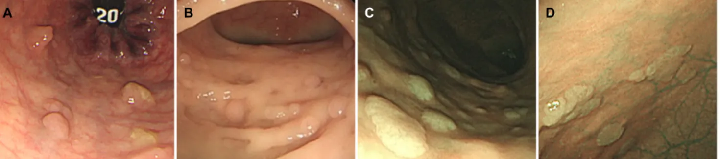
A B C D
Fig. 2. Colonoscopic findings showing multiple polyps with variable sizes in the (A) rectum, (B) sigmoid colon, (C, D) descending colon.
A B
Fig. 3. (A) Colonoscopy finding revealed a 1.4 cm sized polyp in the sigmoid colon. (B) Histologic features of the lesion revealed elongated glands and crypts with serration in the upper- third of the mucosa (H&E, 100).
CASE REPORT
A 61-year-old man visited the Kangwon National University Hospital for a follow-up colonoscopy. He had received an en- doscopic resection for a polyp in the sigmoid colon in a private
clinic one month earlier. The polyp was identified as a well-dif- ferentiated intramucosal adenocarcinoma (pTisNx) (Fig. 1).
The cancer lesion was 1.2 cm in size with a polypoid (Ip Paris classification) type and was located at the proximal sigmoid with lamina propria invasion. No lymphovascular or perineural
Table 1. Summary of Histologic Findings and Treatment in Three Patients Reported as Serrated Adenoma with Adenocarcinoma in the Previous Reports
Study Sex/age Histology Treatment
Yamauchi et al. (2002)3 F/76 Serrated adenoma/Adenocarcinoma Transverse colectomy
Pyleris et al. (2014)4 M/58 Serrated adenoma/Adenocarcinoma Total colectomy
Chino et al. (2016)5 F/77 Serrated adenoma/Adenocarcinoma Surgery
invasion was observed. Other parts of the polyp showed ser- rated adenoma on a pathology review. Many polyps were not- ed at other sites, but they were not removed.
The patient had a 20-year history of diabetes mellitus. He did not complain of any gastrointestinal symptoms and had no family history of colorectal cancer or other malignancies.
He had a 40 pack-year smoking history and social drinking history (one bottle×15 days/month for 40 years).
The physical examination showed that the abdomen was soft and flat, and the bowel sounds were normo-active. No tenderness or rebound tenderness was noted. The chest and abdominal X-ray results were normal. The complete blood count profile showed 7,100 white blood cells/mcL, hemoglobin level of 13.7 g/mcL, and a platelet count of 272 K/mcL. The other laboratory findings were also within the normal ranges, except for his fasting serum glucose level was elevated (135 mg/dL).
Carcinoembryonic antigen and other tumor markers were not checked. No specific findings were observed in abdominal CT.
A colonoscopy performed in the Kangwon National University Hospital revealed multiple polyps (more than 40) of variable sizes throughout the entire colon (Fig. 2). They were distributed mainly from the descending colon to the rectum. Thirteen pol- yps, 1.0 to 2.0 cm in size, were removed by EMR. Histologically, one polyp in the descending colon was a tubular adenoma, while most of them were confirmed histologically as HPs. Some were identified as serrated adenoma (Fig. 3). Two of the HPs might have been sessile serrated polyps. On the other hand, the L- or T-shaped parts of the crypts were too small to be diagnosed as serrated polyps in the histology examination.
DISCUSSION
Although multiple sporadic HPs resemble multiple ad- enomatous polyps endoscopically, they do not show any associ- ation with the development of adenocarcinoma. On the other hand, the progression of mixed hyperplastic adenomatous pol- yps and sessile serrated adenomas (SSA) to adenocarcinoma
has been reported. In particular, SPS showed a high risk of developing adenocarcinoma (Table 1).3-5 The pathological find- ings for typical HP have serrations in the upper portion of the crypts with otherwise normal architectures and a normal proliferative zone at the base of the crypt. SSA have serrations at the base of the crypt with branching and dilation. Traditional serrated adenomas (TSAs) have serrated crypts with cytologic dysplasia.6 SSA and TSAs have additional pathological findings, such as the presence of horizontally oriented crypts, large areas without endocrine cells, focal mucus overproduction, and frequent (focal or diffuse) eosinophilia of the cytoplasm.7 In the present case, the patient had more than 50 polyps in the entire colon distributed mainly from the descending colon to the rectum. Most of them were confirmed histologically as HPs, and some were identified as serrated adenoma. The patient was diagnosed with SPS based on the World Health Organization criteria (II) mentioned above.
In the epidemiology of patients with SPS, there is no sex predominance. The mean age at the time of diagnosis is 55 years, and 10% to 50% of the SPS patients have a family history of colorectal cancer.8 Smoking and obesity have been identified as potential risk factors of SPS.9
The pathogenesis of CRC in SPS involves the serrated path- way instead of the adenoma-carcinoma sequence. The ser- rated pathway is divided into the sessile serrated pathway and the traditional serrated pathway. This might be associated partially with the presence of conventional adenoma. A recent genetic study on the origins of CRC arising in SPS patients showed that these tumors have diverse molecular profiles.10 SSA are characterized by a high rate of BRAF mutations. They are likely to be precursor lesions of most CRCs evolving through the serrated neoplastic pathway in SPS.11 The sessile serrated pathway develops SSA into dysplasia or microsatellite in- stability/stability cancer through a BRAF mutation, CpG island methylator phenotype, and MLH-1 promoter hypermethylation.
The traditional serrated pathway develops TSA with high-grade dysplasia and MSS cancer through a pathway related to
KRAS/BRAF mutations.12 In the present case, the endoscopic finding was a polypoid lesion-like adenoma. A mucosal pit pattern was not identified. Molecular genetics assays were not performed. On the other hand, this focal adenocarcinoma might have developed through the serrated pathway based on the histological findings.
Endoscopically, serrated polyps appear sessile, flat, and similar to the surrounding mucosa and are usually covered by mucus. These special features make their detection more difficult.13 Because of the malignant potential of these le- sions, particularly in the context of SPS, early endoscopic de- tection becomes more important. In this regard, new endo- scopic techniques, such as a magnifying videoscope with a chromoendoscopy, narrow-band imaging, and confocal laser endomicroscopy, are useful. Two recent retrospective studies on SPS were published in Korean. One study reported that the prevalence of SPS was 0.06%, whereas the other study estimated the prevalence at 0.025%.14,15 In western studies, the prevalence of SPS ranged from 0.08% to 0.66%.16-18 The prevalence of SPS was reported to be 8.4% in Japan.19 Magnifying video endoscopy with chromoendoscopy is used for the initial colonoscopies in Japan, which can explain the difference between Japan and other countries. The results of the study in Japan suggest that the prevalence of SPS might be higher than what is known.
Until recently, neither a screening method nor an optimal therapeutic strategy for SPS had been established. Immuno- logical fecal occult blood testing has a high diagnostic accu- racy for the detection of colorectal cancer.20 On the other hand, fecal occult blood tests could be less suitable for screening because serrated adenomas are less likely to bleed than conventional adenomas. Large polyps in SPS should be removed and followed up periodically. In general, polyps with an abnormal shape or larger than 5 mm in size should be re- moved, and repeat colonoscopy should be performed rou- tinely in three years or less. SPS appears to be stricter com- pared to other conditions.21 Bleijenberg et al.22 recommended 2- year surveillance rather than 1-year surveillance to reduce the colonoscopy burden without increasing the risk of ad- vanced neoplasia.
This paper reports a case of SPS that showed both well-differ- entiated adenocarcinoma and serrated adenoma from the same lesion by EMR. Distinguishing it completely as an ad- enomatous polyp was impossible by the gross findings on
colonoscopy. Patients with SPS should undergo regular screen- ing colonoscopies to remove potential premalignant lesions.
ACKNOWLEDGEMENT
We sincerely thank all the staff of the hospital who have contributed to this work.
REFERENCES
1. Dekker E, Bleijenberg A, Balaguer F; Dutch-Spanish-British Serrated Polyposis Syndrome Collaboration. Update on the World Health Organization criteria for diagnosis of serrated poly- posis syndrome. Gastroenterology 2020;158:1520-1523.
2. Edelstein DL, Cruz-Correa M, Soto-Salgado M, et al. Risk of color- ectal and other cancers in patients with serrated polyposis. Clin Gastroenterol Hepatol 2015;13:1697-1699.
3. Yamauchi T, Watanabe M, Hasegawa H, et al. Serrated adenoma developing into advanced colon cancer in 2 years. J Gastroenterol 2002;37:467-470.
4. Pyleris E, Koutsounas IS, Karantanos P. Three colon ad- enocarcinomas arising in a patient with serrated polyposis syn- drome: case report and review of the literature. Viszeralmedizin 2014;30:136-139.
5. Chino A, Nagayama S, Ishikawa H, et al. Cancer emerging from the recurrence of sessile serrated adenoma/polyp resected en- doscopically 5 years ago. Jpn J Clin Oncol 2016;46:89-95.
6. Noffsinger AE, Hart J. Serrated adenoma: a distinct form of non-polypoid colorectal neoplasia?. Gastrointest Endosc Clin N Am 2010;20:543-563.
7. Torlakovic E, Snover DC. Serrated adenomatous polyposis in humans. Gastroenterology 1996;110:748-755.
8. Guarinos C, Sánchez-Fortún C, Rodríguez-Soler M, Alenda C, Payá A, Jover R. Serrated polyposis syndrome: molecular, patho- logical and clinical aspects. World J Gastroenterol 2012;18:
2452-2461.
9. Walker RG, Landmann JK, Hewett DG, et al. Hyperplastic poly- posis syndrome is associated with cigarette smoking, which may be a modifiable risk factor. Am J Gastroenterol 2010;105 :1642-1647.
10. Rosty C, Walsh MD, Walters RJ, et al. Multiplicity and molecular heterogeneity of colorectal carcinomas in individuals with ser- rated polyposis. Am J Surg Pathol 2013;37:434-442.
11. Yamane L, Scapulatempo-Neto C, Reis RM, Guimarães DP.
Serrated pathway in colorectal carcinogenesis. World J Gastro- enterol 2014;20:2634-2640.
12. East JE, Atkin WS, Bateman AC, et al. British Society of Gastroenterology position statement on serrated polyps in the colon and rectum. Gut 2017;66:1181-1196.
13. East JE, Saunders BP, Jass JR. Sporadic and syndromic hyper- plastic polyps and serrated adenomas of the colon: classi- fication, molecular genetics, natural history, and clinical management. Gastroenterol Clin North Am 2008;37:25-46.
14. Kim ER, Jeon J, Lee JH, et al. Clinical characteristics of patients with serrated polyposis syndrome in Korea: comparison with Western patients. Intest Res 2017;15:402-410.
15. Kim HK, Seo KJ, Choi HH, et al. Clinicopathological character- istics of serrated polyposis syndrome in Korea: single center experience. Gastroenterol Res Pract 2015;2015:842876.
16. Biswas S, Ellis AJ, Guy R, Savage H, Madronal K, East JE. High prevalence of hyperplastic polyposis syndrome (serrated poly- posis) in the NHS bowel cancer screening programme. Gut 2013;62:475.
17. Elorza G, Enríquez-Navascués JM, Bujanda L, Larzábal M, Gil Lasa I, Martí L. Phenotype characteristics of patients with colonic serrated polyposis syndrome: a study of 23 cases. Cir Esp 2014;
92:659-664.
18. Moreira L, Pellisé M, Carballal S, et al. High prevalence of ser- rated polyposis syndrome in FIT-based colorectal cancer screen-
ing programmes. Gut 2013;62:476-477.
19. Toyoshima N, Sakamoto T, Makazu M, et al. Prevalence of ser- rated polyposis syndrome and its association with synchronous advanced adenoma and lifestyle. Mol Clin Oncol 2015;3:69-72.
20. Guarinos C, Sánchez-Fortún C, Rodríguez-Soler M, Alenda C, Payá A, Jover R. Serrated polyposis syndrome: molecular, patho- logical and clinical aspects. World J Gastroenterol 2012;18:
2452-2461.
21. Suzuki D, Matsumoto S, Mashima H. A case with serrated poly- posis syndrome controlled by multiple applications of endoscopic mucosal resection and endoscopic submucosal dissection. Am J Case Rep 2017;18:304-307.
22. Bleijenberg AG, IJspeert JE, van Herwaarden YJ, et al.
Personalised surveillance for serrated polyposis syndrome: re- sults from a prospective 5-year international cohort study. Gut 2020;69:112-121.