Biomedical Science Letters 2014, 20(1): 32~38 eISSN : 2288-7415
TNF-α Regulates Potassium Cyanate-induced Apoptosis via NF-κB Activation in HCT 116 Cells
Eun Ju Yang and Jeong Hyun Chang
†Department of Clinical Laboratory Science, Daegu Haany University, Gyeongsan-si 712-715, Korea
Potassium cyanate (KOCN) that is known as an inducer of the protein carbamylation is an inorganic compound and is the conjugate based of cyanic acid (HOCN). Based on these studies, we confirmed that KOCN induces the apoptosis of the human colorectal cancer cell line, HCT 116 cells, by various mitochondrial pathways. To investigate other mechanisms of KOCN-mediated apoptosis, in the present study, we examined KOCN-induced cytokines production in HCT 116 cells and identified the intracellular signaling pathway in these processes. We first demonstrated that KOCN considerably increased the cell apoptosis via intracellular Ca2+ signaling, mitochondrial dysfunction and ROS production. And then we examined TNF-α and IL-1β levels mediated by KOCN in HCT 116 cells. Although IL-1β was not involved in KOCN-mediated HCT 116 cell apoptosis, the release of TNF-α was mediated by KOCN in HCT 116 cells via NF-κB activation. Apoptosis was also enhanced by incubation with supernatants from HCT 116 cells after KOCN treatment and this effect was partially reduced by BAY 11-7085 pre-treated supernatant. Taken together, our results indicate that KOCN-induced apoptosis in HCT 116 cells is dependent on the releases of TNF-α and the increased factors and that the mechanism involves the activation of NF-κB.
Key Words: Potassium cyanate, HCT 116 cells, Apoptosis, TNF-α, NF-κB
INTRODUCTION
Post-translation carbamylation of proteins through cyanate can change the structure of proteins, and modify the activity of enzymes, cofactors, hormones and antibodies (Kuckel et al., 1993; Inoue et al., 2001). Recently, the carbamylated proteins induce cell death in various diseases, including chronic kidney disease, atherosclerosis and coronary artery disease (OK et al., 2005; Apostolov et al., 2011). Potassium cyanate (KOCN) that is known as an inducer of the protein carbamylation is an inorganic compound and is the conjugate based of cyanic acid (HOCN) (Kraus and Kraus, 2001).
These evidences indicate that KOCN induces apoptotic cell death.
Based on these studies, we examined the potential thera- peutic agent of KOCN on cancer diseases in our previous study (Yang and Chang, 2011). In particular, we confirmed the therapeutic effect of KOCN on colorectal cancer-related cells. Colorectal cancer is one of main causes of cancer- related deaths and its prevalence is increasing yearly (Rychahou et al., 2008). Although surgery and various chemotherapies for colorectal cancer have been developed, most response rate is still too low and these therapies have side effects with toxicity (Henriette et al., 2009). Therefore, there is a pressing need for the development of new agent for the treatment of colorectal cancer.
According to the results in our previous study, KOCN induces the apoptosis of HCT 116 cells by disruption of Ca2+ homeostasis and ROS generation and induction of apoptotic proteins in our previous study (Yang and Chang, 2011). The understanding of these processes may be helpful
Original Article
*Received: March 29, 2014 / Revised: March 30, 2014 Accepted: March 30, 2014
†Corresponding author: Jeong Hyun Chang. Department of Clinical Laboratory Science, Daegu Haany University, Gyeongsan-si, 712-715, Korea.
Tel: +82-53-819-1352, Fax: +82-53-819-1269 e-mail: [email protected]
○CThe Korean Society for Biomedical Laboratory Sciences. All rights reserved.
for the development of colorectal cancer therapy. However, although it has been reported that KOCN mediates colorectal cell death, its use for cancer therapy has not been fully investigated.
In the present study, we investigated other mechanisms of KOCN-mediated apoptosis on the human colorectal cancer cell line, HCT 116 cells. In various intracellular mechanisms of biological activity, stimulation of cytokines might play an important role in the cell apoptosis. Therefore, we examined KOCN-induced cytokines production in HCT 116 cells and identified the intracellular signaling pathway in these processes.
MATERIALS AND METHODS
ReagentsPotassium cyanate (KOCN) (Sigma Aldrich, St. Louis, MO) was dissolved in sterile distilled water to prepare the stock solutions (10 mg/ml). RPMI 1640 medium and FBS were purchased from Life Technologies, Inc. (Gaithersburg, MD). 2', 7'-dichlorofluorescein diacetate (DCFDA), Flou-3, and 3', 3'-dihexyloxacabocyanine (DiOC6(3)) were pur- chased from Fluka Chemie GmbH (Steinheim, Switzerland).
Annexin V-fluorescein isothiocyanate (FITC) apoptosis detection kit were purchased from BD biosciences (San Diego, USA). BAX and Bcl-2 ELISA kit were purchased from Abcam Inc. (Cambridge, USA). OptEIA Set human TNF-β and IL-1β were purchased from BD biosciences.
BAY 11-7082 was purchased from SantaCruz Biotechnology, Inc. (CA, USA).
Cell culture
HCT 116 cells were the human colorectal cancer cell line and were purchased from American Type Culture Collection (Rockville, MD, USA). These cells were cultured in RPMI 1640 medium supplemented with 10% heat-inactivated FBS, penicillin (100 U/ml), and streptomycin (100 μg/ml), and were incubated at 37℃ in 5% CO2 incubator.
Intracellular Ca2+ concentration
The changes of intracellular Ca2+ concentration were determined by a fluorescent dye, fluo-3-acetoxylmethyl
(AM). Cells were washed with PBS and incubated with 5 μM Fluo-3-AM for 30 min at 37℃. After incubation, the cells were washed and analyzed with flow cytometry.
Alteration of mitochondrial membrane potential (MMP, ΔΨm)
Mitochondrial membrane potential was determined by the retention of the dye DiOC6(3). The cells resuspended at 1 × 10⁶ cells in PBS and incubated with 50 nM DiOC6(3) for 30 min at 37℃. After incubation, the cells were washed and analyzed with flow cytometry.
ROS production
The cells were resuspended in culture medium supple- mented with/without KOCN. After incubation for the in- dicated time, these cells were washed and were resuspended at 1 × 10⁷ cells/ml in prewarmed PBS, respectively. The cells were 5 μM of DCFDA to label the intracellular ROS and were incubated for 10 min at room temperature.
Labeled cells were immediately observed using fluorescence- activated cell sorting (FACS) analysis (BD Biosciences).
Cell apoptosis
For measurement of the apoptosis, the cells were incubated with the FITC-labeled annexin V and propidium iodide (PI) for 15 min at room temperature. Apoptotic cells were analyzed by flow cytometry using CellQuest software (BD bioscience) and were defined as the cells in the right quadrant that stained positive for annexin V with/without PI.
To analyze, 10,000 events were collected for each sample.
Enzyme-linked immunosorbent assay (ELISA)
Bax and Bcl-2 in the cell lysates were measured with a sandwich ELISA using ELISA kit human Bax and Bcl-2 according to the manufacturer's instructions. TNF-α and IL-1β in the supernatant of the cells were measured with a sandwich ELISA using OptEIATM set human TNF-α and IL-1β according to the manufacturer's instructions. All assays were performed in triplicate. The concentration of each protein was calculated from the standard curve.
Statistical analysis
All data were expressed as mean ± SD. Data were analyzed by Student's t-test using SPSS statistical software
package (Version 10.0, Chicago, IL). A p values less than 0.05 was considered statistically significant.
RESULTS
KOCN induces the apoptosis of HCT 116 cells via mitochondrial pathway
To confirm the results of our previous study (Yang and Chang, 2011), we examined whether the KOCN-induced apoptosis of HCT 116 cells via mitochondrial pathway.
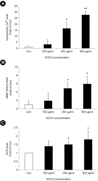
First, to determine the role of Ca2+ signaling in KOCN- induced apoptosis, HCT 116 cells were treated with 100, 250 and 500 μg/ml of KOCN for 48 hours, respectively.
During the period of 48 h, intracellular Ca2+ concentration was elevated by KOCN in a dose-dependent manner (Fig.
1A). During cellular Ca2+ overload, mitochondria take up cytosolic Ca2+, which in turn disrupts the mitochondrial membrane potential (MMP, ΔΨm), and disruption of MMP associated with production of reactive oxygen species (ROS) (Rottenberg and Wu, 1998). In our study, KOCN induced the loss of MMP in a dose-dependent manner (Fig. 1B) and then ROS generation was enhanced by KOCN in a dose-dependent manner (Fig. 1C).
To determine the effect of KOCN on cell death of HCT 116 cells, we examined the apoptosis and necrosis of the cell induced by KOCN. After addition of KOCN in HCT 116 cells, apoptotic cells were considerably increased by KOCN as compared with medium alone (Fig. 2A). KOCN induced the apoptosis of HCT 116 cells in a dose-dependent manner at 48 hours. In processes of KOCN-induced apoptosis, the level of Bax, a pro-apoptotic signal protein, was increased in a dose-dependent manner (Fig. 2B). In contrast, KOCN continuously suppressed the level of Bcl-2, an anti-apoptotic signal protein (Fig. 2B). Bax and Bcl-2 are associated with the membranes of various organelles including mitochondria (Krajewski et al., 1994). These results indicate that KOCN induces the apoptosis of HCT 116 cell via mitochondrial pathway.
KOCN induces the release of TNF-α via NF-κB activation in apoptotic HCT 116 cells
In mitochondrial activation, cytokines are described to
MMP (ΔΨm) level (Fold of Con)
Fig. 1. KOCN-induced mitochondrial dysfunction and ROS production in HCT 116 cells. HCT 116 cells were incubated for 48 h in the absence (Con) and presence of KOCN (100 μg/ml, 250 μg/ml and 500 μg/ml). (A) The intracellular Ca2+ concentration was determined by the fluorescent activity of Fluo-3 AM using flow cytometry as described in the materials and methods section.
(B) The MMP changes were determined by DioC6(3) fluorescence with flow cytometry as described in the materials and methods section. (C) The ROS generation was determined by the DCFDA fluorescence with flow cytometry as described in the materials and methods section. Data are expressed as the means ± SD in three individual experiments. *P < 0.05 and **P < 0.01 were considered a significant difference between the untreated group and KOCN- treated group at same incubation time.
induce ROS production and alter mitochondrial function (Busquets et al, 2003; Schulze-Osthoff et al., 2005). Next, we confirm that whether KOCN is involve in the release of pro-inflammatory cytokine such as TNF-α andIL-1β in HCT 116 cells. As shown in Table 1, KOCN-activated cells showed a significant increase the protein level of TNF-α in a dose-dependent manner, but IL-1β level was not signifi- cantly altered by KOCN. TNF-α is known to activate the ROS-dependent transcription factor NF-κB (Droge, 2001).
Therfore, we investigated the role of NF-κB in TNF-α production in KOCN-induced apoptotic HCT 116 cells.
BAY 11-7085, an inhibitor of NF-κB, considerably blocked the release of TNF-α on KOCN-treated HCT 116 cells (Table 2). These results indicate that the release of TNF-α following KOCN treatment requires the NF-κB pathway.
Induction of HCT 116 cell apoptosis by KOCN is associated with molecules released by KOCN
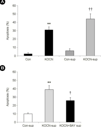
We examined the effect of other molecules induced by KOCN on the apoptosis of HCT 116 cells. To investigate the effect of the other molecules released by KOCN, HCT 116 cells were incubated with KOCN for 48 h, and then the collected cell-free supernatants were added to fresh HCT 116 cells. The supernatant from the KOCN-treated HCT 116 cells led to a significant increase in the apoptosis of fresh HCT 116 cells (Fig. 3A). We also collected the supernatant in KOCN-treated cells after the inhibition of NF-κB activation, and the supernatant was added to fresh HCT 1116 cells. The apoptotic effect of molecules release by KOCN was partially reduced by pretreatment of BAY 11-7085 (Fig. 3B). These results indicate that unknown secretion factors mediated by KOCN have the apoptotic effect in HCT 116 cells and NF-κB plays an important role in the secretion of these molecules from KOCN-treated HCT 116 cells.
DISCUSSION
Cyanate is formed in vivo by breakdown of urea, and only a small portion of urea is decomposed into cyanate.
This cyanate induces the carbamylation of proteins and these carbamylated proteins increases cell death via apoptosis (OK et al., 2005; Apostolov et al., 2011). In our previous study, we focused on the cytotoxic activity of KOCN on the human colorectal cancer cell line, HCT 116 cells. We demonstrated that KOCN considerably increased the apoptosis of HCT 116 cells via intracellular Ca2+ signaling, mitochondrial dysfunction and ROS generation.
Calcium has been recognized as ubiquitous intracellular signal responsible for a number of cellular events, such as growth, proliferation, differentiation, and survival/apoptosis Fig. 2. KOCN-induced apoptosis and the expression of
apoptosis-associated proteins in HCT 116 cells. HCT 116 cells were incubated for 48 h in the absence (Con) and presence of KOCN (100 μg/ml, 250 μg/ml and 500 μg/ml). (A) The apoptosis of these cells was analyzed by measuring the binding of annexin V-FITC and PI using flow cytometry as described in the materials and methods section. The percentage of apoptotic cell in total cell population was included all annexin V binding cells. Data are expressed as the means ± SD in three individual experiments. (B) Cell lysate were collects from harvested cells and were performed ELISA as described in the materials and methods section. White columns represent the expression levels of Bax protein. Black columns represent the expression levels of Bcl-2 protein. The sum of the ratio of each protein expression was to be 1. *P < 0.05 was considered a significant difference of Bax protein between the untreated group and KOCN-treated group. †P < 0.05 was con- sidered a significant difference of Bcl-2 protein between the untreated group and KOCN-treated group.
† ††
KOCN concentration
KOCN concentration
(Clapham, 2007). In apoptotic pathway, calcium concen- tration is controlled by apoptosis-associated proteins. The anti-apoptotic Bcl-2 has been reported to exert its inhibitory effects on apoptosis by blocking the release of cytochrome c and the loss of MMP, and this protein has been acting on the inhibition of cell apoptosis in cancer pathogenesis (Gross et al., 1999). In contrast, Bax, a pro-apoptotic protein, integrates to the outer mitochondrial membrane and causes cytochrome c release. Bax also mediates Ca2+ fluxes and involves in sensitization of mitochondria (Zhe et al., 1999).
In the present study, we confirmed that KOCN-induced apoptosis of the HCT 116 cells causes a rise in the intra- cellular Ca2+ concentration, an alteration of apoptosis- associated protein and the increase of MMP loss (Fig. 1A, 1B and 2). The homeostasis of Ca2+ levels is also modulated by mitochondria membrane potential (MMP, ΔΨm). MMP is altered by opening of permeability transition pore (PTP) in early stage of apoptosis, and it continuously mediates pro- apoptotic signals and ROS generation that mediates DNA damage or activation of the caspase pathway (Desagher and Martinou, 2000; Terasaka et al., 2005; Ishihama et al., 2008).
In these various mitochondrial activations, cytokines are described to induce ROS production and can alter mito- Table 1. The protein levels of TNF-α- and IL-1β-induced by KOCN in HCT 116 cells
KOCN 0 μg/ml 100 μg/ml 250 μg/ml 500 μg/ml
TNF-α (ng/ml) 2.00 ± 0.2 1.98 ± 0.3 3.82 ± 0.1* 6.10 ± 0.5**
IL-1β (ng/ml) 0.90 ± 0.1 0.97 ± 0.1 0.85 ± 0.4 1.16 ± 0.3
The cells were treated with KOCN for 48 h. Data represent the mean ± SD in three individual experiments. *P < 0.05 and **P < 0.01 were considered a significant difference between the untreated group and KOCN-treated group.
Table 2. Involvement of NF-κB in the secretion of TNF-α in KOCN-treated HCT 116 cells
KOCN 0 μg/ml 500 μg/ml 500 μg/ml
Inhibitor - - BAY
TNF-α
(ng/ml) 2.11 ± 0.3 5.93 ± 0.6** 1.99 ± 0.8††
IL-1β
(ng/ml) 1.79 ± 0.1 1.51 ± 0.4 1.44 ± 0.3 The cells were pre-treated for 1 h with and without 10 μM BAY- 11-7085 (BAY). These cells were incubated for 48 h in the presence and absence of KOCN (500 μg/ml). Data represent the mean ± SD in three individual experiments. **P < 0.01 was considered a significant difference between the untreated group and KOCN- treated group. ††P < 0.01 was considered a significant difference between the KOCN-treated group and BAY-pre-treated group.
Fig. 3. The effect of released factor induced by KOCN on cell apoptosis. (A) HCT 116 cells were incubated with 500 μg/ml of KOCN for 48 h. The supernatant was collected and added to the fresh HCT 116 cells for 24 h. **P < 0.01 were considered a significant difference between the untreated group and KOCN- treated group. ††P < 0.01 were considered a significant difference between the con-sup-treated group and KOCN-sup-treated group.
(B) HCT 116 cells were pre-treated for 1 h with and without 10 μM BAY-11-7085 (BAY). These cells were incubated for 48 h in the presence and absence of KOCN (500 μg/ml). The supernatant was collected and added to the fresh HCT 116 cells for 24 h. **P
< 0.01 were considered a significant difference between the con- sup-treated group and KOCN-sup-treated group. †P < 0.05 were considered a significant difference between the KOCN-sup-treated group and KOCN-sup-treated group with pre-treatment of BAY.
All data are expressed as the means ± SD in three individual experiments.
††
†
chondrial function (Busquets et al., 2003; Schulze-Osthoff et al., 2005). TNF-α and IL-1β are the pro-inflammatory cytokines and play a major role in both inflammation and apoptosis (Lao and Chang, 2008). Although, elevated levels of IL-1β are associated with various cell apoptosis and increase of intracellular Ca2+ (Corbett and McDaniel, 1994), IL-1β was not involved in KOCN-mediated HCT 116 cell apoptosis (Table 1). However, the release of TNF-α mediated by KOCN in HCT 116 cells via NF-κB activation (Table 1 and 2). In apoptotic cells, TNF-α is involved in activating caspase cascade and increasing intracellular Ca2+ concen- tration. And TNF-α can induce ROS production in mito- chondria as well as alter mitochondrial function by impairing membrane permeability (Busquets et al, 2003). In signaling mechanisms associated with TNF-α stimulation, TNF-α leads to IKK activation resulting in Iκ-Bα phophorylation and subsequent IκB degradation. This pathway stimulated by TNF-α eventually induces of NF-κB activation. Activation of NF-κB results in enhanced both cell survival and death (Schulze-Osthoff et al., 2005). In this study, KOCN- mediated apoptosis significantly induced the release of TNF-α and the elevated level of TNF-α was inhibited by BAY 11-7085, NF-κB specific inhibitor (Table 2). Although TNF-α might play as a major molecule in KOCN-induced apoptosis, we next evaluated the effects of other unmeasured factors on apoptosis. Apoptosis was increased by incubation with supernatants from HCT 116 cell after KOCN treatment.
Interestingly, the apoptotic effect of molecules released by KOCN was partially reduced by BAY 11-7085 pre-treated supernatant (Fig. 3B). Taken together, our results indicate that KOCN-induced apoptosis in HCT 116 cells is occurred via the mitochondrial pathway that is dependent on the releases of TNF-α and the increased factors. And these mechanisms involve the activation of NF-κB. Our findings suggest that the KOCN has a potent apoptosis-inducing activity and it may be useful for various cancer therapies.
REFERENCES
Apostolov EO, Ray Debarti, Alovuia WM, Mikhailova MV, Wang X, Basnakian AG, Shah SV. Endonuclease G mediates endothelial cell death induced by carbamylated LDL. Am J
Physiol Heart Circ Physiol. 2011. 300: H1997-H2004.
Busquets S, Aranda X, Ribas-Carbo M, Azcon-Bieto J, Lopez- Soriano FJ, Argiles JM. Tumor necrosis factor-alpha uncouples respiration in isolated rat mitochondria. Cytokine. 2003. 22:
1-4.
Clapham DE. Calcium signaling. Cell. 2007. 131: 1047-1058.
Corbett JA, McDaniel ML. Reversibility of interleukin-1 beta- induced islet destruction and dysfunction by the inhibition of nitric oxide synthase. Biochem J. 1994. 299: 719-724.
Desagher S, Martinou SC. Mitochondria as the central control point of apoptosis. Trends Cell Biol. 2000. 10: 369-377.
Droge W. Free radicals in the physiological control of cell function.
Physiol Rev. 2002. 82: 47-95.
Gross A, McDonnell JM, Korsmeyer SJ. Bcl-2 family members and the mitochondria in apoptosis. Genes Dev. 1999. 13:
1899-1911.
Henriette TL, Guchelaar HJ, Gelderblom H. Pharmacogenetics in chemotherapy of colorectal cancer. Best Pract Res Clin Gastroenterol. 2009. 23: 257-273.
Inoue EN, Nagano I, Ichinohasama R, Asato N, Kondo Y, Iinuma K. Bimodal effects of platelet-derived growth factor on rat mesangial cell proliferation and death, and the role of lysophosphatidic acid in cell survival. Clin Sci (Lond). 2001.
101: 11-19.
Ishihama M, Toyooka T, Ibuki Y. Generation of phosphorylated histone H2AX by benzene metabolites. Toxicol in Vitro. 2008.
22: 1861-1868.
Krajewski S, krajewski M, Shabaik A, miyashita T, Wang HE, Reed JC. Immunohistochemical determination of in vivo distribution of Bax, a dominant inhibitor of Bcl-2. 1994.
American J Pathol. 145: 1326-1336.
Kraus LM, Kraus AP. Carbamylation of amino acids and proteins in uremia. Kidney Int. 2001.78: S102-S107.
Kuckel CL, Lubit BW, Lambooy PK, Farnsworth PN. Methyl- isocyanate and actin polymerization: The in vitro effects of carbamylation. Biochim Biophys Acta. 1993. 1162: 143-148.
Lao Y, Chang DC. Mobilization of Ca2+ from endoplasmic reticulum to mitochondria plays a positive role in the early stage of UV- or TNF alpha0induced apoptosis. Biochem Biophys Res Commun. 2008. 373: 42-47.
OK E, Basnakian AG, Apostolov EO, Barri YM, Shah SV.
Carbamylated low-density lipoprotein induces death of endothelial cells: A link to atherosclerosis in patients with kidney disease. Kidney Int. 2005. 68: 173-178.
Rottenberg H, Wu S. Quantitative assay by flow cytometry of the
mitochondrial membrane potential in intact cells. Biochim Biophys Acta. 1998. 1404: 393-404.
Rychahou PG, Kang J, Gulhati P, Doan HQ, Chen LA, Xiao SY, Chung DH, Evers BM. Akt2 overexpression plays a critical role in the establishment of colorectal cancer metastasis. Proc Nat Acad Sci U S A. 2008. 105: 20315-20320.
Schulze-Osthoff K, Bakker AC, Vanhaesebroeck B, Beyaert R, Jacob WA, Fiers W. Cytotoxic activity of tumor necrosis factor is mediated by early damage of mitochondrial functions.
Evidence for the involvement of mitochondrial radical gene-
ration. J Biol Chem. 2005. 267: 5317-5323.
Terasaka H, Kadoma Y, Sakagami H, Fujisawa S. Cytotoxicity and apoptosis-inducing activity of bisphenol A and hydroquinone in HL-60 cells. Anticancer Res. 2005. 3B: 2241-2247.
Yang EJ, Chang JH. Potassium cyanate induces apoptosis of human colorectal cancer cell via mitochondrial pathway. J Exp Biomed Sci. 2011. 17: 177-184.
Zhu L, Ling S, Yu XD, Venkatesh LK, Subramanian T, Chinnadurai G, Kuo TH. Modulation of mitochondrial Ca2+ homeostasis by Bcl-2. J Biol Chem. 1999. 274: 33267-33273.