Introduction
Scapular posterior tilt (SPT) is important in the prevention of scapular dyskinesis, which is pathologic alterations in scapular alignment related to imbalance of muscle activation or lengths resulting in abnormal scapular movement and shoulder pain during ele-
vation of the arm (Kibler and Mcmullen, 2003; Kibler and Sciascia, 2010), and for correction of rounded shoulder posture (RSP), forward scapular position, and humeral head anterior gliding (Escamilla et al, 2009; Ha et al, 2012; Lee et al, 2015; Solem-Bertoft et al, 1993). The serratus anterior (SA) and lower tra- pezius (LT) muscles are activated during SPT, and Corresponding author: Heon-seock Cynn [email protected]
Effects of Verbal Cue for Scapular Depression During Scapular Posterior Tilt Exercise on Scapular Muscle Activities and Clavicular Tilt
Angle in Subjects With Rounded Shoulder Posture and Upper Trapezius Myofascial Pain
Sil-ah Choi1, MSc, PT, Heon-seock Cynn1,2, PhD, PT, A-reum Shin1, BPT, PT, Da-eun Kim1, BHSc, PT
1Applied Kinesiology and Ergonomic Technology Laboratory, Dept. of Physical Therapy, The Graduate School, Yonsei University,
2Dept. of Physical Therapy, College of Health Science, Yonsei University
Abstract
1)Background: Scapular posterior tilt (SPT) is important in the prevention of abnormal scapular movement and pain during elevation of the arm. However, previous studies have overlooked increased upper trapezius (UT) muscle activity interrupting the normal force couple of scapular motion and compensation of levator scapulae (LS) muscle activated simultaneously with UT during SPT exercise.
Objects: The purpose of this study was to compare the effects of modified SPT with depression exercise versus SPT exercise on serratus anterior (SA), lower trapezius (LT), UT, and LS muscle activities and the clavicular tilt angle, in subjects with rounded shoulder posture (RSP) and myofascial pain in the UT muscle region.
Methods: Eighteen subjects with RSP were recruited and randomly allocated to 2 groups; 9 in the SPT group and 9 in the SPT with depression group. All subjects met the specific RSP criteria and had myofascial pain of UT region. Depending on the allocated group, subjects performed the assigned SPT exercise and EMG data were recorded during the each exercise. Clavicular tilt angle was defined as the angle between the line joining the medial and lateral end of the clavicle and a horizontal line.
Results: The SA muscle activity was significantly greater in SPT with depression than with SPT exercise (p<.05). The UT, LS muscle activity and the clavicular tilt angle was significantly lower in SPT with depression than with SPT exercise (p<.05).
Conclusion: These findings were insightful because the potential risk of pain from overactivation of the UT and LS was considered, in contrast with SPT exercise. SPT with depression exercise can be implemented as an effective method to facilitate scapular muscle activity for stability and to prevent myofascial pain in the neck and shoulder.
Key Words: Levator scapulae; Myofascial pain; Rounded shoulder posture; Scapular posterior tilt;
Upper trapezius.
are the primary muscles used to stabilize the medial border and the inferior angle of the scapula on the thoracic wall (Ekstrom et al, 2003; Neumann, 2002).
Several previous studies have investigated strength- ening exercises for the SA and LT muscles to treat excessive scapular anterior tilt or winging (Arlotta et al, 2011; Hardwick et al, 2006; Park et al, 2013;
Pontillo et al, 2007).
In particular, the arm lift exercise in prone, back- ward rocking, or standing positions has been com- monly used for SPT in physical therapy programs.
When elevating the arm, normal scapular movement occurs in combination with upward rotation, external rotation, and posterior tilting (Escamilla et al, 2009;
Ludewig et al, 1996), and scapular depression and slight adduction also occur at the end-range of scap- ular upward rotation (Sahrmann, 2002). During scap- ular upward rotation, force couples of the SA, LT, and upper trapezius (UT) occur, described as muscles pulling in different directions to accomplish the same motion in a synergistic manner (Neumann, 2002).
However, previous studies have overlooked over- activation or dominant recruitment of UT muscles during SPT exercise. Several studies have reported that poor shoulder postures, including RSP or for- ward scapular position, demonstrated decreased SA and LT activity and increased UT activity interrupt- ing the normal force couple of scapular motion (Kibler and Mcmullen, 2003; Lewis et al, 2005; Michener et al, 2003). Moreover, previous studies have not considered levator scapulae (LS) muscle compensation activated simultaneously with UT in scapular elevation to complete the arm lift during SPT exercise. If it is just focusing on arm lift to complete the SPT ex- ercise, scapular elevation excessively occurs and LS muscle would overactivate to help UT muscle for scapular elevation. Consequently, the scapula would be elevated with downward, not upward rotation.
These unbalanced force couples of scapular motion or compensation could cause scapular dyskinesia and tissue overuse, increasing the risk of pain and injury (Sahrmann, 2002). Generally, chief complaints in the
neck and shoulder region have originated from mus- cular soreness or referred pain and have involved the UT and LS muscles, resulting in myalgia, myositis, and myofascial pain syndrome (Azevedo et al, 2008;
Kannan, 2012; Olson et al, 2000).
The UT and LS muscle function as scapular ele- vation and excessive activation or increased tightness of UT and LS are responsible for neck and shoulder pain. For these reasons, the present study introduced a modified SPT with depression exercise to relieve myofascial pain of the UT muscle region. Thus, the purpose of this study was to compare the effects of modified SPT with depression exercise versus SPT exercise on SA, LT, UT, and LS muscle activities and the clavicular tilt angle, in subjects with RSP and myofascial pain in the UT muscle region. It was hypothesized that SPT with depression exercise would increase the SA and LT muscle activity, and decrease the UT and LS muscle activity and clav- icular tilt angle, compared with the effect of SPT exercise, in subjects with RSP and myofascial pain in the UT muscle region.
Methods
Subjects
A power analysis was performed with G*power software ver. 3.1.2 (Franz Faul, University of Kiel, Kiel, Germany) using the results of a pilot study in- volving 5 subjects. The calculation of sample size was carried out for a power of .80, alpha level of .05, and effect size of .82. This provided the neces- sary sample size of 12 subjects for this study. From the initial 25 subjects, 7 were eliminated from the study (4 did not met the RSP criteria and 3 had no taut band in UT region). Eighteen subjects with RSP were recruited and randomly allocated to 2 groups; 9 in the SPT group and 9 in the SPT with depression group. The randomization was conducted using se- lection from a table of random numbers in Microsoft Excel (Microsoft Corp., Redmond, WA, USA). More
female subjects were recruited than males, it is con- sistent with the previous studies that mechanical neck pain has a lifetime prevalence of 45—54% in the general population, and up to 30% of men and 50% of women (Dziedzig et al. 2005; Fernandez et al.
2006). The demographic characteristics and in- formation of the 18 subjects, which include gender, age, height, weight, body mass index, severity of RSP, tenderness grading scale score, and visual ana- log scale (VAS) score are shown in Table 1.
All subjects met the specific RSP criteria and had myofascial pain of UT region. RSP was indicated by a distance ≥2.5 ㎝ from the posterior aspect of the acromion to the table, in the supine position (Sahrmann, 2002). Myofascial pain was confirmed through examination of active trigger point (TrP) be- cause myofascial pain symptoms involve pain in spe- cific TrPs on compression (Duyur Çakıt et al, 2009;
Kanna, 2012). An active UT TrP was defined as a palpable taut band, hypersensitive tender spot within the taut band, and referred pain in a pattern specific for UT TrP1 or TrP2 (Oliveira-Campelo et al, 2013;
Gemmell et al, 2008; Simons et al, 1999). TrP1 is lo- cated in the posterolateral aspect of the neck (top of the trapezius) and TrP2 is located in more posterior and horizontal line of UT (Simons et al, 1999). In addition, the tenderness grading scale (Hubbard, 1993) was used for assessing soft tissue tenderness and documenting patient responses to provocation.
The grading is as follows: 0- No tenderness, 1- Tenderness to palpation without grimace or flinch, 2-
Tenderness with grimace or flinch to palpation, 3- Tenderness with withdrawal (Jump sign), and 4- Withdrawal (Jump sign) to non-noxious stimuli (i.e., superficial palpation). All measurements were per- formed on the dominant side used by subjects when eating and writing, and were performed by a phys- ical therapist with 3 years of clinical experience in musculoskeletal treatment.
Subjects were excluded if they had a history of surgery or existing pathology of the shoulder and el- bow, dysfunction that substantially limited shoulder and elbow motion, current complaint of numbness or tingling in the upper extremity limiting activity, and any congenital postural abnormalities (Cole et al, 2013; Lee et al, 2015). We also excluded those who were taking long-term corticosteroid and having un- dergone any therapy for their neck pain within the past month before this study (Gemmell et al, 2008;
Meseguer et al, 2006). In this study, there were no eliminated subjects due to the exclusion criteria.
Prior to collecting data, the examiner informed the subjects of the study procedures and each subject provided written informed consent. The study proto- col was approved by the Yonsei University Wonju Institutional Review Board (approval number:
1041849-201704-BM-017-01).
Procedures
Subjects underwent a familiarization period for the assigned SPT exercise until a proper performance
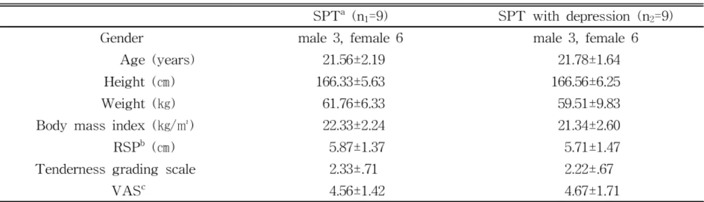
SPTa (n1=9) SPT with depression (n2=9)
Gender male 3, female 6 male 3, female 6
Age (years) 21.56±2.19 21.78±1.64
Height (㎝) 166.33±5.63 166.56±6.25
Weight (㎏) 61.76±6.33 59.51±9.83
Body mass index (㎏/㎡) 22.33±2.24 21.34±2.60
RSPb (㎝) 5.87±1.37 5.71±1.47
Tenderness grading scale 2.33±.71 2.22±.67
VASc 4.56±1.42 4.67±1.71
ascapular posterior tilt, brounded shoulder posture,cvisual analog scale.
Table 1. The demographic characteristics and information of the subjects (N=18)
capability was achieved. Five minutes after the fa- miliarization, subjects performed the assigned SPT exercise for 5-sec twice, with 1-min of rest between the two trials to avoid muscle fatigue. The examiner made certain that all the subjects were comfortable, and data collection was ceased immediately when subjects failed to perform or maintain the stand- ardized position during the exercise.
1) Scapular posterior tilt exercise
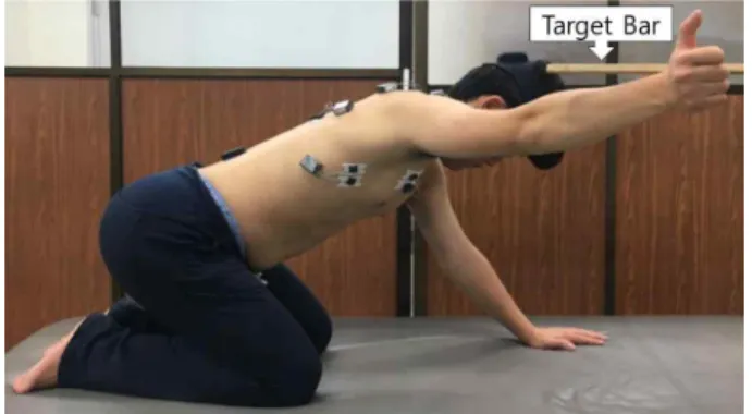
Subjects were positioned in the quadruped position, which is on all fours with hands under shoulders and knees under hips, both at 90°. To assess cranio- cervical, thoracolumbar, and lumbopelvic stability, Myomotion motion analysis system (Noraxon Inc., Scottsdale, AZ, USA) was used. Three inertial meas- urement sensors were placed on the posterior head, upper thorax, and lumbar spine and calibrated for a value of 0° with the head, trunk, and pelvis in a straight line in the quadruped position. The subjects were then instructed to move the hips backward slowly while keeping the head, trunk, and pelvis in a straight line and looking downward at this time. The sensors provided 3D angular orientation; the examiner continuously monitored whether the subjects main- tained a value of 0° (within ±5° error tolerance), and advised the subjects of incorrect position to prevent compensation by thoracolumbar flexion or pelvic pos- terior tilt. Subsequently, a wooden bar was placed at a target height for 145° of shoulder abduction to en- sure consistent arm lifting; the subjects then lifted the dominant arm (thumb up towards the ceiling and elbow in extension) until the radial border of the wrist lightly touched the target bar while maintaining the opposite arm with full extension to support the position (Lee at al, 2015; Ha et al, 2012)(Figure 1).
2) Scapular posterior tilt with depression exercise SPT with depression exercise followed the same procedure as SPT exercise, with the addition of a scapular depression. Subjects lifted the dominant arm to 145° of shoulder abduction and were verbally cued
to “bring your scapula down as far as possible.” At this time, subjects would maintain the radial border of the wrist in slight contact with the target bar (Figure 2).
3) Surface electromyography and data processing Electromyographic (EMG) data were collected using a wireless TeleMyo DTS (Noraxon Inc., Scottsdale, AZ, USA), and Myo-Research Master Edition 1.06 XP software (Noraxon Inc., Scottsdale, AZ, USA) was used for analyzing EMG data. The EMG signals were sampled at 1,000 ㎐. A band pass filter was used between 20 and 450 ㎐ and a notch filter was preset to reject 60 ㎐. The raw data were processed into the root mean square with a window of 50 ㎳.
Two surface electrodes with an inter-electrode distance of 2 ㎝ were positioned on the SA, LT, UT, and LS muscles. Two electrodes were placed in the middle of each muscle belly and parallel to the mus- cle fibers. The electrode sites were shaved and rub- bing alcohol was used to reduce skin impedance. For Figure 2. Scapular posterior tilt with depression
exercise.
Figure 1. Scapular posterior tilt exercise.
recordings of the SA, the electrodes were placed be- low the axillary area, at the level of the inferior tip of the scapula, and medial to the latissimus dorsi.
The electrodes for the LT were placed at a point 5
㎝ inferior to the scapular spine, beside the medial border of the scapula at a 55° oblique angle. For re- cordings of the UT, the electrodes were placed slightly laterally one-half the distance between the cervical spine at C7 and the acromion. The electro- des for the LS were placed between the anterior margin of the UT and the posterior margin of the sternocleidomastoid (Criswell, 2010).
Normalization was needed to minimize variables between different recording sites and subjects. The maximum voluntary isometric contraction (MVIC) normalization method was applied for each muscle examined, and MVIC was recorded with a manual muscle test (MMT) in the positions (Hislop and Montgomery, 2007). To collect MVIC data, subjects maintained the MMT position of each muscle being examined against manual resistance, for 5-sec, twice.
The middle 3-sec contraction, excluding 1-sec each from the beginning and the end, was used for data analysis for each muscle, and the mean value of the middle 3-sec contraction of two trials was considered the MVIC for each muscle.
All EMG data during the assigned type of SPT exercise were recorded for 5-sec twice and calcu- lated from the middle 3-sec isometric phase, exclud- ing each 1-sec from the beginning and the end. The mean value of the middle 3-sec contraction of two trials for each SPT type was used for data analysis.
4) Clavicular tilt angle
The subjects stood straight with neutral forearm and resting hand position at the sides. The examiner marked two landmarks with round stickers at the midpoint of the end of the medial clavicle and the midpoint of the end of the lateral clavicle (Ha et al, 2013). Then, a digital camera captured a photo- graphic image 2 m away from the subject’s frontal side. Clavicular tilt angle was defined as the angle
between the line joining the medial and lateral end of the clavicle and a horizontal line from the medial end of the clavicle (Akel et al, 2008) and was calcu- lated automatically using ImageJ software (National Institutes of Health, Bethesda, MD, USA) (Figure 3).
Statistical Analysis
The Kolmogorov-Smirnov Z-test was used to as- sess normal distribution. An independent t-test was used to identify the homogeneity of demographic characteristics and to compare SA, LT, UT, and LS muscle activities and clavicular tilt angle between the two groups. Statistical significance was set at .05.
All statistical analyses were performed using SPSS 18 (SPSS Inc., Chicago, IL, USA).
Results
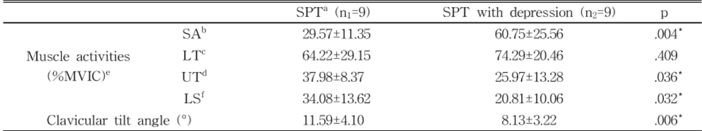
All dependent variables were found to approximate a normal distribution (Kolmogorov-Smirnov Z-test, p>.05). We found no significant differences in age, height, weight, body mass index, RSP, tenderness grading scale score, and VAS score between the two groups (p>.05). The SA muscle activity was sig- nificantly greater in SPT with depression than with SPT exercise (p<.05). However, there was no stat- istically significant difference in LT activity between the two groups (p>.05). The UT and LS muscle ac- tivity was significantly lower in SPT with depression than with SPT exercise (p<.05). The clavicular tilt
Figure 3. Measurement of clavicular tit angle.
angle was significantly lower in SPT with depres- sion than with SPT exercise (p<.05) (Table 2).
Discussion
This study compared the effect of SPT with de- pression exercise with that of SPT exercise, on SA, LT, UT, and LS muscle activities and clavicular tilt angle, in subjects with RSP and myofascial pain in the UT muscle region. In SPT with depression ex- ercise, SA muscle activity significantly increased, and UT and LS muscle activity and clavicular tilt angle significantly decreased. To our knowledge, this is the first study to compare scapular muscle activities and clavicular tilt angle using modified SPT with depres- sion exercise versus SPT exercise.
SA muscle activity was significantly greater with SPT with depression than with SPT exercise (by 105.44%), supporting the research hypothesis. These findings were in agreement with those of previous studies on various types of SPT exercise to strengthen SA and LT, stabilize the scapula, and treat shoulder dysfunction (Ekstrom et al, 2003; Ha et al, 2012; Lee et al, 2015). SA muscle activity is optimal above 90° of shoulder elevation, and the greater the increase in shoulder elevation, the greater the SA muscle activity (Ekstrom et al, 2003, 2005;
Hardwick et al, 2006). Ekstrom et al. (2003) reported that SA muscle activity was greater above 120° than below 80° of shoulder elevation in the scapular plane.
Hardwick et al. (2006) found that SA muscle was
more facilitated at 140° of humeral elevation than at 90° and 120°. Moreover, SA facilitates scapular pro- traction, upward rotation, abduction, and depression, ultimately holding the scapula to the thoracic wall (Ekstrom et al, 2004; Escamilla et al, 2009; Neumann, 2002). In this study, scapular depression was addi- tionally applied during SPT exercise, and such scap- ular depression effectively elicited greater SA muscle activity compared with SPT. Smith et al. (2006) confirmed scapular depression produced the largest SA muscle activity (47% MVIC) and suggested scapular depression and protraction exercises could be safely performed to improve scapulothoracic alignment. Also, McCabe et al. (2007) reported scap- ular depression produced moderate LT activity and minimal UT activity so showed high ratio of LT/UT activity.
UT and LS muscle activity was significantly low- er during SPT with depression than with SPT ex- ercise (by 31.62% and 41.17%, respectively), support- ing the research hypothesis. These findings have clinical implications because during SPT exercise, in- dividuals with RSP would just focus on shoulder el- evation without controlling UT and LS muscle activ- ity, resulting in myofascial shoulder pain (Azevedo et al, 2008; Kannan et al, 2012; Olson et al, 2000). This could be explained by previous findings that abnor- mal posture, such as RSP and forward head posture, showed unbalanced scapular muscle activities (Page et al, 2009; Ludewig and Braman, 2011). RSP is as- sociated with upper crossed syndrome and the scap- ular elevation seen in upper crossed syndrome facili-
SPTa (n1=9) SPT with depression (n2=9) p
Muscle activities (%MVIC)e
SAb 29.57±11.35 60.75±25.56 .004*
LTc 64.22±29.15 74.29±20.46 .409
UTd 37.98±8.37 25.97±13.28 .036*
LSf 34.08±13.62 20.81±10.06 .032*
Clavicular tilt angle (°) 11.59±4.10 8.13±3.22 .006*
ascapular posterior tilt, bserratus anterior, clower trapezius, dupper trapezius, emaximal voluntary isometric contraction,
flevator scapulae, *p<.05.
Table 2. Comparison of measurement variables between scapular posterior tilt exercise and scapular posterior
tilt with depression exercise. (N=18)
tates the UT and LS (Page et al, 2009). Forward head posture is related to decreased SA activity and increased UT activity (Thigpen et al, 2010; Weon et al, 2010). Ludewig and Braman (2011) reported that synergistic dominance of the UT over insufficient SA and inhibited LT during scapular upward rotation resulted in limited overhead movement, subacromial impingement, and overuse shoulder injuries. Moreover, LS can be compensated or contracted simultaneously with UT during shoulder exercise programs, such as rowing, shoulder abduction, and shrug exercises (Moseley et al, 1992; Lee et al, 2016). Furthermore, Szeto et al. (2002) reported that excessive stiffness of the LS contributes to an increased compressive and shear force on the cervical spine during active neck movement. In this study, addition of a scapular depression phase to scapular upward rotation facili- tated scapular depressors and reciprocally inhibited scapular elevators during SPT with depression (significantly increased SA activity versus sig- nificantly reduced UT and LS activity). Although variety of SPT exercises were performed in many previous studies (Ebaugh et al, 2005; Ha et al, 2012;
Hardwick et al, 2006; Lee et al, 2015), the results only examined the primary muscle of scapular stabil- ity such as SA and LT and did not confirm UT and LS so it could not suggest the best method for scapular dyskinesis because the unexpected risk or side-effect of neck and shoulder pain occur.
The previous studies identified that normalization of MVIC in injured populations may be influenced by pain and result in overestimation of muscle activity (Ettinger et al, 2016). However, the results showed different data according to each muscle and it was unpredictable. Also Kofler (2003) explained muscle alterations in the presence of pain can be dependent on task or organism. The %MVIC is more sensitive to contractions that require more effort and decreases its sensitivity at low levels of effort (Criswell, 2010).
In addition, clavicular tilt angle was significantly lower in SPT with depression than with SPT exercise (by 28.38%), supporting the research hypothesis.
Decreased clavicular tilt angle indicates that ex- cessive or unnecessary scapular elevation by the overactive UT and LS did not occur and the scapula was placed in a more optimal alignment (neutral po- sition rather than elevation). This result was sup- ported by UT and LS muscle activities in the pres- ent study. This study showed that SPT group had greater UT and LS muscle activities and clavicular tilt angle than SPT with depression group. It could mean that the higher UT and LS muscle activities are, the greater clavicular tilt angle occurs. Using the 3D analysis, Ludewig et al. (2009) and McClure et al. (2004) reported the clavicular tilt angle was about 5.9° in subjects without scapular abnormality and 4°
in subjects with shoulder impingement respectively.
These data are different from the present study be- cause this study used 2D photographic analysis and recruited the subjects with RSP. On the other hand, Ha et al. (2013) verified goniometric and photographic measurements of clavicular tilt angle had high reli- ability and validity against the radiographic findings and reported the clavicular tilt angle of healthy sub- jects was about 10° in goniometric and 8° in photo- graphic measurement. Although the change in clav- icular tilt angle was small in this study, the result could be meaningful because clavicular tilt angle in subjects with RSP was closer to the normal range.
This study has several limitations. First, it had a cross-sectional design. Future longitudinal studies are necessary to determine the long-term effects of SPT with depression on scapular muscle activities and clavicular position in subjects with RSP and my- ofascial pain of the UT region. Second, the results could not be generalized to the patient population be- cause healthy young subjects with only one specific RSP criteria, an average grade 2 tenderness score, and an average VAS score of 4 participated. Therefore, a sample population with a broad age range and mod- erate to severe symptoms should be investigated in future studies. Third, this study had relatively low statistical power because it designed experiment of independent two groups so future studies need re-
peated-measurement design to further differentiate between the two exercise protocols. Finally, the more precise measurement devices such as 3D or real-time measuring system, digital pressure algometry, or ul- trasonography will be need to improve measurement errors in future studies.
Conclusion
The purpose of this study was to compare the ef- fects of SPT with depression exercise with SPT ex- ercise on SA, LT, UT, and LS muscle activities and clavicular tilt angle, in subjects with RSP and my- ofascial pain in the UT region. In SPT with depres- sion exercise, SA muscle activity significantly in- creased, and UT and LS muscle activity and clav- icular tilt angle significantly decreased. These find- ings were insightful because the potential risk of pain from overactivation of the UT and LS was considered, in contrast with SPT exercise. Therefore, SPT with depression exercise can be implemented as an effective method to facilitate scapular muscle ac- tivity for stability and to prevent myofascial pain in the neck and shoulder.
References
Akel I, Pekmezci M, Hayran M, et al. Evaluation of shoulder balance in the normal adolescent pop- ulation and its correlation with radiological parameters. Eur Spine J. 2008;17(3):348-354.
Arlotta M, LoVasco G, McLean L. Selective recruit- ment of the lower fibers of the trapezius muscle.
J Electromyogr Kinesiol. 2011;21(3):403-410.
https://doi.org/10.1016/j.jelekin.2010.11.006
Azevedo DC, de Lima Pires T, de Souza Andrade F, et al. Influence of scapular position on the pres- sure pain threshold of the upper trapezius mus- cle region. Eur J Pain. 2008;12(2):226-232.
Cole AK, McGrath ML, Harrington SE, et al. Scapular
bracing and alteration of posture and muscle activity in overhead athletes with poor posture.
J Athl Train 2013;48(1):12-24. https://doi.org/
10.4085/1062-6050-48.1.13
Criswell E. Cram’s introduction to surface electro- myography. 2nd ed. Sudbury, MA, Jones and Bartlett Publishers. 2010:268-297.
Duyur Çakit B, Genç H, Altuntaş V, et al. Disability and related factors in patients with chronic cer- vical myofascial pain. Clin Rheumatol. 2009;28(6):
647-654. https://doi.org/10.1007/s10067-009-1116-0 Dziedzig K, Hill J, Lewis M, et al. Effectiveness of manual therapy or pulsed shortwave diathermy in addition to advice and exercise for neck dis- orders: a pragmatic randomised controlled trail in physical therapy clinics. Arthritis Rheum 2005;
53:214-222.
Ebaugh DD, McClure PW, Karduna AR. Three-di- mensional scapulothoracic motion during active and passive arm elevation. Clin Biomech (Bristol, Avon). 2005:20(7):700-709.
Ekstrom RA, Donatelli RA, Soderberg GL. Surface electromyographic analysis of exercises for the trapezius and serratus anterior muscles. J Orthop Sports Phys Ther 2003;33(5):247-258.
https://doi.org/10.1016/j.jelekin.2004.09.006
Ettinger L, Weiss J, Shapiro M, et al. Normalization to Maximal Voluntary Contraction is Influenced by Subacromial Pain. J Appl Biomech. 2016;
32(5):433-440.
Escamilla RF, Yamashiro K, Paulos L, et al.
Shoulder muscle activity and function in com- mon shoulder rehabilitation exercises. Sports Med. 2009;39(8):663-685.
Fernandez de las Penas C, Alonso-Blanco C, Miangolarra JC. Myofascial trigger points in sub- jects presenting with mechanical neck pain: A blinded, controlled study. Man Ther 2006;12:29-33.
Gemmell H, Miller P, Nordstrom H. Immediate effect of ischaemic compression and trigger point pressure release on neck pain and upper tra- pezius trigger points: A randomised controlled
trial. Clin Chiropr. 2008;11(1):30-36.
Ha SM, Kwon OY, Cynn HS, et al. Comparison of electromyographic activity of the lower trapezius and serratus anterior muscle in different arm-lifting scapular posterior tilt exercises. Phys Ther Sport. 2012;13(4):227-232. https://doi.org/
10.1016/j.ptsp.2011.11.002
Ha SM, Kwon OY, Weon JH, et al. Reliability and validity of goniometric and photographic meas- urements of clavicular tilt angle. Man Ther.
2013;18(5):367–371. https://doi.org/10.1016/j.math.
2012.12.006
Hardwick DH, Beebe JA, McDonnell MK, et al. A comparison of serratus anterior muscle activa- tion during a wall slide exercise and other tra- ditional exercises. J Orthop Sports Phys Ther.
2006;36(12):903-910.
Hislop H, Montgomery J. Daniels and Worthingham’s Muscle Testing: Techniques of manual examination.
8th ed. St. Louis, MO: Saunders, Elsevier. 2007:
64-79.
Hubbard DR. Myofascial trigger points show sponta- neous needle EMG activity. Spine (Phila, Pa. 1976).
1993;18(13):1803-1807.
Kannan P. Management of myofascial pain of upper trapezius: A three group comparison study. Glob J Health Sci. 2012;4(5):46-52. https://doi.org/
10.5539/gjhs.v4n5p46
Kibler WB, McMullen J. Scapular dyskinesis and its relation to shoulder pain. J Am Acad Orthop Surg. 2003;11(2):142-151.
Kibler WB, Sciascia A. Current concepts: Scapular dyskinesis. Br J Sports Med.2010;44(5):300–305.
https://doi.org/10.1136/bjsm.2009.058834
Kofler M. Functional organization of exteroceptive inhibition following nociceptive electrical finger- tip stimulation in humans. Clin Neurophys.
2003;114(6):973-980.
Lee JH, Cynn HS, Choi WJ, et al. Various shrug exercises can change scapular kinematics and scapular rotator muscle activities in subjects with scapular downward rotation syndrome.
Hum Mov Sci. 2016;45:119-129. https://doi.org/
10.1016/j.humov.2015.11.016
Lee JH, Cynn HS, Yoon TL, et al. The effect of scapular posterior tilt exercise, pectoralis minor stretching, and shoulder brace on scapular alignment and muscles activity in subjects with round-shoulder posture. J Electromyogr Kinesiol.
2015;25(1):107-114. https://doi.org/10.1016/j.jelekin.
2014.10.010
Lewis JS, Wright C, Green A. Subacromial impinge- ment syndrome: The effect of changing posture on shoulder range of movement. J Orthop Sports Phys Ther. 2005;35(2):72-87.
Ludewig PM, Braman, JP. Shoulder impingement:
Biomechanical considerations in rehabilitation.
Man Ther. 2011;16(1):33-39.
Ludewig PM, Cook, TM, Nawoczenski DA. Three- dimensional scapular orientation and muscle ac- tivity at selected positions of humeral elevation.
J Orthop Sports Phys Ther. 1996;24(2):57-65.
Ludewig PM, Phadke V, Braman JP, et al. Motion of the shoulder complex during multiplanar humeral elevation. J Bone Joint Surg Am 2009;91(2):
378-389.
McCabe RA, Orishimo KF, McHugh MP, et al Surface electromygraphic analysis of the lower trapezius muscle during exercises performed be- low ninety degrees of shoulder elevation in healthy subjects. N Am J Sports Phys Ther.
2007;2(1):34-43.
McClure PW, Bialker J, Neff N, et al. Shoulder func- tion and 3-dimensional kinematics in people with shoulder impingement syndrome before and after a 6-week exercise program. Phys Ther 2004;84(9):832-848.
Michener LA, Pidcoe PE, Frith AM. Reliability and validity of scapular muscle strength testing in patients with shoulder pain and functional loss.
Med Sci Sport Exer. 2003;35(5):S242.
Moseley JB, Jobe FW, Pink M, et al. EMG analysis of the scapular muscles during a shoulder re- habilitation program. Am J Sports Med. 1992;
20(2):128-134.
Neumann DA. Kinesiology of the Musculoskeletal System: Foundations for physical rehabilitation.
1st ed. St Louis, Mosby, 2002:114-125.
Oliveira-Campelo NM, de Melo CA, Alburquerque- Sendín F, et al. Short-and medium-term effects of manual therapy on cervical active range of motion and pressure pain sensitivity in latent myofascial pain of the upper trapezius muscle:
A randomized controlled trial. J Manipulative Physiol Ther. 2013;36(5):300-309.
Olson SL, O’Connor DP, Birmingham G, et al.
Tender point sensitivity, range of motion, and perceived disability in subjects with neck pain. J Orthop Sports Phys Ther. 2000;30(1):13-20.
Page P, Frank CC, Lardner R. Assessment and Treatment of Muscle Imbalance: The janda approach. 1st ed. Champaign, IL, Human Kinetics.
2009:52-55.
Park KM, Cynn HS, Yi CH, et al. Effect of iso- metric horizontal abduction on pectoralis major and serratus anterior EMG activity during three exercises in subjects with scapular winging.
J Electromyogr Kinesiol. 2013;23(2):462-468.
https://doi.org/10.1016/j.jelekin.2012.11.013
Pontillo M, Orishimo KF, Kremenic IJ, et al.
Shoulder musculature activity and stabilization during upper extremity weight-bearing activities.
N Am J Sports Phys Ther. 2007;2(2):90-96.
Sahrmann SA. Diagnosis and Treatment of Movement Impairment Syndrome. 1st ed. St. Louis, Mosby, 2002:210-227.
Simons DG, Travell JG, Simons LS, et al. Myofascial Pain and Dysfunction: The trigger point manual.
2nd ed. Baltimore, Williams and Wilkins. 1999:
285-288.
Smith J, Dahm DL, Kaufman KR, et al. Electromyo- graphic activity in the immobilized shoulder gir- dle musculature during scapulothoracic exercises.
Arch Phys Med Rehabil. 2006;87(7):923-927.
Solem-Bertoft E, Thuomas KA, Westerberg CE. The influence of scapular retraction and protraction on the width of the subacromial space: An MRI study. Clinical Orthopaedics and Related Research.
1993;296:99-103.
Szeto GP, Straker L, Raine S. A field comparison of neck and shoulder postures in symptomatic and asymptomatic office workers. Appl Ergon. 2002;
33(1):75-84.
Thigpen CA, Padua DA, Michener LA, et al. Head and shoulder posture affect scapular mechanics and muscle activity in overhead tasks. J Electromyogr Kinesiol. 2010;20(4):701-709.
https://doi.org/10.1016/j.jelekin.2009.12.003
Weon JH, Oh JS, Cynn HS, et al. Influence of for- ward head posture on scapular upward rotators during isometric shoulder flexion. J Bodyw Mov Ther. 2010;14(4):367-374. https://doi.org/10.1016/
j.jbmt.2009.06.006
This article was received August 10, 2017, was reviewed August 10, 2017, and was accepted September 11, 2017.