J. Exp. Biomed. Sci. 11 (2005) 51–56
Human Papillomavirus Testing with Hybrid Capture II and DNA Chip
Jee-Aee Im
1, Moon-Jung Shim
2, Yong-Suk Ryang
3and Duk-Chul Lee
4†1
Department of Laboratory Medicine, MizMedi Hospital,
2Department of Clinical Pathology, Ansan College,
3
Department of Biomedical Laboratory Science and Institute of Health Science, College of Health Science, Yonsei University,
4Department of Family Medicine, Young-dong Severance Hospital, Korea
The detection of high-risk human papilloma virus (HPV) allows us to predict the presence and future development of cervical intraepithelial lesion. In this study, we compared Hybrid Capture II and DNA chip methods for detection of HPV in cervical swab samples. And we evaluated the clinical efficacy and diagnostic performance of HPV DNA chip and Hybrid Capture II for detecting HPV in cervical neoplastic lesions. Seventy four patients were classified into three groups according to their histologic diagnosis: Group I (nonspecific chronic cervicitis), Group II (low-grade squamous intraepithelial lesion (SIL); koilocytosis, and mild dysplasia), and Group III (high-grade SIL;, moderate, severe dysplasia and in situ carcinoma). Cytologic diagnosis were based on the Bethesda System. Hybrid Capture II and DNA chip methods were performed to detect HPV. In 41 of the 74 cervical samples (55.4%), HPV DNAs were detected by Hybrid Capture II. In Group III, HPV-positive cases were detected in 15 (20.3%) of 74 patients by Hybrid Capture II.
25 patients with ASCUS cytology were histopathologically examined: 9 cases (36%) were Group II. In 18 patients with low-grade SIL cytology, 13 cases (72.2%) were Group II and 3 cases (16.7%) were Group III. 12 cases (92.3%) were Group III of 13 patients with high-grade SIL cytology. The sensitivity of each test was 82% in Hybrid Capture II and 53.9% in DNA chip test. And the specificity was 74.3%, 85.7% in Hybrid Capture II and DNA chip. In conclusion, Hybrid Capture II test is more sensitive than DNA chip in detecting women with cervical neoplastic lesions. Especially, in diagnosing of ASCUS, Hybrid Capture II test is more sensitive. Therefore, Hybrid Capture II test for cancer-associated HPV DNA is a viable option in the management of women with ASCUS.
Key Words: Human papilloma virus (HPV), Hybrid Capture II, DNA chip
서 론
인유두종 바이러스 (human apilloma virus, HPV)가 자궁경 부암 발생과 밀접한 관계가 있으며 자궁경부암 환자 암조직 의 70~90%에서 인유두종 바이러스가 검출된 것으로 보고 되어 있다 (Munoz, 2000). 인유두종 바이러스는 사람의 편평 상피를 침범하는 직경 55 nm 크기의 약 8 kb의 genome을 가 진 double strand circular DNA 바이러스로 자궁경부암의 95%
이상에서 발견된다. 현재까지 약 120종의 아형 (subtype)이 알 려져 있으며, 이중 40여 종의 아형이 자궁경부암과 관련된다.
인유두종 바이러스 아형은 지역에 따라 각기 다른 분포를
보이며 감염 후 암 발생까지의 기간도 아형에 따라 3~15년 까지 다양하다 (Gissman et al., 1977; Matsukura and Sugase, 2001). 생식기에 감염되는 인유두종 바이러스 아형은 인유두 종 바이러스 16과 18처럼 침윤암 세포에서 발견되는 고위험 군과 상피내 위험도가 낮은 병변 부위 세포에서 주로 발견되 는 인유두종 바이러스 6, 11, 42, 43 및 44 등의 저위험군으 로 나누고 있다 (Lorincz et al., 1992).
Bethesda system에서는 인유두종 바이러스의 감염을 분류 체계 중 CIN 1에 속하도록 분류하고, 자궁 경부 상피내 병 변을 침윤암으로 진행될 위험이 높은 고등급 상피내 병변 (High-grade squamous intraepithelial lesion, HSIL)과 침윤암으 로의 진행위험이 적은 저등급 상피내 병변 (Low-grade squa- mous intraepithelial lesion, LSIL)으로 나누어서 전암 병변에 대한 새로운 세포진단학적 분류체계를 세웠다 (National Can- cer Institute Workshop, 1992). 그러나 여전히 세포진 검사는 많 은 문제점을 안고 있으며, 특히 ASCUS (Atypical squamous cells of undetermined significance)나 저등급 상피내 병변을 보
*논 문 접 수: 2005년 2월 1일 수정재접수: 2005년 2월 16일
†교신저자: 이덕철, (우) 135-720 서울시 강남구 도곡동 146-92, 영동 세브란스 병원 가정의학과
Tel: 02-2007-1359, Fax: 02-2007-1358 e-mail: [email protected]
이는 경우에는 자궁경부의 병변이 임상적인 의미가 없는 경 우가 많아서 정기적인 세포진 검사를 반복하거나, 질확대경 검사 및 자궁경부의 조직 검사를 권유하는 것이 일반적으로 행해지는 처치이다. 그러나 이들 검사들은 침습적이며 비용 의 과다, 환자의 불안감 등이 문제점으로 지적되고 있어서 (McNeil, 1995; Sun et al., 1995), 고위험군 인유두종 바이러스 검사가 ASCUS 진단에 있어 임상적 가치가 있는지 많은 연 구가 진행되고 있다.
인유두종 바이러스를 검출하는 방법으로는 세포진 검사 및 병리소견에 의한 검사, hybrid capture, Southern blot, dot blot, 효소중합연쇄반응 (polymerase chain reaction; PCR), DNA chip 등이 있다. 이 중 세포진 및 병리소견에 의한 방법은 바 이러스를 직접 검사하는 방법이 아니기 때문에 검출율과 특 이성이 낮은 방법이며, Southern blot법은 조작과정이 복잡하 고, 효소중합연쇄반응은 인유두종 바이러스의 아형을 알기 위해서는 여러번 검사를 해야 하므로 선별 검사로는 hybrid capture법과 DNA Chip법이 유용한 방법으로 생각되고 있다 (Wick, 2000; Koss, 2000).
본 연구는 인유두종 DNA chip (HPV DNA chip test)과 Hy- brid Capture II 검사를 비교하고, 세포 검사 결과에서 ASCUS 로 진단된 환자에서 인유두종 바이러스 검사의 임상적 유용 성을 알아보고자 하였다.
재료 및 방법
1. 연구 대상자
2004년 6월 16일부터 8월 12일까지 한 여성전문병원 산부 인과를 내원하여 자궁경부암 세포진 검사, 인유두종 바이러 스 Hybrid Capture II 검사와 DNA chip 검사를 받은 여성 중 과거에 자궁경부 병변과 관련하여 치료를 받은 경험이 없 는 여성 74명을 대상으로 하였다. 이 중 세포진 검사 결과 ASCUS (atypical squamous cell undetermined significance)이거 나 저등급 상피내 병변 (low grade squamous intraepithelial lesion; LSIL) 이상의 등급을 보이는 경우에 질확대경 검사 (colposcopy)를 시행하였다. 대상군을 질확대경 검사 결과에 따라 3개의 군으로 나누었는데, 정상이거나 비특이 만성 감 염 (non-specific inflammation)을 보인 34명을 1군 (group 1), koilocytosis와 mild dysplasia를 포함한 저등급 상피내 병변을 보인 24명을 2군 (group 2), moderate dysplasia, severe dysplasia, 상피내 암 (carcinoma in situ)로 구성되는 고등급 상피내 병 변을 보인 16명을 3군 (group 3)으로 분류하였다.
2. 검체채취
자궁경부 세포진 검사를 위해서 환자의 자궁경부 내막 (endocervix)을 360°로 3회 원을 그리듯 회전시켜 세포를 채
취하여 검체를 슬라이드에 도말한 다음 95% 에탄올에 고정 하였다. Hybrid Capture II와 DNA chip 검사를 위하여 각각의 kit 내에 들어 있는 사이토브러쉬 (cytobrush)를 이용하여 자 궁경부와 외구의 세포를 충분히 채취한 다음 수송배지 (tran- sport medium)에 즉시 담구고 검사실로 이송하였다.
3. 검사방법 1) Hybrid capture II
Hybrid Capture II (Digene Co., Gaithersburg, MD, USA)는 저 위험군 5종 (6, 11, 42, 43, 44)과 고위험군 13종 (16, 18, 31, 33, 35, 39, 45, 51, 52, 56, 58, 59, 68)의 인유두종 바이러스 검사가 가능한데 본 연구에서는 고위험군에 대한 probe를 사용하였 다. 검사방법은 denaturation 시약과 검체를 혼합한 다음 65℃
항온조에 45분간 방치한다. 13가지 고위험 인유두종 바이러 스에 type-specific RNA probe와 검체를 혼합한 후 65℃에서 60분간 방치한다. RNA-DNA hybrid가 포함된 용액을 micro- plate 표면에 부착시킨 후 알카라인 인산효소 (alkaline phosp- hatase)와 교잡체 특이 항체 (hybrid specific antibody)를 넣고 화학발광물질을 넣고 15분간 배양한 다음 DML 2000 Lumi- nometer (Digene Co., Gaithersburg, MD, USA)를 이용하여 측 정한다. 대조검체에 대한 relative light unit (RLU)값이 1.0 이 상인 경우를 양성으로 하였다.
2) DNA chip
인유두종 바이러스 검출을 위한 DNA chip (Biomedlab, Seoul, Korea)은 고위험군 15종 (HPV-16, 18, 31, 33, 35, 39, 45, 51, 52, 56, 58, 59, 66, 68, 69)과 저위험군 7종 (HPV-6, 11, 34, 40, 42, 43, 44)으로 구성되어 있다. DNA 분리 kit (Bioneer, Daejeon, Korea)를 이용하여 DNA를 분리한 다음 GPd5+/
Gp6d+ primers (GP5d+, 5'-tttkttachgtkgtdgatacyac-3'; GP6d+, 5'- gaaahata aaytgyaadtcataytc-3'; k, g/t; h, t/a/c; d, a/t/g; y, t/c)를 사 용하여 인유두종 바이러스를 증폭 시켰다. DNA 증폭 여부를 판단하기 위한 control로 beta-globin을 이용하였고 GPPC03/
PC04 primers (PC03, 5'-acacaactgtgttcactagc-3'; PC04, 5'-caac- ttcatccacgtt cacc-3')로 증폭하였다. 증폭된 DNA에 Cy5-dUTP (NEN® Life Science Products, Inc., Boston, USA)을 붙인 후 3N NaOH solution (10% v/v)으로 DNA를 denaturation 시킨다.
검체와 6X SSPE (saline-sodium phosphate-EDTA buffer, Sigma, St. Louis, MO, USA)와 0.2% SDS로 구성된 교잡용액 (hybri- dization solution)을 혼합한 다음 DNA chip에 분주한 뒤 40℃
에서 2시간 동안 교잡 (hybridization) 시킨다. 3X SSPE로 세 척한 다음 DNA chip Scanner (GenePix Pro3.0, Axon Instruments, Inc., Union City, CA, USA)를 이용하여 결과를 판독하였다.
4. 통계방법
통계방법은 SAS 통계 팩키지를 이용하여 각 군별로 Hy-
brid Capture II와 DNA chip 결과를 Mantel-Haenszel 카이제 곱 검정법을 이용하였으며, P값이 0.05 이하인 경우 유의한 것으로 간주하였다.
결 과
1. 각 군별 세포진 검사 결과
각 군별로 자궁암 세포진 검사의 결과를 보면, 1군의 경우 는 35명 중 정상 또는 자궁경부 만성 염증의 경우가 17명 (48.6%), ASCUS가 16명 (45.7%), 저등급 상피내 병변이 2명 (5.7%)이었고, 2군의 경우는 23명 중 ASCUS 9명 (39.1%), 저등급 상패내 병변 13명 (56.5%), 고등급 상피내 병변 1명 (4.4%)이었고 3군의 경우는 16명 중 정상 또는 자궁경부 만성 염증의 경우가 1명 (6.2%), 저등급 상피내 병변 3명 (18.8%), 고등급 상피내 병변 12명 (75%)이였다 (Table 1).
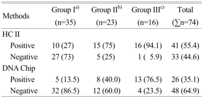
2. 조직학적 소견과 Hybrid Capture II와 DNA chip 결과 조직학적 소견에서의 인유두종 바이러스 고위험군에 대한 각각의 결과를 보면 1군의 경우는 전체 37명 중 Hybrid Cap- ture II에서 양성 10명 (27%), 음성 27명 (73%)이었고 DNA chip에서는 양성 5명 (13.5%), 음성 32명 (86.5%)이었다. 2군 의 경우는 20명 중 Hybrid Capture II에서 양성 15명 (75%), 음성 5명 (25%)이었고 DNA chip의 경우는 양성 8명 (40%), 음성 12명 (60%)이었다. 3군의 경우는 17명 중 Hybrid Cap- ture II에서 양성 16명 (94.1%), 음성 1명 (5.9%)이었고 DNA chip에서는 양성 13명 (76.5%), 음성 4명 (23.5%) 으로 조직 학적 병변의 악성도가 심할수록 인유두종 바이러스 고위험 군에 높은 양성률을 보였다 (Table 2) (P<0.0001).
3. ASCUS에서 각 검사 결과의 비교
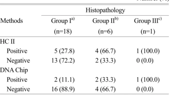
자궁암 세포진 검사에서 ASCUS가 나온 경우 질확대경 검 사와 Hybrid Capture II 그리고 DNA chip의 결과를 비교해 보면, 질확대경 검사에서 정상 또는 만성 자궁내 염증의 경 우가 18명 이었고, 이 중 Hybrid Capture II에 양성인 경우가 5명 (27.8%), 음성 13명 (72.2%)이었고, DNA chip은 양성 2 명 (11.1%), 음성 16명 (88.9%)이었다. 질확대경 검사에서 저 등급 상피내 병변을 보인 경우는 6명 이었고 이중 Hybrid Capture II에서 양성 4명 (66.7%), 음성 (33.3%)이었고, DNA chip은 양성 2명 (33.3%), 음성 4명 (66.7%)이었다. 그리고 질확대경 검사에서 고등급 상피내 병변의 소견을 보인 경우 는 1명 이었는데 이 때 Hybrid Capture II와 DNA chip 검사 모두에서 양성을 보였다 (Table 3).
4. Hybrid Capture II와 DNA chip 결과 일치 여부 Hybrid Capture II와 DNA chip 검사법 모두 인유두종 바이 러스 고위험군에 양성인 경우가 25명, 모두 음성인 경우가 32명으로 57명의 결과가 일치하였고 두 검사간 결과가 다른 경우를 보면, Hybrid Capture II는 양성이나 DNA chip 검사가 음성인 경우가 16명이었는데 이 중 조직학적 검사에서 정상 또는 만성 자궁경부 염증의 경우가 5명, 저등급 상피내 병변 의 경우가 7명, 고등급 상피내 병변의 경우가 4명이었다. 그 리고 DNA chip은 양성이나 Hybrid Capture II에 음성인 경우 가 1명으로 조직학적 검사에서 고등급 상피내 병변의 경우 였다. 인유두종 바이러스의 검출률은 Hybrid Capture II에서 유의하게 높게 나타났다 (Table 4) (P<0.001).
5. 두 검사법의 임상적 유용성 비교
저등급 상피내 병변 (group 2) 이상의 소견을 보일 경우를 인유두종 바이러스 양성으로 간주하였을 때 (Kim et al., 2003), Table 1. Histopathologica) group by cytologic classification
Number (%) Group Ib) Group IIc) Group IIId) Total
(n=35) (n=23) (n=16) (∑n=74) Cytology
Normal,
Benign 17 (48.6) 0 ( 0.0) 1 ( 6.2) 18 (24.3) ASCUSe) 16 (45.7) 9 (39.1) 0 ( 0.0) 25 (33.8) LSILf) 2 ( 5.7) 13 (56.5) 3 (18.8) 18 (24.3) HSILg) 0 ( 0.0) 1 ( 4.4) 12 (75.0) 13 (17.6) a: Histophathologic means colposcopic study.
b: Group I : nonspecific chronic cervicitis.
c: Group II : koilocytosis, mild dysplasia.
d: Group III: moderate dysplasia, severe dysplasia, carcinoma in situ.
e: ASCUS : atypical squamous cell undetermined significance.
f: LSIL : low grade squamous intraepithelial lesion.
g: HSIL : high grade squamous intraepithelial lesio.
Table 2. Results of HPV testing by Hybrid Capture II and DNA chip in different groups
Number (%) Group Ia) Group IIb) Group IIIc) Total Methods
(n=35) (n=23) (n=16) (∑n=74) HC II
Positive 10 (27) 15 (75) 16 (94.1) 41 (55.4) Negative 27 (73) 5 (25) 1 ( 5.9) 33 (44.6) DNA Chip
Positive 5 (13.5) 8 (40.0) 13 (76.5) 26 (35.1) Negative 32 (86.5) 12 (60.0) 4 (23.5) 48 (64.9) a: Group I : nonspecific chronic cervicitis.
b: Group II : koilocytosis, mild dysplasia.
c: Group III: moderate dysplasia, severe dysplasia, carcinoma in situ.
Hybrid Capture II와 DNA chip 검사의 민감도 (sensitivity)는 각각 83.8%, 56.8%, 특이도 (specificity)는 각각 73%, 86.5%, 양성예측도 (positive predictive value)는 각각 75.6%, 80.8%, 음성예측도 (negative predictive value)는 각각 81.8%, 66.7%
였다 (Table 5). 민감도와 음성예측도는 Hybrid Capture II 검 사법이 우수하고 특이도와 양성예측도는 DNA chip 검사가 우수하였다.
고 찰
인유두종 바이러스 감염이 자궁경부암과 관련이 있다고 보고한 이후 많은 연구가 활발히 진행되어 왔으며 유전형에 따라 자궁경부암 발병에 차이가 있음을 알 수 있었다 (Van den Brule et al., 1991; Lorincz et al., 1992; Zur Hausen, 1994;
Bosch et al., 1995). Koutsky 등 (1992)의 보고에 따르면 고위 험군 인유두종 바이러스가 자궁경부 평편상피내 병변을 일 으키는데 8~11배의 상대적 위험이 있다고 하였다. 최근 인 유두종 바이러스의 생식기 감염이 자궁경부암 및 전암 병변 의 발생과 밀접하게 연관된 것으로 알려지면서 인유두종 바 이러스를 진단할 수 있는 선별 검사법에 대한 많은 연구가 진행되고 있는데 Hybrid Capture II 법과 DNA chip 법이 효 용성 측면에서 유용한 방법으로 알려져 있다 (Godfroid et al.,
1998). Hybrid capture 검사법은 미국 FDA (Food and Drug Association) 공인을 받은 검사로서 자궁경부 도말 세포에서 추출한 DNA로부터 chemiluminescent 검출을 이용한 sand- witch capture, molecular hybridization 방법에 근거하여 인유 두종 바이러스를 발견하는 단순한 방법으로 방사성 동위원 소를 사용하지 않으면서도 Southern hybridization과 비교하여 그 일치도가 90%에 이른다고 한다 (Impramin, 1992). 또한 이 방법은 바이러스 양성 반응을 상대적인 양으로 알 수 있 어 병소 부위와 진행을 예측하는 데도 도움을 주며 빠른 시 간 내 검사가 가능하고 일반실험실에서도 시행할 수 있다 (Ho et al., 1995). 그러나 감염된 인유두종 바이러스가 고위 험군에 속하는지 저위험군에 속하는지만 알 수 있고 각각 의 유전형을 판별이 불가능하다는 단점이 있다. 반면 인유두 종 DNA chip 검사는 바이러스의 유전자형을 구분할 수 있으 며 중복 감염에 대한 판독이 가능하다는 장점이 있다 (Kim et al., 2003).
본 연구에서 저등급 상피내 병변 (group 2) 이상의 소견을 보일 경우를 인유두종 바이러스 양성으로 간주하였을 때, Hybrid Capture II와 DNA chip 검사의 민감도 (sensitivity)는 각각 83.8%, 56.8%, 특이도 (specificity)는 각각 73.0%, 86.5%, 양성예측도 (positive predictive value)는 각각 75.6%, 80.8%, 음성예측도 (negative predictive value)는 각각 81.8%, 66.7%
였다. Kim 등 (2003)은 자궁경부 병변에 대한 예민도는 Hy- brid Capture II가 94.9%, DNA chip이 93.7%라고 하여 본 연 구의 결과와 같았다. 한편, Kwon 등 (2002)의 연구에 의하면 침윤성 암을 포함한 고등급 병변에서 DNA chip의 민감도는 86.8%였고, Hybrid Capture II 검사의 민감도는 71.7%로 DNA chip의 민감도가 더 높게 나타났다. 그리고 특이도는 DNA chip 검사법이 34.7%, Hybrid Capture II 법이 71.4%로 Hybrid Capture II가 높았다고 보고하여 본 연구와 다른 결과를 보였 다. Mc Lachlin 등 (2000)은 Hybrid Capture II의 음성예측도 는 93%로 인유두종 바이러스 검사와 세포진 검사의 병행으 로 추적 관찰기간의 연장으로 비용절감의 가능성을 제시하 였고, 한편 DNA chip 검사법의 특이도가 낮음으로 질확대경 검사 등 추가 검사에 따른 비용과 환자의 고통이 우려될 수 있다고 하였다. 자궁경부 세포진 검사에서 ASCUS로 나타난 Table 3. Comparison of histopatholoy, Hybrid Capture II and
DNA Chip in ASCUS
Number (%) Histopathology
Group Ia) Group IIb) Group IIIc) Methods
(n=18) (n=6) (n=1) HC II
Positive 5 (27.8) 4 (66.7) 1 (100.0) Negative 13 (72.2) 2 (33.3) 0 (0.0) DNA Chip
Positive 2 (11.1) 2 (33.3) 1 (100.0) Negative 16 (88.9) 4 (66.7) 0 (0.0) a: Group I : nonspecific chronic cervicitis.
b: Group II : koilocytosis, mild dysplasia.
c: Group III: moderate dysplasia, severe dysplasia, carcinoma in situ.
Table 4. Comparison between HC II and DNA chip test DNA Chip
HC II
Negative Positive Total
Negative 32 1 33
Positive 16 25 41
Total 48 26 74
Table 5. Comparison of screening efficiency between Hybrid Capture II and DNA Chip
Sensitivity Specificity PPVa) NPVb) Methods
(%) (%) (%) (%)
Hybrid Capture II 83.8 73.0 75.6 81.8 DNA Chip 56.8 86.5 80.8 66.7 a: PPV : positive predictive value.
b: NPV: negative predictive value.
경우에 더욱 진행된 병변을 갖고 있거나 추후 상피내암 또는 자궁경부암으로 진행될 가능성이 높은 환자들을 발견하는데 도움이 된다는 보고가 있다 (Nindil, et al., 1998).
Solomon 등 (2001)의 연구에 의하면 ASCUS 환자의 56.6%
에서 Hybrid Capture II 검사에 양성을 보였고 CIN 이상에 서는 민감도가 95.5%라고 하였다. 본 연구에서는 25명의 ASCUS 중 Hybrid Capture II 검사에 양성률이 10명 (40%), 3군에서는 양성율이 100%였다. DNA chip의 경우는 25명 중 5명 (20%)가 양성이었고 3군에서는 100%의 양성률을 보여 ASCUS 세포진 검사에서 고위험 HPV 감염의 진단이 세포 진 검사와 질확대경 검사의 중간단계의 검사로서 자궁경부 암으로 진행될 위험성이 증가된 여성을 선별하는데 유용하 게 쓰일 수 있음을 시사하였다. 995명의 ASCUS를 대상으로 기존의 반복 세포진 검사가 CIN2/3+를 검출할 수 있는 민감 도는 75.8%이었으나 Hybrid Capture II에 의한 인유두종 바 이러스 검사의 민감도는 89.2%가 되어 반복 세포진 검사방 법으로 인유두종 바이러스 검사의 민감도에 도달하려면 6개 월 간격으로 두 번의 반복세포진 검사가 필요하다고 하였다 (Manos et al., 1999).
Manos 등 (1999)은 ASCUS 환자를 추적할 때 인유두종 바이러스 검사를 바로 실시하여 양성인 경우 질확대경 검사 를 실시하고 인유두종 검사에서 음성인 경우 6개월 후 다시 세포진 검사를 반복하게 되면 CIN2/3+를 검출할 수 있는 민 감도가 이론적으로 97.3%까지 올릴 수 있는 반면 세포진 만 으로 추적한다면 6개월 간격으로 6번의 반복 세포진 검사가 요구되기 때문에 인유두종 바이러스 검사를 이용한 추적 프 로그램이 질확대경 회수와 검진 받는 횟수를 줄이고 추적에 실패한 환자의 숫자를 줄여서 비용 효과 면에서 우수하다고 하였다.
결론적으로, Hybrid Capture II 검사와 DNA chip 검사는 각각의 장단점이 있어 검사의 목적에 따라 서로 보완적으로 사용하면 많은 도움이 되리라 생각되며, 자궁 세포진 검사에 서 ASCUS인 경우 고위험 인유두종 바이러스 DNA 검사를 병합 시행하여 고위험군을 선별적으로 조직 검사하는 것이 불필요한 조직 검사를 줄임과 동시에 고위험 병변을 빨리 발 견하여 치료하는데 유용하리라 사료된다.
REFERENCES
Bosch FX, Manos MM, Munoz N. International biological study in cervical cancer study group. Prevalence of human papi- llomavirus in cervical cancer: a worldwide perspective. J Natl Cancer Inst. 1995. 87: 796-802.
Gissman H, Pfister H, zur Hausen H. Human papillomavirus (HPV): Characterization of 4 different isolates. Virology 1977.
76: 569-580.
Godfroid E, Heinderyckx M, Mansy F, Fayt I, Noel JC, Thiry L.
Detection and identification of human papilloma viral DNA, types 16, 18, and 33, by a combination of polymerase chain reaction and a colorimetric solid phase capture hybridization assay. J Virol Methods. 1998. 75: 69-81.
Ho GY, Burk RD, Klein S, Kadish AS, Chang CJ, Palan P, Basu J, Tachezy R, Lewis R, Romney S. Persistent genital human papillomavirus infection as a risk factor for persistent cervi- cal dysplasia. J Natl Cancer Inst. 1995. 20: 1365-1371.
Impramin C. Abstract presented at 92nd General meeting of the Am Soci Microbiol. 1992.
Kim CJ, Jeong JK, Park M, Park TS, Park TC, Namkoong SE, Park JS. HPV oligonucleotide microarray-based detection of HPV genotypes in cervical neoplastic lesions. Gynecol Oncol.
2003. 89: 210-217.
Koss LG. Human papillomavirus testing as a screening tool for cervical cancer. JAMA. 2000. 283: 2525-2555.
Koutsky LA, Holmes KK, Critchlow CW. A cohort study of the risk of cervica lintraepithelial neoplasia grade 2 or 3 in rela- tion to papillomavirus infection. N Engl J Med. 1992. 327:
1272-1278.
Kwon HS, Kim YT, Kim JW, Kim SH. Comparison of oligonu- cleotide microarray-based test with hybrid capture-based test for detecting carcinogenic HPV in patients with CIN and invasive cervical cancer. Gyncol Oncol. 2002. 13: 327-335.
Lorincz AT, Reid R, Jenson AB, Greenberg MD, Lancaster W, Kuman RJ. Human papillomavirus infection of the cervix:
relative risk associations of 15 common anogenital types.
Obster Gynecol. 1992. 79: 328-337.
McNeil C. Getting a handle on ASCUS: a new trial could show how. J Natl Cancer Inst. 1995. 87: 787-790.
Manos MM, Kinney WK, Hurley LB, Sherman ME, Shieh-Ngai J, Kurman RJ, Ransley JE, Fetterman BJ, Hartinger JS, Mc- Intosh KM, Pawlick GF, Hiatt RA. Indentifying women with cervical neoplasia using human papillomavirus DNA testing for equivocal Papanicolaou results. JAMA. 1999. 281: 1605 -1610.
Matsukura T, Sugase M. Relationships between 80 human papi- llomavirus genotypes and different grades of cervical intrae- pithelial neoplasia: association and causality. Virology. 2001.
283: 139-147.
Mc Lachlin CM, Alanen KW, Elit LM, Smith EA, Kerkvliet NA.
Hybrid capture human papillomavirus testing as an adjunt to the follow-up of patients with ASCUS and LGSIL pap smear:
A study of a screening population. Obstet Gyneocol Surv.
2000. 55: 286-288.
Munoz N. Human papillomavirus and cancer: the epidemiological evidence. J Clin Virol. 2000. 19: 1-5.
National Cancer Institute Workshop. The 1988 Bethesda System for reporting cervical/vaginal cytological diagnoses. National Cancer Institute Workshop. JAMA. 1989. 262: 931-934.
Nindil I, Lorincz A, Mielzynska I, Petry U, Baur S, Kirchmayr R, Michels W, Schneider A. Human papillomavirus detection in cervical intraepithelial neoplasia by the second-generation hybrid capture microplate test, comparing two different cervi- cal specimen collection methods. Clin Diagn Virol. 1998. 10:
49-56.
Solomon D, Schiffman MH, Tarone R. Comparison of three management strategies for patients with atypical squamous cells of undetermined significance: Baseline results from from
a randomized trial. J Natl Cancer Inst. 2001. 93: 293-299.
Sun XW, Ferenczy A, Johnson D, Koulos JP, Lungu O, Richart RM, Wright TC Jr. Evaluation of the Hybrid Capture human papillomavirus deoxyribonucleic acid detection test. Am J Obstet Gynecol. 1995. 173: 1432-1437.
Van den Brule AJC, Walboomers AJM, Du Maine M. Difference in prevalence of human papillomavirus genotypes in cyto- morphologically normal smears is associated with a history of cervical intraepithelial neoplasia. Int J Cancer. 1991. 48:
404-408.
Wick MJ. Diagnosis of human papillomavirus gynecologic infec- tions. Clin Lab Med. 2000. 20: 271-287.
Zur Hausen H. Molecular pathogenesis of cancer of the cervix and its causation by specific human papillomavirus types.
Curr Top Microbial Immunol. 1994. 186: 131-156.