Journal of Bacteriology and Virology 2014. Vol. 44, No. 2 p.197 – 205 http://dx.doi.org/10.4167/jbv.2014.44.2.197
The Molecular Epidemiology of Circulating Group A Rotavirus in Gwangju Metropolitan City, Korea: 2008~2012
Hye-Young Kee1, Min Ji Kim1, Sun Hee Kim1, Dong-Ryong Ha1, Eun Sun Kim1, Hye Sook Jeong2, Jae Keun Chung1 and Bong Joo Lee3*
1Health and Environment Institute of Gwangju; 2National Institute of Health, Korea Centers for Disease Control and Prevection, Chungcheongbuk-do; 3College of Veterinary Medicine, Chonnam National University,
Gwangju, Korea
Group A rotaviruses are a major cause of acute gastroenteritis in young children worldwide. For the proper management of rotavirus infections, knowledge of the distribution of G and P genotypes including detection of emerging genotype is crucial. Therefore, the aim of this study is to describe epidemiological changes in rotavirus gastroenteritis in Gwangju metropolitan city, South Korea. Stool samples were collected from 14,314 patients with diarrhea, who visited hospitals in Gwangju from 2008 to 2012. Samples were screened for rotavirus with Enzyme-Linked Immunosorbent Assay (ELISA) method and rotavirus P (VP4), G (VP7) genotypes were determined by reverse-transcription polymerase chain reaction. And we performed nucleotide sequencing analysis. Among a total of 14,314 samples investigated 1,982 samples (13.8%) were ELISA positive. Genotyping of Rotavirus was performed using 526 rotavirus samples. The most prevalent circulating G genotype was G1 (40.5%), followed by G2 (27.6%), G3 (19.4%), G9 (9.7%), G4 (2.5%) and G12 (0.4%).
The predominant type of P genotypes was P[8] (69.6%), followed by P[4] (27.8%) and P[6] (2.3%). In this study, 13 G-P combinations were detected. From 2008 to 2010, G1P[8] was the most prevalent, followed by G3P[8]. Whereas, 2011 and 2012, G2P[4] was the most common, followed by G1P[8]. Rotavirus gastroenteritis is a common disease associated with significant morbidity, mortality and economic burden. Ongoing rotavirus surveillance to understand the distribution of G and P genotypes will be critical for the development of effective prevention measurements.
Key Words: Group A rotavirus, VP4, VP7, RT-PCR, Genotype, Gwangju metropolitan city
INTRODUCTION
로타바이러스는 영아와 어린이에서 설사를 일으키는 가장 중요한 원인체로서 심한 탈수증으로 사망에 이르기 도 하며 (1), 전체 장염 중에서 약 45%를 차지할 정도로 발생률이 높다. 특히 후진국에서는 로타바이러스로 인한
설사가 해마다 1억 3천만명이 발생하고 45만명이 사망하 는 등 경제 · 사회적 측면에서 큰 피해를 주는 것으로 알 려져 있다 (2).
로타바이러스는 분류학적으로 Reoviridae과, Rotavirus 속에 속하고 바이러스 입자는 70 nm 크기이며, 정 20면 체의 외각을 가지며 3층의 capsid 구조 내에 11개의 분절 된 double-stranded RNA 게놈을 갖고 있다. 11개의 분절된
197
Original Article
Received: February 6, 2014/ Revised: June 5, 2014/ Accepted: June 10, 2014
*Corresponding author: Bong Joo Lee. College of Veterinary Medicine, Chonnam National University 300 Yongbongdong, Buk-gu, Gwangju, 500-757, Korea.
Phone: +82-62-530-2850, Fax: +82-62-530-2878, e-mail: [email protected]
**This research as a part of the laboratory investigations of acute diarrhea, managed by the National Institute of Health, Korea Centers for Disease and Prevention and supported by Health & Environment Institute of Gwangju Metropolitan city.
○CCThis is an Open Access article distributed under the terms of the Creative Commons Attribution Non-Commercial License (http://creativecommons.org/license/by-nc/3.0/).
198 H-Y Kee, et al.
게놈은 VP1, VP2, VP3, VP4, VP6, VP7의 구조 단백질과 NSP1, NSP2, NSP3, NSP4, NSP5의 비구조 단백질로 이루 어지며, VP6는 많은 로타바이러스주가 공통적으로 갖고 있는 보존적 항원결정기를 갖기 때문에 항원을 검출하기 위한 진단에 주로 이용된다. 로타바이러스는 VP6 단백질 에 대한 항체를 이용하여 혈청형이 A~G까지 7개의 혈청 군으로 분류되고, 사람에서 설사를 일으키는 모든 로타바 이러스성 설사는 group A 로타바이러스에 의한 것으로 알려져 있다 (3).
Group A 로타바이러스 단백질 중에서 VP4와 VP7은 바이러스의 중화항체 형성과 외피를 구성하는 중요한 단 백질로 VP7은 G type (Glycoprotein), VP4 단백질은 P type (protease-sensitive prtotein)으로 분류할 수 있다 (3~6). 이 와 같은 분류방법으로 현재 G type 27개와 P typ3 35개가 알려졌으며, G type과 P type이 계속적으로 보고되고 있다 (7).
바이러스의 전파는 주로 분변에 오염된 음용수, 손과 음식물 등을 매개로 한 경구감염이 대부분이나, 구토물이 나 비말에 의한 호흡기 분비물에서도 바이러스 입자가 검출되어 공기전염도 가능하다. 로타바이러스의 감염 대 상은 주로 6~24개월의 영 · 유아이며 지역에 관계없이 전 세계에서 발생하는 것으로 알려져 있다 (8). 계절적으로 는 추운 계절에 주로 발생한다고 알려져 있으나, 계절적 인 요인에 관해서는 구체적으로 밝혀진 바 없으며 가정 에서의 낮은 습도가 바이러스의 생존을 높인다는 보고도 있지만 여러 역학적인 상황에서 볼 때 로타바이러스 감 염과의 상관 관계는 관찰되지 않았다고 보고하였다 (9).
또한 Santos 등은 전 세계의 어린이들에서 발생한 로 타바이러스 유전자형 조사결과 G1P[8], G2P[4], G3P[8], G4P[8] 등 4종의 유전자형이 주로 나타났으며, 이들 주요 유전자형이 대륙간에도 다양한 차이가 있음을 보고하였 다 (10). 이와 같이 로타바이러스는 유전자형의 다양성으 로 인하여 해마다 유전자형이 변화하고 새로운 로타바이 러스 유전자형이 계속적으로 출현하는 것으로 알려져 있 어, 지역별, 시기별로 유행하는 로타바이러스의 혈청형이 나 유전자형에 대한 지속적인 연구가 필요한 실정이다.
따라서 본 연구에서는 지난 2008년부터 5년 동안 광주 광역시에서 발생한 급성 위장관염 환아로부터 검출된 로 타바이러스의 발생현황을 살펴보고, G와 P 유전자형에 대 한 분포조사를 통해 지역 내에서 유행하는 로타바이러스 유전자형을 파악하고자 하였다. 이를 통하여 로타바이러
스 백신 후보물질 개발에 필요한 데이터 제공과 로타바 이러스 백신 변이주에 대한 기초자료로 활용하고자 한다.
MATERIALS AND METHODS
시료 채취 및 전처리
2008~2012년까지 광주광역시 소재 종합 병 · 의원에 급 성설사로 입원 또는 내원한 환아로부터 수집된 14,314건 의 대변 검체를 이용하였다. 대변 1 g에 phosphate buffered saline (PBS, 7.2, Sigma, St, Louis, MO, USA)을 혼합하여 10배 희석액을 만든 후, 4℃에서 20분간 3,000 rpm으로 원 심분리하여 상층액을 시험에 사용하였다.
로타바이러스 항원 검사
로타바이러스 항원 검사는 바이러스 항원 검출 Kit (BioTracer Rotavirus ELISA kit, Biofocus, Korea)를 이용하였 다. 항체가 코팅된 각각의 well에 100 μl의 대변 상층액과 HRP conjugated anti rotavirus 항체를 첨가한 후 37℃에서 60분간 반응시켰으며, 반응시킨 well은 5회 세척 후 기질 액을 첨가하여 실온에서 15분간 반응시킨 다음 50 μl의 반응정지액을 가한 후 450 nm에서 판독(Hydroflex, Tecan, Austria)하였다.
로타바이러스 유전자형 분석 시료
14,314건의 대변 검체 중 ELISA 실험을 통해 1,982건의 로타바이러스 항원 양성 검체를 확보하였다. 이들 중에서 로타바이러스 G (VP7) 및 P (VP4) 유전자형에 대한 염기 서열 분석을 위해 2008~2011년의 양성 검체 1,922건 중 25%의 검체를 연도별, 월별, 연령별로 적절히 안배하여 선별한 건수와 2012년의 양성 검체를 더한 526건에 대하 여 유전자형 분석을 실시하였다.
RNA 추출
바이러스의 RNA 추출은 viral nucleic acid prep kit (Greenmate, Korea)을 사용하였고, 추출된 RNA는 -70℃에 보관하면서 VP7와 VP4 유전자 검출용 template로 사용하 였다.
RT-PCR에 의한 VP7, VP4 유전자 증폭
VP7과 VP4 유전자 분석용 primer는 Gentsch 등 (11)이 보고한 VP4 유전자에 특이적인 MY-Con3 (5'-TGG CTT
CGC CAT TTT ATA GAC A-3')와 Con2 (5'-ATT TCG GAC CAT TTA TAA CC-3') primer와 Iturriza-Gomara 등 (12)이 보고한 특이적인 primer로 VP7-F (5'-ATG TAT GGT ATT GAA TAT ACC AC-3')와 VP7-R (5'-AAC TTG CCA CCA TTT TTT CC-3')가 포함된 RT-PCR kit (Accupower Hotstart RT/PCR premix kit, Bioneer, Korea)를 사용하였다. 실험방 법은 VP4와 VP7용 kit를 95℃에서 5분간 가열한 후, 바 로 냉각한 RNA 추출액 20 μl씩을 혼합하여 42℃에서 40분간 cDNA를 합성하였다. 94℃에서 15분간 변성시킨 후 94℃에서 1분, 50℃에서 1분, 72℃에서 1분 동안 35 cycle을 반복하고, 72℃에서 7분간 확장하였다. PCR 산물 은 1.5% agarose gel에서 전기영동하여 ethidium bromide (EtBr)로 염색하여 분석하였다.
염기서열 분석
VP7과 VP4 유전자에 대한 PCR 반응산물은 Qiaquick Gel Extraction kit (Quigen GmbH, Hilden, Germany)로 정제 하였다. 정제된 DNA를 주형으로 각각의 유전자형에 특 이적인 primer를 사용하여 ABI PRISM BigDye terminator (Applied Biosystems, Roster city, CA, USA)로 sequencing reaction을 하였고, 반응산물은 BigDye XTerminator purifi- cation kit (Applied Biosystems, USA)로 정제한 후 Automated DNA sequencer (ABI prism 3100, Applied Biosystems, USA)
로 염기서열 분석을 하였다. 염기서열의 결정은 Lasergene software (DNASTAR Inc., Madison, Wi, USA) 프로그램을 이용하였다.
RESULTS
로타바이러스의 검출현황
2008년부터 2012년까지 광주광역시의 급성설사환자를 대상으로 로타바이러스 양성률을 조사한 결과는 Fig. 1과 같다. 로타바이러스 양성은 검사건수 14,314건 중 1,982건 (13.8%)이었으며, 연도별 양성건수는 2008년 3,079건 중 413건(13.4%), 2009년 3,202건 중 456건(14.2%), 2010년 2,897건 중 428건(14.8%), 2011년 2,921건 중 525건(18.0%), 2012년 2,215건 중 160건(7.2%)으로 나타났다.
월별 로타바이러스 양성률은 Fig. 2와 같다. 즉, 1월 23.1%, 2월 30.0%, 3월 29.4%, 4월 27.1%로 양성률이 20%
이상 유지되었으며, 5월부터 18.2%로 감소하였고 6월부 터 12월까지 10% 미만의 양성률을 보였다. 연도별로 양 성률이 높은 시기는 2008년 2월 39.1%, 3월 32.8%, 2009년 2월 27.1%, 3월 29.6%, 2010년에는 4월 30.6%, 5월 33.3%, 2011년에는 1월 38.4%, 2월 40.5%의 양성률을 나타냈으 며, 2012년에는 3월 17.7%, 4월 18.6%의 양성률을 보였다.
Figure 1. Yearly detection rate of group A rotavirus infection in Gwangju metropolitan city during 2008~2012. Total of 14,314 samples investigated 1,982 samples (13.8%) were ELISA positive. Yearly detection rate were 13.4%, 14.2%, 14.8%, 18.0%, and 7.2%, respectively.
200 H-Y Kee, et al.
로타바이러스의 연령별 검출률
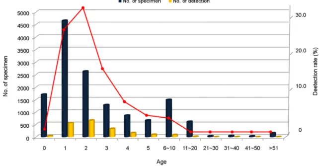
연령별 로타바이러스 검출률은 Fig. 3으로서 0세 2.2%, 1세 28.1%, 2세 33.6%, 3세 16.8%, 4세 8.3%, 5세 4.9%로 5세 이하가 대부분을 차지하였고, 6~10세 4.3%, 11세 이상 은 1% 미만으로 검출되었다.
로타바이러스의 유전자형 분포조사
VP7 및 VP4 유전자 증폭을 위한 RT-PCR 결과 ELISA 양성 검체 526건에 대한 G 및 P형 분석을 위하 여 각각 VP7 및 VP4 유전자 증폭을 위한 RT-PCR을 실 시한 결과, 876 bp와 881 bp 크기의 PCR product를 526건 모두에서 확인하였다.
VP7 유전자 염기서열의 분석
VP7 유전자 염기서열을 분석한 결과 6개의 G형을 확인 하였다(Fig. 4). 유전자형의 분포현황은 G1이 213건(40.5%) 으로 가장 높은 비중을 차지하였고, 다음으로는 G2 145건 (27.6%), G3 102건(19.4%), G9 51건(9.7%), G4 13건(2.5%) 그리고 G12 2건(0.4%) 순으로 나타났다.
연도별 확인결과, 2008년은 G1 37건(46.8%), G3 18건
(22.8%), G2 13건(16.5%), 2009년은 G1 45건(45%), G3 32건 (32%), G2 9건(9.0%), G9 9건(9.0%), 2010년은 G1 63건 (64.3%), G3 21건(21.4%), G2 9건(9.2%)를 차지하여 G1, G3, G2 유전자형 순으로 분포하였다. 그러나 2011년부터 는 G2 50건(41.3%), G1 39건 (32.2%), G3 11건(9.1%), 2012 년 G2 64건 (50%), G1 29건(22.7%), G3 20건(15.6%)으로 나타나 G2, G1, G3 유전자형으로 변화를 보였다.
VP4 유전자 염기서열의 분석
VP4 유전자 염기서열을 분석한 결과 5개의 P형을 확 인하였다(Fig. 5). 유전자형의 분포현황은 P[8]이 366건 (69.6%)으로 가장 높게 나타났으며, P[4] 146건(27.8%), P[6] 12건(2.3%) 그리고 P[9]와 P[19] 유전자형이 각각 1건(0.2%)으로 확인되었다.
연도별 확인결과 2008년은 P[8] 66건(83.5%), P[4] 12건 (15.2%), P[6] 1건(1.3%), 2009년은 P[8] 86건(86%), P[4] 9건 (9.0%), P[6] 4건(4.0%), 2010년은 P[8] 85건(86.7%), P[4] 10 건(10.2%), P[6] 2건(2.0%)을 차지하였다. 그러나 2011년은 P[8] 68건(56.2%), P[4] 50건(41.3%), P[6] 3건 (2.5%)을 차지 하여 P[4] 유전자형이 전년에 비해 큰 폭으로 상승하였으 며, 2012년에는 P[4] 65건 (50.8%), P[8] 61건(47.7%), P[6]
Figure 2. The Monthly detection rate of group A rotavirus infections in Gwangju metropolitan city during 2008~2012. The highest percentages were observed from Jan to April, and percentages showed a marked reduction after May in 2008~2011.
2건(1.6%)으로 나타나 P[4] 유전자형이 가장 높은 분포를 보였다.
G 및 P 유전자 조합형의 분포
G와 P 유전자형의 조합형 분석결과는 G1P[8]형이 212 건(40.3%)으로 가장 높은 분포를 보였으며, G2P[4]형이 142건(27.0%), G3P[8]형은 100건(19.0%), G9P[8]형은 49건
(9.3%), G4P[6]형이 11건(2.1%) 순으로 나타났다(Fig. 6, Table 1). 연도별 확인 결과로는 2008년 G1P[8]형이 37건 (46.8%), G3P[8]형 18건(22.8%), 2009년 G1P[8]형이 44건 (44%), G3P[8]형 32건(32%), 2010년 G1P[8]형이 63건 (64.3%), G3P[8]형 19건(19.4%)으로 유행양상이 비슷하였 다. 그러나 2011년부터 G2P[4]형이 50건(41.3%), G1P[8]
형이 39건(32.2%)으로 나타났고, 2012년에는 G2P[4]형이 Figure 3. Age distribution of group A rotavirus detected 1,982 patients with rotavirus gastroenteritis in Gwangju metropolitan city between 2008 and 2012. Rotavirus was detected mostly in children 5 years (93.7%), and the peak age incidence was 1~2 years.
Figure 5. Frequencies (%) of group A rotavirus P-genotypes among 526 samples in Gwangju metropolitan city from 2008 to 2012. P typing of the VP4 showed that P[8] was the most pre- dominant circulating genotype (69.6%), followed by P[4] (27.8%), P[6] (2.3%).
Figure 4. Frequencies (%) of group A rotavirus G-genotypes among 526 samples in Gwangju metropolitan city from 2008 to 2012. G typing of the VP7 showed that G1 (40.5%), the most predominant circulating genotype (69.6%), followed by G2 (27.6%), G3 (19.4%), G9 (9.7%).
202 H-Y Kee, et al.
Table 1. Distribution of group A rotavirus G & P genotypes detected acute gastroenteritis in Gwangju metropolitan city from 2008 to 2012
Year Genotype
No. of samples (%)
2008 2009 2010 2011 2012 Total
G1P[4] 0 1 (1.0) 0 0 0 1 (0.2)
G1P[8] 37 (46.8) 44 (44.0) 63 (64.3) 39 (32.2) 29 (22.7) 212 (40.3)
G2P[4] 12 (15.2) 8 (8.0) 9 (9.2) 50 (41.3) 63 (49.2) 142 (27.0)
G2P[8] 1 (1.3) 1 (1.0) 0 0 1 (0.8) 3 (0.6)
G3P[4] 0 0 1 (1.0) 0 0 1 (0.2)
G3P[8] 18 (22.8) 32 (32.0) 19 (19.4) 11 (9.1) 20 (15.6) 100 (19.0)
G3P[19] 0 0 1 (1.0) 0 0 1 (0.2)
G4P[6] 1 (1.3) 4 (4.0) 1 (1.0) 3 (2.5) 2 (1.6) 11 (2.1)
G4P[8] 0 0 0 2 (1.7) 0 2 (0.4)
G9P[4] 0 0 0 0 2 (1.6) 2 (0.4)
G9P[8] 10 (12.7) 9 (9.0) 3 (3.1) 16 (13.2) 11 (8.6) 49 (9.3)
G12P[6] 0 0 1 (1.0) 0 0 1 (0.2)
G12P[9] 0 1 (1.0) 0 0 0 1 (0.2)
Total 79 (100) 100 (100) 98 (100) 121 (100) 128 (100) 526 (100)
Figure 6. Distribution of group A rotavirus G & P genotypes detected acute gastroenteritis in Gwangju metropolitan city from 2008 to 2012. The most prevalent types in 2008~2010 was G1P[8], followed by G3P[8]. Whereas, 2011 and 2012, G2P[4] was the most common, followed by G1P[8].
63건 (49.2%), G1P[8]형이 29건(22.7%)의 분포를 보였다.
DISCUSSION
본 연구는 지난 5년간(2008~2012) 광주광역시에 소재 한 병 · 의원에 입원 또는 내원한 급성설사환자를 대상 으로 로타바이러스 발생현황과 유전자형에 대한 분포를 조사한 결과이다. 연도별 검출률을 보면 2008년 13.4%, 2009년 14.2%, 2010년 14.8%, 2011년 18.0%로 꾸준히 증 가추세를 보였으나, 2012년에는 7.2%로 크게 감소하였다.
이 같은 결과는 국가 예방접종 프로그램 대상이 아님에 도 예방 접종률 상대평가에서 2007년 27.7%에서 2010년 60.8%로 접종률이 상승했다는 보고와 같이 백신 접종률 증가로 인한 효과로 추측된다 (13).
월별 로타바이러스의 검출양상을 살펴본 결과 2012년 을 제외하고 2008~2011년까지는 1월에서 4월까지 30%
정도의 양성률을 보였고 5월부터는 감소하는 경향을 나 타냈다. 일반적으로 온대지방은 로타바이러스 감염증이 늦가을부터 초겨울까지 유행한다고 알려져 있었으나 (14), 이번 연구결과는 Seo 등, Suzuki 등이 90년대 말부터 로 타바이러스 감염증이 초봄에 다발한다는 보고와 비슷하 였다 (15, 16). 또한 Konno 등이 보고한 12월~2월의 발생 률이 66%를 보이고, 봄철인 3~5월 경에는 56%의 발생 양상을 보인다는 보고와도 유사하였다 (17). 연령별 검 출률은 0세 2.2%, 1세 28.1%, 2세 33.6%, 3세 16.8%, 4세 8.3%, %, 5세 4.9%로 분포하였고, 5세 이하가 93.7%로 대 부분을 차지하였으며 특히 1~2세에서 61.7%의 검출률을 보임으로서 영 · 유아에서 설사를 일으키는 중요한 원인체 가 로타바이러스임을 확인할 수 있었다.
유전자형 분포 조사결과 VP7은 G type이 6종으로 확 인되었고, 주로 G1 (40.5%), G2 (27.6%), G3 (19.4%), G9 (9.7%), G4 (2.5%), G12 (0.4%)의 순으로 유전자형이 분포 하였다. 연도별 확인결과 2008~2010년은 G1>G3>G2>G9 순으로 유전자형이 분포하였으나, 2011년 G2>G1>G9>G3, 2012년에는 G2>G1>G3>G9 순으로 유전자형이 변화하였 다. 이는 Kim 등 (18)이 2006~2007년에 우리지역에서 조 사한 G1>G3>G4>G2의 결과와 비교하여 같은 지역임에 도 불구하고 시기별로 유행하는 유전자형이 다르게 분포 함을 알 수 있었다. 그리고 Choi 등 (19)은 인천지역에서 2005~2010년까지 조사결과 G1>G4>G3>G2>G9의 분포현 황을 보고하였고, Shim 등 (20)은 서울, 인천, 강원도 지역
에서 2008~2010년까지 조사결과 G1>G3>G4>G9>G2의 결과를 보고함으로써 연도와 지역별로 유전자형의 분포 결과가 상이함을 확인하였다. 또한 2006년 이후의 우리지 역에 대한 연구결과에서도 VP7에 의한 혈청형은 G1형이 가장 많이 분포하는 유전자형이었으나 2011년부터 G2형 으로 변화하는 양상을 보이므로 출현 가능성이 있는 새 로운 유전자형에 대해서도 관심을 가져야 할 것이다. 또 한 이란 (21), 방글라데시 (22), 프랑스와 말레이시아 (23) 등에서 실시한 로타바이러스 유전자형 분포조사에서는 가장 우세한 유전자형이 G4형임을 보고하여 본 연구결과 와 다른 결과를 나타냈다. G9 유전자형은 주요 유전자형 은 아니지만 3.1~12.7%를 차지하여 매년 검출되었고, 이 는 사람뿐만 아니라 양이나 돼지에서도 검출되는 유전자 형으로 알려져 있으므로 지속적인 조사가 필요할 것으로 생각된다 (24).
VP4에 대한 혈청형인 P type은 5종의 유전자형을 확인 하였으며, P[8] (69.6%), P[4] (27.8%), P[6] (2.3%), P[9]와 P[19] (0.2%)의 분포를 나타냈다. 그러나 연도별 분포현황 에서는 2011년에 P[4] 유전자형의 분포율이 41.3%로 증가 하기 시작하여 2012년에는 50.3%를 차지함으로써 가장 높은 분포를 보였다. P 유전자형 분포조사에 대해 Kim 등 (18), Choi 등 (19), Shim 등 (20)은 P[8]>P[6]>P[4] 유전자 형이 주로 분포한다고 보고하였으며, 가장 우세한 유전자 형은 P[8]형으로 동일한 결과를 보였으나, P[4]형과 P[6]
형에 대한 분포현황은 약간 상이하였다. 이처럼 로타바이 러스 유전자형 분포현황에 있어서도 지역마다 차이를 보 이므로 꾸준한 조사가 수행되어야 할 것이라 생각되었다.
G와 P 조합형 조사결과 13개의 유전자형이 확인되었고, G1P[8] (40.3%), G2P[4] (27.0%), G3P[8] (19.0%), G9P[8]
(9.3%), G4P[6] (2.1%) 순으로 주요 유전자형이 분포하였 다. G1P[8] 유전자형의 세계적 분포현황 조사에서 미국, 유럽, 호주 같은 선진국의 경우 70% 이상, 남미는 30%, 아프리카에서는 23%의 분포율을 보인다고 보고하였고 (10), Gentsch 등도 세계적으로 분포하는 우세한 유전자형 이 G1P[8], G2P[4], G3P[8], G4P[8], G9P[8]이었음을 보고 하여 본 연구결과와 유사한 경향을 나타내었다 (25). G와 P 조합형에 대한 연도별 분포에서는 2008년 G1P[8]>
G3P[8]>G2P[4]>G9P[8]>G4P[6], 2009년 G1P[8]>G3P[8]>
G9P[8]>G2P[4]>G4P[6], 2010년에는 G1P[8]>G3P[8]>G2P[4]
>G9P[8], 2011년 G2P[4]>G1P[8]>G9P[8]>G3P[8]>G4P[6]로 확인되었다. 그 중 가장 우세한 경향을 보이는 유전자형
204 H-Y Kee, et al.
이 G1P[8]에서 G2P[4]로 변화하기 시작하여, 2012년에는 G2P[4]>G1P[8]>G3P[8]>G9P[8]>G4P[6], G9P[4]의 결과를 보였다. Shim 등 (20)은 2008~2010년까지 서울, 인천, 강 원도 지역에서 G1P[8]>G3P[8]>G4P[6]>G9P[8] 유전자형 의 분포를, Choi 등 (19)은 2005년부터 2010년까지 인천지 역에서 G1P[8]>G4P[6]>G3P[8]>G2P[4]형의 분포현황을 보 고하여 주요 유전자형은 본 연구결과와 동일하였으나 분 포율에서 차이를 보였다. 그러나 Kim 등 (26)은 서울지역 의 유전자형에 대한 조사 결과 2007~2009년까지 G9P[8]>
G1P[8]>G3P[8]이 유행하였고, 2009~2010년의 조사에서는 G4P[6]>G3P[8]>G1P[8]을 주요 유전자형으로 보고하였다.
그 중 G3P[8] 유전자형이 11.7%에서 28%로 크게 상승하 였고, 최근의 연구결과 G4P[6] 유전자형이 가장 높은 비 율로 분포함을 보고하여 본 연구결과와 차이가 있었다.
이상의 결과로 광주지역에서 2008~2012년까지 유행한 G type은 G1형에서 최근 G2형으로 변화하였으며, P type 은 P[8]형이 꾸준히 우세하였으나 2011년부터 P[4]형이 증가하고 있음을 확인하였다. 또한 P와 G 조합형은 주로 G1P[8] 유전자형이 40% 이상 분포하였으나, 2011년부터 G2P[4]형의 검출률이 50% 정도로 증가함으로써 유전자 형이 다양하게 변화함을 확인할 수 있었다.
본 연구에서 로타바이러스 백신 접종여부에 대한 조사 결과를 확인하지 못한 것이 제한점으로 생각되나, 연구기 간 내의 G1~G4 유전자형 분포율이 89.9%로 높기 때문 에 백신 접종으로 로타바이러스 감염증 예방이 가능할 것으로 보이며 향후 설사질환 감시사업을 수행할 때 백 신 접종 여부를 확인하여 백신 접종 유무와 로타바이러 스 감염증간의 상관관계를 파악해 보는 것도 좋은 연구 과제가 될 것으로 생각된다.
이상과 같은 결과로 광주광역시에서 유행하고 있는 로 타바이러스의 유전자형이 다양함을 확인하였으며, 현재 의 백신에는 포함되지 않았으나 발병빈도가 높은 G9과 P[4] 유전자형이 포함된 새로운 백신 개발에 필요한 역학 적 자료로서 활용가치가 높을 것으로 생각된다. 그리고 새로운 로타바이러스의 출현에 대비한 신속한 진단법의 확립과 지역사회에서 유행하는 로타바이러스 유전자형에 대한 분포조사를 위해 실험실 감시체계를 강화해야 할 것으로 사료된다.
REFERENCES
1)Kapikian AZ, Flores J, Midthun K, Hoshino Y, Green KY, Gorziglia M, et al. Strategies for the development of a rotavirus vaccine against infantile diarrhea with an update on clinical trials of rotavirus vaccines. Adv Exp Med Biol 1989;
257:67-89.
2) Parashar UD, Burton A, Lanata C, Boschi-Pinto C, Shibuya K, Steele D, et al. Global mortality associated with rotavirus disease among children in 2004. J Infect Dis 2009;1:S9-S15.
3) Estes MK, Kapikian AZ, Knipe DM, Griffin DE, Lamb RA, Straus SE, et al. Rotaviruses. In: Fields. Virology. Fifth ed.
Philadelphia: Lippincott Williams & Wilkins; 2007. p. 1917-74.
4)Ciarlet M, Estes MK. Interactions between rotavirus and gastrointestinal cells. Curr Opin Microbiol 2001;4:435-41.
5) Gorziglia M, Larralde G, Kapikian AZ, Chanock RM. Antigenic relationships among human rotaviruses as determined by outer capsid protein VP4. Proc Natl Acad Sci U S A 1990;87:7155-9.
6)Green KY, Sears JF, Taniguchi K, Midthun K, Hoshino Y, Gorziglia M, et al. Prediction of human rotavirus serotype by nucleotide sequence analysis of the VP7 protein gene. J Virol 1988;62:1819-23.
7) Matthijnssens J, Ciarlet M, McDonald SM, Attoui H, Bányai K, Brister JR, et al. Uniformity of rotavirus strain nomenclature proposed by the Rotavirus Classification Working Group (RCWG). Arch Virol 2011;156:1397-413.
8) Barnett B. Viral gastroenteritis. Med Clin North Am 1983;67:
1031-58.
9) Ansari SA, Springthorpe VS, Sattar SA. Survival and vehicular spread of human rotaviruses: possible relation to seasonality of outbreaks. Rev Infect Dis 1991;13:448-61.
10) Santos N, Hoshino Y. Global distribution of rotavirus serotypes/
genotypes and its implication for the development and implemention of an effective rotavirus vaccine. Rev Med Virol 2005;15:29-56.
11) Gentsch JR, Glass RI, Woods P, Gouvea V, Gorziglia M, Flores J, et al. Identification of Group A rotavirus gene 4 types by polymerase chain reaction. J Clin Microbiol 1992;30:1365-73.
12)Gόmara MI, Cubitt D, Desselberger U, Gray J. Amino acid substitution within the VP7 protein of G2 rotavirus strains associated with failure to serotype. J Clin Microbiol 2001;39:
3796-8.
13)Choe YJ, Yang JJ, Park SK, Choi EH, Lee HJ. Comparative Estimation of Coverage between National Immunization Program Vaccines and Non-NIP Vaccines in Korea. J Korean Med Sci 2013;28:1283-8.
14) Brandt CD, Kim HW, Rodriguez WJ, Arrobio JO, Jeffries BC, Parrott RH. Rotavirus gastroenteritis and weather. J Clin Microbiol 1982;16:478-82.
15)Seo JK, Sim JG. Overview of rotavirus infections in Korea.
Pediatr Int 2000;42:406-10.
16) Suzuki H, Sakai T, Tanabe N, Okabe N. Peak rotavirus activity shifted from winter to early spring in Japan. Pediatr Infect Dis J 2005;24:257-60.
17) Konno T, Suzuki H, Katsushima N. Influence of temperature and relative humidity on human rotavirus infection in Japan. J Infect Dis 1983;147:125-8.
18) Kim SH, Cheon DS, Jeong HS, Kim MJ, Kee HY, Seo JJ, et al. Distribution of group A rotavirus genotypes circulating in Gwangju, Korea. J Bacteriol Virol 2009;39:21-8.
19)Choi HJ, Oh BY, Lee MY, Koh YJ, Gong YW, Hur MJ, et al.
The prevalence and distribution of the P and G Genotypes of a group A rotavirus detected in acute gastroenteritis patients from Incheon. J Life Sci 2012;22:600-4.
20) Shim JO, Thai Than V, Ryoo E, Lim I, Yoon Y, Kim K, et al.
Distribution of Rotavirus G and P genotypes approximately
two years following the introduction of rotavirus vaccines in South Korea. J Med Virol 2013;85:1307-12.
21) Kargar M, Akbarizadeh AR. Prevalence and molecular geno- typing of group A rotaviruses in Iranian children. Indian J Virol 2012;23:24-8.
22) Ahmed MU, Alam MM, Chowdhury NS, Haque MM, Shahid N, Kobayashi N, et al. Analysis of human rotavirus G sero- type in Bangladesh by enzyme-linked immunosorbent assay and polymerase chain reaction. J Diarrhoeal Dis Res 1999;17:
22-7.
23) Saravanan P, Ananthan S, Ananthasubramanian M. Rotavirus infection among infants and young children in Chennai, South India. Indian J Med Microbiol 2004;22:212-21.
24) Kapikian AZ, Hoshino Y, Chanock RM. Rotaviruses. In: Fields, Knipe DM, Howley RM, Griffin DE, et al (eds). Virology.
Philadelphia: Lippincott Williams and Wilkins; 2001. p. 1787 -825.
25)Gentsch JR, Laird AR, Bielfelt B, Griffin DD, Banyai K, Ramachandran M, et al. Serotype diversity and reassortment between human and animal rotavirus strains: implications for rotavirus vaccine programs. J Infect Dis 2005;192:146-59.
26) Kim EJ, Kim MS, Chae YZ, Cheon DS. Genotype of grouop A rotavirus isolated in acute gastroenteritis patients and groundwater in Seoul, Korea. Kor J Microbiol 2011;47:323-7.