nerve damage, and non-union fractures2. These disfigurement and dysfunction contribute to acute and long-term psycho- logical problems as well to social and economic burdens8,10,11 since these detrimental outcomes are largely due to lack of full restoration of function and aesthetics found in the avail- able treatments.
Due to the complexity of CMF injuries, the affected hard and soft tissues within the wound environment are unsuitable to support proper healing4,12,13. Moreover, management of CMF injuries is extremely challenging and involves a multi- disciplinary team of professionals for the treatment of facial bone fractures, dentoalveolar trauma, and soft tissue injuries as well as associated injuries, mainly to the head and neck regions3,11,14,15. Major bone and soft tissue reconstruction often requires the use of autografts or allografts. Although autografts—considered the “gold standard”—and allografts are very attractive for their resorption, mechanical properties and immunological characteristics, both approaches have multiple limitations related to tissue availability, donor site morbidity and infection4,16-19. Since major drawbacks for us- ing autografts and allografts include the need to manually sculpture the grafts in the desired shape4,20, synthetic alterna- tives using additive manufacturing have become an attractive option4,12,17.
I. Introduction
Although the patterns of incidence and their causes have changed over the decades, craniomaxillofacial (CMF) injuries still occur worldwide. The most common causes include traf- fic and sports-related accidents, assaults, falls, civilian war- fare1-3, as well as diseases, congenital disorders and surgery4. CMF injuries are typically characterized by bone fractures in the frontal, orbital, nasal, maxillary and mandibular regions2 and soft tissue damage such as complex lacerations, tissue avulsions, nerve and vessel injuries, and burns5-9. These com- plex injuries can compromise vital structures. Consequences of CMF injuries are disfigurement and dysfunction, includ- ing compromised airway, hemorrhaging, infection, scarring,
Joo L. Ong
Department of Biomedical Engineering, College of Engineering, The University of Texas at San Antonio, One UTSA Circle, San Antonio, TX 78249, USA
TEL: +1-210-458-7208 FAX: +1-210-458-5515 E-mail: [email protected]
ORCID: http://orcid.org/0000-0003-3330-2390
This is an open-access article distributed under the terms of the Creative Commons Attribution Non-Commercial License (http://creativecommons.org/
licenses/by-nc/4.0/), which permits unrestricted non-commercial use, distribution, and reproduction in any medium, provided the original work is properly cited.
CC
Three-dimensional printing for craniomaxillofacial regeneration
Laura Gaviria, Joseph J. Pearson, Sergio A. Montelongo, Teja Guda, Joo L. Ong
Department of Biomedical Engineering, College of Engineering, The University of Texas at San Antonio, San Antonio, TX, USA
Abstract(J Korean Assoc Oral Maxillofac Surg 2017;43:288-298)
Craniomaxillofacial injuries produce complex wound environments involving various tissue types and treatment strategies. In a clinical setting, care is taken to properly irrigate and stabilize the injury, while grafts are molded in an attempt to maintain physiological functionality and cosmesis. This often requires multiple surgeries and grafts leading to added discomfort, pain and financial burden. Many of these injuries can lead to disfigurement and resultant loss of system function including mastication, respiration, and articulation, and these can lead to acute and long-term psychological impact on the patient. A main causality of these issues is the lack of an ability to spatially control pre-injury morphology while maintaining shape and function.
With the advent of additive manufacturing (three-dimensional printing) and its use in conjunction with biomaterial regenerative strategies and stem cell research, there is an increased potential capacity to alleviate such limitations. This review focuses on the current capabilities of additive manufacturing platforms, completed research and potential for future uses in the treatment of craniomaxillofacial injuries, with an in-depth discussion of regeneration of the periodontal complex and teeth.
Key words: Three-dimensional printing, Periodontium, Hydroxyapatite, Biomaterials
[paper submitted 2017. 8. 31 / accepted 2017. 9. 11]
Copyright © 2017 The Korean Association of Oral and Maxillofacial Surgeons. All rights reserved.
availability. All of these limitations have been investigated over the last two decades in order to address stringent per- formance and safety concerns12,19,22,27. As a consequence, 3D printing technology has become more popular in tissue en- gineering and has found many applications in the fabrication of custom implants for the reconstruction of CMF defects26. This has allowed for precise adaptation of the implant to the region of implantation, reducing surgical times and leading to lesser chances for infection, faster recovery and better cos- mesis15,18,19,25,28. 3D printing has also been introduced into the surgical field as a tool for pre-surgical planning25,29, allowing surgeons to review and interact with the anatomical models, thereby facilitating the understanding of the morphology and making it easier to perform complex surgeries in less time18,24,27,30,31. The uses of 3D printing for preoperative plan- ning have been previously described in literature26,32. While these planning techniques have expanded the knowledge in both the scientific and medical communities, the use of 3D printing towards tissue regeneration focuses on the need for specific biomaterial-based printing rather than rapid prototyp- ing for surgical guidance.
1. 3D printed biomaterials for CMF repair
Early 3D printing research focused on the use of metals and ceramics12 for bone tissue engineering. Ceramic scaf- folds have been 3D printed and tested in vitro under static and dynamic conditions, achieving high printing resolution, structural mechanical support and cell growth20,23,33. Today, 3D printing applications are investigated not only for bone reconstruction but also for replacement of soft tissues, using a variety of synthetic and biological materials including met- als, ceramics and polymers12,18,20,22-24,27-29,33. Although most known biomaterials can be processed using 3D printing, extensive optimization of processing and post processing As mentioned before, the primary goals of CMF repair are
restoration of aesthetics and function, both requiring precise pre-surgical planning as well as prostheses and implants fabricated in very unique geometries and sizes15,18,20,21. In the past decade, techniques such as additive manufacturing (e.g., three-dimensional [3D] printing) have been explored for tis- sue engineering purposes, especially for dental and CMF repair. This review focuses on tissue regenerative strategies for the CMF as a whole along with a focused discussion on 1) the regeneration of the periodontium and teeth within the oral cavity, and 2) providing an outlook on the advantages and limitations of current additive manufacturing, treatments and tissue regenerative research. The ability to harness the successes of tissue regeneration within specific regions of the CMF, such as the periodontium and teeth, could lead to a combined approach for regeneration of a larger region. This review will also discuss that potential and the ability of 3D printers to create a platform for manufacturing rather than the multiple manual manufacturing techniques currently used.
II. 3D Printing Technology for CMF Surgery
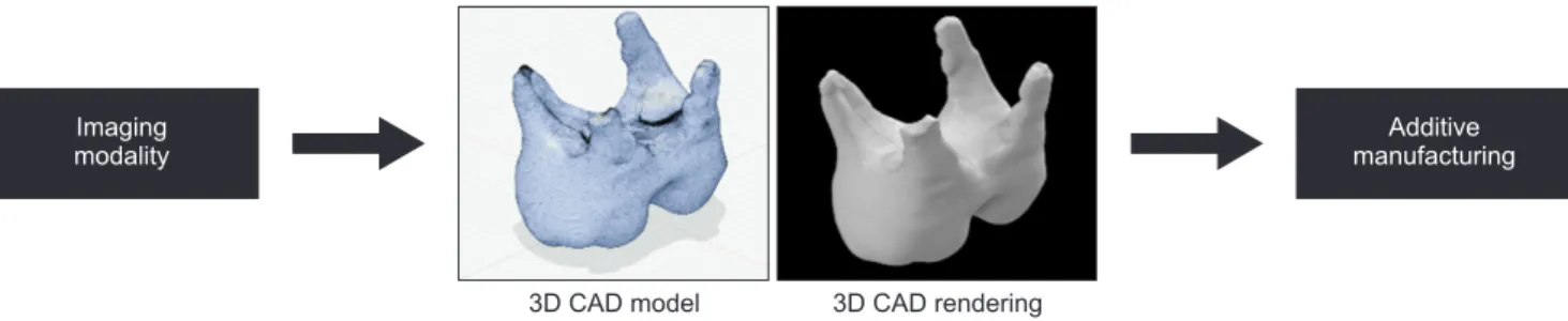
Additive manufacturing techniques, such as 3D printing, use the process of joining materials to create objects from digital 3D model data20. For biomedical applications, 3D printing can be used for the fabrication of complex scaffold shapes that are specific to patients using computer aided de- sign (CAD) and advanced medical imaging techniques such as magnetic resonance imaging and computed tomography (CT)12,18,19,22-29.(Fig. 1)
Although many industries have benefited from the devel- opment of additive manufacturing technologies since the mid-1980s, their applications in the biomedical field have been slow due to technical challenges such as limited ac- curacy, low mechanical properties and lack of biomaterial
Imaging modality
Additive manufacturing
3D CAD model 3D CAD rendering
Fig. 1. The advent of additive manufacturing allows for the use of medical and research based imaging modalities to create three-dimen- sional (3D) computer aided design (CAD) models. These models can be rendered for visual enhancement and surgical simulation or the models can be converted to proper code for additive manufacturing into a graft, prototype or surgical model.
Laura Gaviria et al: Three-dimensional printing for craniomaxillofacial regeneration. J Korean Assoc Oral Maxillofac Surg 2017
biocompatibility properties, ease of use, cost and degradation kinetics24. The most used polymers in 3D printing for hard and soft tissues are polylactic acid (PLA), poly(caprolactone) (PCL), polyether ether ketone (PEEK)24.
4) Composites
Although the initial 3D printing focused on pure materials, composite materials appear to be a most promising approach for the improvement and optimization of biomaterials at the engineering level22. The main goal of using composite inks is to enhance ink properties such as synthesis, printability, mechanics and bioactivity24. Commercial 3D printers can be adapted for co-printing of polymer blends (polymer-based composites) and hydrogel-based composites24. Other alterna- tives to improve mechanical and biological properties have been to add powdered ceramics as well as metals to pure polymers or polymer blends which can be printed using 3D printing nozzles17,22-24,34.
III. Advantages and Limitations of 3D Printing Technology
3D printing offers outstanding possibilities in many aspects when compared to other methods, because it is more precise, faster, easily produced and cost-effective in a limited number of cases16,26, eliminating highly specialized manual labor. 3D printing also offers advantages such as high versatility and capability to print complex designs20,27 using a large variety of biomaterials that can be printed individually or in combi- nation18,21,22.
Although industrial 3D printers, such as Stratasys Polyjet printers, have reached extremely high resolution (~16 µm) in the past few years4,16,21,23, the use of 3D printing technology for implantable biomedical devices is still severely limited by available printable materials that cannot compete with tradi- tional biomedical treatments. The main challenges are the use of processing methods required to work with materials that are not easily printed12,23,24,35 with the use of organic solvents and high processing temperatures which can harm and reduce the working life of 3D printers that are not specifically opti- mized for those very narrow uses23,35. In summary, the main issues to be addressed in 3D printing of biomaterials are:
• The feasibility of low temperature 3D printing, especially for ceramic materials to make them more stable (control shrinkage) with the potential of incorporating biomolecules and polymers12,17,34.
parameters are needed to produce complex structures (e.g., interconnected porosity) with structural integrity, high quality and safety (e.g., sterility)12,14,22. The following sections will describe the different types of materials used in CMF repair and approaches for 3D printing them.
1) Titanium
Titanium has a long history as a bone implant material because of its biocompatibility, strength to weight ratio and osteoconductive properties. In cranioplasty, titanium has been used in the form of sheets and meshes prefabricated using 3D printing techniques such as direct metal laser sintering15. Dental and CMF implants, plates and screws have been fab- ricated using titanium4 and although the use of this material has proven to be useful and clinically established, titanium implants cannot be replaced by ingrowing bone or function as a carrier for bioactive molecules14,18.
2) Ceramics
Ceramics are commonly used in biomedical applications due to their high stiffness and bioactivity. Currently ceramic- based inks are available for direct 3D printing to fabricate pa- tient specific bone grafts for dental and CMF repair applica- tions4,24. The most popular ceramics are calcium phosphates such as tri-calcium phosphates (TCP) and hydroxyapatite (HA) because of their excellent bioactivity, osteoconductivity, similarity to the mineral component of bone and bioresorp- tive properties12,16,17,19,22,34. Previous studies have demonstrated their suitability for the build-up of 3D printed structures with resolutions of ~50 µm16 as well as structures with controlled open pores that are capable of increasing osteoconduction in vivo12. Also, evidence has shown the printability of a combina- tion of TCP and bioactive glass which can be compositionally optimized for tailored biodegradation16,19. Extensive research of 3D printing parameters such as powder packing, drop pen- etration, particle size, and calcium phosphate ratios has to be done for optimization of the 3D printed constructs17,24,34,35.
3) Polymers
Blends of natural and synthetic polymeric biomaterial inks are adequate for printing scaffolds used in medical applica- tions and can be customized for individual needs and applica- tions in the CMF region22. In general, synthetic polymers are often poorly soluble in aqueous media, meaning that organic solvents must be used which raises concerns related to bio- compatibility and large scale production of implants. None- theless, synthetic polymers are of great interest due to their
muscles, vessels, ligaments, tendons and nerves in the CMF complex23. To such end, the following sections focus on spe- cific treatments and approaches for regeneration of the peri- odontium and teeth within the oral cavity.
IV. Regeneration of the Periodontium
Periodontal disease afflicts approximately half of the popu- lation over thirty years of age in the United States. Genetic, environmental, dermatological and hematological factors all influence the high prevalence of this disease36. Around 30%
of the cases are characterized by moderate periodontitis with mild and severe cases about even. However, the number of patients with moderate to severe periodontitis increase to 64% after 65 years37. This reduces the function of the peri- odontium (combined cementum, periodontal ligament [PDL]
and alveolar bone) which is to secure the teeth to the man- dible. In severe cases of periodontitis, the periodontium is de- stroyed and ultimately causing tooth loss38,39. This destruction also leads to various complications and medical intervention, highlighting the need for a viable tissue regenerative ap- proach.
1. The periodontium and strategies for regeneration
The complexity of tissue regeneration of the periodontium lie within the tissues of which it is comprised. The periodon- tal structure begins with the PDL which is unique in its shape and function. The web-like PDL connects the alveolar bone root to the cementum of the tooth providing tensile strength in a gap less than half of a millimeter38 and support for masti- cation. Unlike other ligament attachments to bone throughout
• The development of aqueous binder solutions used in scaffold fabrication to avoid the use of organic solvents that can compromise not only the biocompatibility of the scaffold but also the lifespan of the printer heads. This idea has been gaining attention because of its significant contribution to large-scale manufacturing12,17,34,35.
• Achievement of high resolution and accurate porous interconnected structures with adequate mechanical and degradation properties. This approach can be optimized us- ing composite biomaterial blends, as well as post processing treatments12,14,16,22-24.
Overcoming the above mentioned technological limita- tions will finally lead to the incorporation of cells and growth factors/drugs to 3D printed scaffolds, since most of the cur- rently used processing techniques cannot sustain the viability of cells and biomolecules after printing. This approach can tremendously impact the performance of the 3D printed con- structs by balancing mechanical, biological, drug delivery and degradation properties12,20,22,30. Future advancements in this field can be based on “multi-color” or “multi-component”
printing, where each ink can be positioned on a precise loca- tion, offering the potential to simultaneously arrange multiple types of cells, deposit multiple extra cellular matrix materials, and exert point-to-point control over bioactive agents for bio- logical tissue manufacturing23. However, this approach only relies on the modification of current 3D printing machines and processing temperatures in order to maintain adequate conditions for cells and biomolecules23. To date, some bio- printing technologies have been introduced and investigated in order to achieve this purpose20, providing new insights into the future of complex tissue regeneration for bone, cartilage,
Fig. 2. The periodontium complex is comprised of cementum (A), the peri- odontal ligament and alveolar bone.
These all have distinct porosity and strength. Additive manufacturing allows for different porosities and strengths (B, C) and the ability to create variations within the same grafts (D).
Laura Gaviria et al: Three-dimensional printing for craniomaxillofacial regeneration. J Korean Assoc Oral Maxillofac Surg 2017
A
B
C
D
Cementum
Periodontal ligament
Alveolar bone
ing platforms such as the EnvisonTec 3D Bioplotter have broadened the capacity to 3D print a variety of biomaterials through ink development, and the continued reduction in cost of these platforms will make these technologies more readily available and should further expand the additive manufactur- ing tactics to periodontium regeneration. The following are current approaches using several materials, growth factors and cells.
2. Materials, manufacturing and cells in periodontium regeneration
Several studies performed for the regeneration of the peri- odontum are summarized in Table 1. A recent study created Mg-calcium-silicate cements with varying amounts of Mg which were seeded with PDL cells and evaluated for both odontogenesis and angiogenesis, which is a vital component the axial skeletal in which the ligament generally forms one
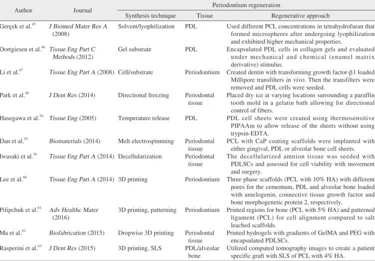
insertion point on the bone, the PDL lines the entire surface with multiple small fibrous units being inserted at varying angles. Connective tissue and vascularization are intertwined with the PDL40. Tissue regenerative approaches may seek to form a viable PDL structure, but most incorporate the entire periodontal complex or periodontium to combat the detach- ment of tissue caused by periodontitis41. The overall necessity for such approaches arises from the increasing concern of periodontitis described above and the lack of periodontium in current dental implants which will be discussed later in this review. Regenerative approaches for the periodontium have included growth factors, various cell types and materials that seek to provide adequate porosity and mechanics for the varying tissue types42. Moreover, additive manufacturing uses line spacing, line thickness and resolution to change mechan- ics and porosity.(Fig. 2) The advent of additive manufactur-
Table 1. An overview of various regenerative approaches discussed in this review and the diverse additive and other manufacturing tech- niques
Author Journal Periodontium regeneration
Synthesis technique Tissue Regenerative approach
Gerçek et al.45 Oortgiesen et al.46 Li et al.47 Park et al.48 Hasegawa et al.54
Dan et al.55 Iwasaki et al.56 Lee et al.60
Pilipchuk et al.61
Ma et al.62 Rasperini et al.63
J Biomed Mater Res A (2008)
Tissue Eng Part C Methods (2012) Tissue Eng Part A (2008) J Dent Res (2014) Tissue Eng (2005)
Biomaterials (2014) Tissue Eng Part A (2014) Tissue Eng Part A (2014)
Adv Healthc Mater (2016)
Biofabrication (2015) J Dent Res (2015)
Solvent/lyophilization Gel substrate Cell/substrate Directional freezing Temperature release
Melt electrospinnning Decellularization 3D printing
3D printing, patterning
Dropwise 3D printing 3D printing, SLS
PDL PDL Periodontium
Periodontal tissue PDL
Periodontal tissue Periodontal tissue Periodontium
Periodontium
Periodontal tissue PDL/alveolar bone
Used different PCL concentrations in tetrahydrofuran that formed microspheres after undergoing lyophilization and exhibited higher mechanical properties.
Encapsulated PDL cells in collagen gels and evaluated under mechanical and chemical (enamel matrix derivative) stimulus.
Created dentin with transforming growth factor-β1 loaded Millipore transfilters in vivo. Then the transfilters were removed and PDL cells were seeded.
Placed dry ice at varying locations surrounding a paraffin tooth mold in a gelatin bath allowing for directional control of fibers.
PDL cell sheets were created using thermosensitive PIPAAm to allow release of the sheets without using trypsin-EDTA.
PCL with CaP coating scaffolds were implanted with either gingival, PDL or alveolar bone cell sheets.
The decellularized amnion tissue was seeded with PDLSCs and assessed for cell viability with movement and surgery.
Three phase scaffolds (PCL with 10% HA) with different pores for the cementum, PDL and alveolar bone loaded with amelogenin, connective tissue growth factor and bone morphogenetic protein 2, respectively.
Printed regions for bone (PCL with 5% HA) and patterned ligament (PCL) for cell alignment compared to salt leached scaffolds.
Printed hydrogels with gradients of GelMA and PEG with encapsulated PDLSCs.
Utilized computed tomography images to create a patient specific graft with SLS of PCL with 4% HA.
(PDL: periodontal ligament, PCL: poly(caprolactone), PIPAAm: poly(N-isopropylacrylamide), EDTA: ethylenediaminetetraacetic acid, CaP:
calcium phosphate, PDLSCs: stem cells from the periodontal ligament, 3D: three-dimensional, HA: hydroxyapatite, GelMA: gelatin methacryloyl, PEG: polyethylene glycol, SLS: selective laser sintering)
Laura Gaviria et al: Three-dimensional printing for craniomaxillofacial regeneration. J Korean Assoc Oral Maxillofac Surg 2017
ity at lower temperatures and allows the cells and extrcellular matrix to detach without using trypsin54. These sheets can be incorporated into porous scaffolds or electrospun meshes55. One study evaluated cell sheets formed from PDL, alveolar bone and gingival cells on a PCL scaffold with melt electro- spun bone and electrospun PDL sections. The alveolar and PDL cell sheets produced periodontal regeneration whereas the gingival cell sheet did not55. PDLSC therapy has also been employed by seeding PDLSCs on decellularized amniotic membranes for transplantation56. These cells can be combined with growth factors delivered in scaffolds to enhance regen- eration. The main concern is being able to spatiotemporally control the delivery of cells and growth factors. Although the complexity of the periodontium and current techniques make this difficult42, additive manufacturing technology has the ability to improve the spatiotemporally control of tissue regeneration.
3. Additive manufacturing in periodontium regeneration
The continued advancement of additive manufacturing has allowed for printing of more materials and the printing of the same materials in conditions more relevant to tissue regenera- tion57. This section gives an overview of the many advantages of additive manufacturing for periodontium regeneration.
The first example is a PCL/HA scaffold of composite mate- rial manufactured in a layer-by-layer fashion by 3D printing using a 3D model created from laser scanning. Then, the scaffolds were infiltrated with growth factors such as stromal cell-derived factor-1 (SDF-1) and bone morphogenetic pro- tein-7 (BMP-7)58 in a collagen gel solution, showing signifi- cantly higher cell infiltration and angiogenesis59. Other study printed PCL/HA composite scaffolds using the EnvisionTec 3D Bioplotter which is a pneumatic-based system that allows the user to vary parameters based on the solution viscos- ity. The scaffolds were triphasic in that the design changed mesh size for all three components of the periodontal com- plex with the alveolar bone and cementum having a smaller, stiffer mesh compared to the PDL. The scaffolds were loaded with poly(lactide-co-glycolide) (PLGA) microspheres with recombinant human amelogenin, connective tissue growth factor and bone morphogenetic protein 2 (BMP-2) in the cementum, PDL and alveolar bone sections respectively. In vivo evaluation with DPSCs found proper expression of bone and cementum tissues and alignment of collagen fibers in the PDL region60. Other examples are the use of selective laser sintering to produce a PCL/HA scaffold with grooved pat- in the periodontal complex43 co-existing with the PDL be-
tween the alveolar bone and cementum. It was discerned that higher Mg content provided higher angiogenic expression and may be an option for future studies44. Another group ly- ophilized PCL in tetrahydrofuran to synthesize microspheres which could successfully maintain of PDL cells45. PDL cells have also been incorporated into a collagen gel delivery system and stimulated via mechanical and chemical means.
The unilateral loading alone increased alignment and cell number while the combination of mechanical stimulus with Emdogain (a protein-based stimulus for periodontal regen- eration) did not produce improved results46. In another study, PDL cells were cultured on dentin that was regenerated on readily available Millipore transfilters loaded with trans- forming growth factor-β1 to ascertain the ability of dentin to regenerate PDL tissue47. A unique approach to manufacturing periodontal scaffolds came from directional freezing fol- lowed by lyophilization. The approach used paraffin molds of the tooth and socket to form a gelatin periodontal complex which was frozen directionally by placing the ice at different regions surrounding the mold. This allowed for variation in the gelatin surface and the formation of a PDL template of fibers frozen in different directions which were lyophilized48. Other manufacturing techniques used in PDL regeneration are electrospinning and melt electrospinning which provide random fibrous meshes as platforms for regeneration. These manufacturing techniques, materials and mechanical stimuli provide a solid base with which to model future studies.
Moreover, these techniques combined with cells such as stem cells from the periodontal ligament (PDLSCs), alveolar bone stem cells (ABSCs), and dental pulp stem cells (DPSCs), and other primary cells49-51, growth factors and enhanced spatio- temporal control using additive manufacturing could lead to a regenerative solution for the periodontal complex. Also, as discussed earlier, the importance of vascularization in the re- generative process is essential when postulating cell type and culture environment43, and as such, PDLSCs have also been evaluated for angiogenic response. When seeded with endo- thelial cells (ECs), the PDLSCs increased the expression of vascular endothelial growth factor compared to ECs alone52. On collagen gels, PDLSCs can also differentiate towards an osteoblastic lineage to form alveolar bone53 which connects with the PDL.
One tactic for employing the regenerative capacity of cells is to create cell sheets from cells of each tissue type. This can be achieved by releasing the cell sheet from poly(N- isopropylacrylamide) (PIPAAm) which changes hydrophilic-
terning for cell alignment61 compared to the previously dis- cussed pneumatic approach, whereas another additive manu- facturing technique uses a dropwise gel printing method with cell encapsulation. Using dropwise gel printing technique, one study printed PDLSCs dropwise in gelatin methacryloyl/
polyethylene glycol (PEG) hydrogels with a gradient of the two hydrogel components (ratios for 0:5 to 5:0 across a well plate) and reported that the lower ratios of PEG increased cell viability, area and proliferation62.
Some additive manufacturing approaches have moved be- yond the periodontium to include a layer of native dentin to determine if the regenerated periodontium could form a func- tion junction with the dentin of the tooth. The use of a native tissue also has the capacity to reduce immunogenic response once implanted. Studies have employed this dentin technique leading up to and following the first human trial of a 3D printed periodontium which was completed in 2015 with a PCL-based scaffold with specific bone and PDL sections and a burst release of platelet-derived growth factor (PDGF). The implant was removed at 12 months showing that the long PCL degradation timeline was not advantageous and that other materials, degradation profiles and inductive factors should be explored63. Moreover, additive manufacturing ap- proaches for the regeneration of the periodontium and teeth combined can be very beneficial in this field. Next section of this review will discuss those approaches.
V. Regeneration of Teeth
The tooth is a complex organ formed by a variety soft (dental pulp) and hard tissues (dentin, enamel, and cemen- tum)64 which, in conjunction, maintain the physiological and biological environment39. Enamel cannot regenerate itself in an adult tooth65. If eroded, enamel can leave the tooth ex- posed and may lead to the need for tooth replacement with an implant or graft. These options for adult patients stress the importance of dental care and the ability to intervene with periodontium focused tissue regeneration as discussed above prior to the need for implants.
1. Dental implants, imaging and additive manufacturing
Dental implants are usually designed to allow for osteoin- tegration and increased aesthetics. The process of implanta- tion of synthetic implants takes months, with time taken for metal post integration after bone grafting and prior to final insertion of the abutment and synthetic crown. However, this
standard implant does not provide or seek to replace the PDL.
The abutment screw is designed to create a preload that often determines the success of the implant through load sharing and osteointegration. If the post does not integrate into the al- veolar bone, the implant can loosen. Nonetheless, preloading of the screw to certain values and subsequent loading after initial positioning has been shown to increase success66.
New imaging techniques such as cone-beam CT and 3D modeling can be utilized not only for manufacturing scaf- folds, but also for prototyping and preparing the implant site.
The surgeon can use a stereolithographic additive (SLA) manufacturing technique to make a tooth prototype and shape the alveolar bone to fit the tooth that is to be transplanted.
This reduces the transplantation time and helps maintain the vasculature and cells in the tooth67. The prototypes can be directly printed through SLA manufacturing or resin cast into a 3D printed wax negative68. These techniques provide an outlook on what additive manufacturing can offer in tooth regeneration. These same strategies can be used to take im- ages of the teeth of a specific patient and 3D print a tooth to those exact specifications. However, this would not restore function because the wax and resin prints currently used can- not form a functional replacement tooth. However, with the advancement of strategies to print a tooth with biomaterials, growth factors and cells could harness this approach and cre- ate a functional solution. Additive manufacturing approaches towards tooth regeneration are explored below.
2. Additive manufacturing in tooth regeneration
Currently, tooth 3D printing is performed by different 3D printing technologies and extrusion methods69,70. Different biomaterials such as collagen sponge71,72, agarose73, alginate74, hyaluronan-chondroitin copolymers75, poly-glycolic acid (PGA), PLA76,77, and fibrin78 have been paired with dental stem cells to regenerate different components of teeth includ- ing dental pulp79-81, dentin82,83 crown71,84 and roots85-87. From the wide variety of materials, ceramics such as HA and TCP are obvious candidates for regeneration of the tooth, alveolar bone complex due to their known osteoconductive proper- ties88.
3D printing techniques can manufacture scaffolds in the exact shape and size of the missing tooth using imaging the contralateral existing tooth18,89,90. Other materials such as 3D printed alumina coated with HA70, silica-β-TCP, zinc oxide- β-TCP91 and printable composites such as PLGA-β-TCP69, as well as PCL-β-TCP92 and PCL-HA59 have also been evalu-
ated for 3D printed regeneration of the tooth/alveolar bone complex. In 2010, Kim et al.59 were the first team to dem- onstrate tissue ingrowth (including PDL) in an anatomically correct 3D bioprinted tooth scaffolds in vivo.
The ultimate goal in the development of 3D printed tooth scaffolds would be the incorporation of stem cells since this is an area that attracts great interest from the regenerative medicine viewpoint93-95. The ability of stem cells to differenti- ate into different cell types makes them a viable candidate for therapies that could result in the regeneration of the different tissues that form the tooth complex96,97. Different types of stem cells have been investigated for their potential in tooth regeneration. DPSCs, as mentioned in periodontium regen- eration, have also been identified to be capable of forming a structure similar to dentin lined by odontoblast-like cells surrounding a tissue comparable to dental pulp98. Stem cells from human exfoliated deciduous teeth (SHEDs) have the ability to differentiate into odontoblasts, osteoblasts and adi- pocytes99 and are easily accessible100. When compared to DP- SCs, SHEDs were shown to have a higher proliferation rate and differentiation capacity in vitro as well as a potentially higher mineralization capacity101. In addition, stem cells from apical papilla (SCAPS) have also been isolated from extract- ed wisdom teeth displaying greater potential for proliferation, stemness, and dentin regeneration than DPSCs102.
Since embryonic stem cell research has raised ethical con- cerns103, most efforts have been focused on differentiating stem cells obtained from adult tissues or inducing embryonic- like pluripotency on other cells. The combinational use of these cell types together with the above additive manufactur- ing techniques comprised of osteoconductive biomaterials, medical imaging and 3D modelling has the potential to pro- duce a patient specific tissue regenerative approach for teeth.
VI. Conclusion
CMF defects caused by disease, surgery or trauma are complex in nature and involve repair of many different tissue types with unique properties and intricate geometries. There- fore, CMF surgery not only represents a challenge for CMF surgeons, but it also poses a multifaceted design problem to fabricate a complex, 3D biomedical tissue regenerative alter- native to current treatments23. In recent years various tissue engineering approaches for CMF repair have been explored.
Traditionally, the biomedical field has relied on manually fabricated scaffolds for hard and soft tissue. However, recent developments have adapted 3D printing into an increasingly
common technique to fabricate scaffolds and devices for CMF applications due to its potential to provide patient-spe- cific designs, high structural complexity, and relatively rapid, fully-automated fabrication at a low-cost24,27. Moreover, the long-term goal of 3D printing in tissue engineering will be to develop printable biomaterial inks capable of creating safe and reproducible scaffolds with tunable mechanical, biologi- cal and degradation properties22,23,34. To achieve that, 3D printers need to continue to be re-designed with the specific capabilities needed for multi-component printing of bioma- terials, viable cells and biomolecules in order to mimic the physiological environment and enhance tissue repair4,22-24.
As shown in this review, there have been strides in CMF repair, especially in periodontium and tooth regeneration in large part due to the advancement in additive manufacturing techniques. The periodontium scaffold combined with native dentin slices63 gives an outlook of the potential combination of multiple tissue additive manufacturing strategies to regen- erate complex defects with many tissue types. Although, 3D printing holds great overall promise due to its diverse ap- plicability in routine and complex cases of dental and CMF surgery and planning20,27, there are still many technical chal- lenges to overcome before it can be recognized as a common biofabrication technique in medicine24,26,29,104.
Conflict of Interest
No potential conflict of interest relevant to this article was reported.
ORCID
Laura Gaviria, http://orcid.org/0000-0003-3495-8762 Joseph J. Pearson, http://orcid.org/0000-0003-0054-5732 Sergio A. Montelongo, http://orcid.org/0000-0001-8923- 0096
Teja Guda, http://orcid.org/0000-0002-3218-2916 Joo L. Ong, http://orcid.org/0000-0003-3330-2390
References
1. Gadre KS, Halli R, Joshi S, Ramanojam S, Gadre PK, Kunchur R, et al. Incidence and pattern of cranio-maxillofacial injuries: a 22 year retrospective analysis of cases operated at major trauma hos- pitals/centres in Pune, India. J Maxillofac Oral Surg 2013;12:372- 2. Henderson R. Maxillofacial injuries [Internet]. Leeds, UK: Patient, 8.
2014 [cited 2017 Jun 5]. Available from: patient.info/doctor/maxil- lofacial-injuries.
3. Gassner R, Tuli T, Hächl O, Rudisch A, Ulmer H. Cranio-maxillo- facial trauma: a 10 year review of 9,543 cases with 21,067 injuries.
J Craniomaxillofac Surg 2003;31:51-61.
4. Hikita A, Chung UI, Hoshi K, Takato T. Bone regenerative medi- cine in oral and maxillofacial region using a three-dimensional printer. Tissue Eng Part A 2017;23:515-21.
5. Lew TA, Walker JA, Wenke JC, Blackbourne LH, Hale RG. Char- acterization of craniomaxillofacial battle injuries sustained by United States service members in the current conflicts of Iraq and Afghanistan. J Oral Maxillofac Surg 2010;68:3-7.
6. Hale RG, Lew T, Wenke JC. Craniomaxillofacial battle injuries:
injury patterns, conventional treatment limitations and direction of future research. Singapore Dent J 2010;31:1-8.
7. Chan RK, Siller-Jackson A, Verrett AJ, Wu J, Hale RG. Ten years of war: a characterization of craniomaxillofacial injuries incurred during operations Enduring Freedom and Iraqi Freedom. J Trauma Acute Care Surg 2012;73(6 Suppl 5):S453-8.
8. Brown Baer PR, Wenke JC, Thomas SJ, Hale CR. Investigation of severe craniomaxillofacial battle injuries sustained by U.S. ser- vice members: a case series. Craniomaxillofac Trauma Reconstr 2012;5:243-52.
9. Carano RA, Filvaroff EH. Angiogenesis and bone repair. Drug Dis- cov Today 2003;8:980-9.
10. Alsberg E, Hill EE, Mooney DJ. Craniofacial tissue engineering.
Crit Rev Oral Biol Med 2001;12:64-75.
11. Kraft A, Abermann E, Stigler R, Zsifkovits C, Pedross F, Kloss F, et al. Craniomaxillofacial trauma: synopsis of 14,654 cases with 35,129 injuries in 15 years. Craniomaxillofac Trauma Reconstr 2012;5:41-50.
12. Bose S, Vahabzadeh S, Bandyopadhyay A. Bone tissue engineering using 3D printing. Materials Today 2013;16:496-504.
13. Mehta M, Schmidt-Bleek K, Duda GN, Mooney DJ. Biomaterial delivery of morphogens to mimic the natural healing cascade in bone. Adv Drug Deliv Rev 2012;64:1257-76.
14. Klammert U, Gbureck U, Vorndran E, Rödiger J, Meyer-Marcotty P, Kübler AC. 3D powder printed calcium phosphate implants for reconstruction of cranial and maxillofacial defects. J Craniomaxil- lofac Surg 2010;38:565-70.
15. Parthasarathy J. 3D modeling, custom implants and its future per- spectives in craniofacial surgery. Ann Maxillofac Surg 2014;4:9- 16. Bergmann C, Lindner M, Zhang W, Koczur K, Kirsten A, Telle R, 18.
et al. 3D printing of bone substitute implants using calcium phos- phate and bioactive glasses. J Eur Ceram Soc 2010;30:2563-7.
17. Inzana JA, Olvera D, Fuller SM, Kelly JP, Graeve OA, Schwarz EM, et al. 3D printing of composite calcium phosphate and col- lagen scaffolds for bone regeneration. Biomaterials 2014;35:4026- 18. Cohen A, Laviv A, Berman P, Nashef R, Abu-Tair J. Mandibular 34.
reconstruction using stereolithographic 3-dimensional printing modeling technology. Oral Surg Oral Med Oral Pathol Oral Radiol Endod 2009;108:661-6.
19. Gross BC, Erkal JL, Lockwood SY, Chen C, Spence DM. Evalua- tion of 3D printing and its potential impact on biotechnology and the chemical sciences. Anal Chem 2014;86:3240-53.
20. Farré-Guasch E, Wolff J, Helder MN, Schulten EA, Forouzanfar T, Klein-Nulend J. Application of additive manufacturing in oral and maxillofacial surgery. J Oral Maxillofac Surg 2015;73:2408-18.
21. Xiao K, Zardawi F, van Noort R, Yates JM. Developing a 3D colour image reproduction system for additive manufacturing of facial prostheses. Int J Adv Manuf Technol 2014;70:2043-9.
22. Butscher A, Bohner M, Hofmann S, Gauckler L, Müller R. Struc- tural and material approaches to bone tissue engineering in powder- based three-dimensional printing. Acta Biomater 2011;7:907-20.
23. Chia HN, Wu BM. Recent advances in 3D printing of biomaterials.
J Biol Eng 2015;9:4.
24. Guvendiren M, Molde J, Soares RM, Kohn J. Designing biomateri-
als for 3D printing. ACS Biomater Sci Eng 2016;2:1679-93.
25. Liu YF, Xu LW, Zhu HY, Liu SS. Technical procedures for tem- plate-guided surgery for mandibular reconstruction based on digital design and manufacturing. Biomed Eng Online 2014;13:63.
26. Schwam ZG, Chang MT, Barnes MA, Paskhover B. Applications of 3-dimensional printing in facial plastic surgery. J Oral Maxillo- fac Surg 2016;74:427-8.
27. Dawood A, Marti Marti B, Sauret-Jackson V, Darwood A. 3D printing in dentistry. Br Dent J 2015;219:521-9.
28. Malik HH, Darwood AR, Shaunak S, Kulatilake P, El-Hilly AA, Mulki O, et al. Three-dimensional printing in surgery: a review of current surgical applications. J Surg Res 2015;199:512-22.
29. Rengier F, Mehndiratta A, von Tengg-Kobligk H, Zechmann CM, Unterhinninghofen R, Kauczor HU, et al. 3D printing based on imaging data: review of medical applications. Int J Comput Assist Radiol Surg 2010;5:335-41.
30. Cillo JE Jr, Basi D, Peacock Z, Aghaloo T, Bouloux G, Dodson T, et al. Proceedings of the American Association of Oral and Maxil- lofacial Surgeons 2015 Research Summit. J Oral Maxillofac Surg 2016;74:429-37.
31. Velasco I, Vahdani S, Ramos H, Guzman J. Clinical application of desktop three-dimensional printing technology in ablative and re- constructive maxillofacial surgery. Oral Surg Oral Med Oral Pathol Oral Radiol 2017;123:e23-4.
32. Thomas DJ, Azmi MABM, Tehrani Z. 3D additive manufacture of oral and maxillofacial surgical models for preoperative planning.
Int J Adv Manuf Technol 2014;71:1643-51.
33. Leukers B, Gülkan H, Irsen SH, Milz S, Tille C, Schieker M, et al.
Hydroxyapatite scaffolds for bone tissue engineering made by 3D printing. J Mater Sci Mater Med 2005;16:1121-4.
34. Khalyfa A, Vogt S, Weisser J, Grimm G, Rechtenbach A, Meyer W, et al. Development of a new calcium phosphate powder-binder system for the 3D printing of patient specific implants. J Mater Sci Mater Med 2007;18:909-16.
35. Zhou Z, Buchanan F, Mitchell C, Dunne N. Printability of calcium phosphate: calcium sulfate powders for the application of tissue engineered bone scaffolds using the 3D printing technique. Mater Sci Eng C Mater Biol Appl 2014;38:1-10.
36. Pihlstrom BL, Michalowicz BS, Johnson NW. Periodontal dis- eases. Lancet 2005;366:1809-20.
37. Eke PI, Dye BA, Wei L, Thornton-Evans GO, Genco RJ. Preva- lence of periodontitis in adults in the United States: 2009 and 2010.
J Dent Res 2012;91:914-20.
38. Wang J, Zhang R, Shen Y, Xu C, Qi S, Lu L, et al. Recent advances in cell sheet technology for periodontal regeneration. Curr Stem Cell Res Ther 2014;9:162-73.
39. Tatullo M, Marrelli M, Shakesheff KM, White LJ. Dental pulp stem cells: function, isolation and applications in regenerative medicine. J Tissue Eng Regen Med 2015;9:1205-16.
40. Nanci A, Bosshardt DD. Structure of periodontal tissues in health and disease. Periodontol 2000 2006;40:11-28.
41. Kim JH, Park CH, Perez RA, Lee HY, Jang JH, Lee HH, et al. Ad- vanced biomatrix designs for regenerative therapy of periodontal tissues. J Dent Res 2014;93:1203-11.
42. Ivanovski S, Vaquette C, Gronthos S, Hutmacher DW, Bartold PM.
Multiphasic scaffolds for periodontal tissue engineering. J Dent Res 2014;93:1212-21.
43. Jakab K, Norotte C, Marga F, Murphy K, Vunjak-Novakovic G, Forgacs G. Tissue engineering by self-assembly and bio-printing of living cells. Biofabrication 2010;2:022001.
44. Chen YW, Hsu TT, Wang K, Shie MY. Preparation of the fast set- ting and degrading Ca-Si-Mg cement with both odontogenesis and angiogenesis differentiation of human periodontal ligament cells.
Mater Sci Eng C Mater Biol Appl 2016;60:374-83.
45. Gerçek I, Tigli RS, Gümüşderelioglu M. A novel scaffold based on formation and agglomeration of PCL microbeads by freeze-drying.
J Biomed Mater Res A 2008;86:1012-22.
46. Oortgiesen DA, Yu N, Bronckers AL, Yang F, Walboomers XF, Jansen JA. A three-dimensional cell culture model to study the mechano-biological behavior in periodontal ligament regeneration.
Tissue Eng Part C Methods 2012;18:81-9.
47. Li Y, Jin F, Du Y, Ma Z, Li F, Wu G, et al. Cementum and peri- odontal ligament-like tissue formation induced using bioengineered dentin. Tissue Eng Part A 2008;14:1731-42.
48. Park CH, Kim KH, Rios HF, Lee YM, Giannobile WV, Seol YJ.
Spatiotemporally controlled microchannels of periodontal mimic scaffolds. J Dent Res 2014;93:1304-12.
49. Eleuterio E, Trubiani O, Sulpizio M, Di Giuseppe F, Pierdomenico L, Marchisio M, et al. Proteome of human stem cells from peri- odontal ligament and dental pulp. PLoS One 2013;8:e71101.
50. Horst OV, Chavez MG, Jheon AH, Desai T, Klein OD. Stem cell and biomaterials research in dental tissue engineering and regen- eration. Dent Clin North Am 2012;56:495-520.
51. Liu B, Song YW, Jin L, Wang ZJ, Pu DY, Lin SQ, et al. Silk struc- ture and degradation. Colloids Surf B Biointerfaces 2015;131:122- 52. Yeasmin S, Ceccarelli J, Vigen M, Carrion B, Putnam AJ, Tarle 8.
SA, et al. Stem cells derived from tooth periodontal ligament en- hance functional angiogenesis by endothelial cells. Tissue Eng Part A 2014;20:1188-96.
53. Alves LB, Mariguela VC, Grisi MF, Souza SL, Novaes Junior AB, Taba Junior M, et al. Expression of osteoblastic phenotype in peri- odontal ligament fibroblasts cultured in three-dimensional collagen gel. J Appl Oral Sci 2015;23:206-14.
54. Hasegawa M, Yamato M, Kikuchi A, Okano T, Ishikawa I. Human periodontal ligament cell sheets can regenerate periodontal liga- ment tissue in an athymic rat model. Tissue Eng 2005;11:469-78.
55. Dan H, Vaquette C, Fisher AG, Hamlet SM, Xiao Y, Hutmacher DW, et al. The influence of cellular source on periodontal regen- eration using calcium phosphate coated polycaprolactone scaffold supported cell sheets. Biomaterials 2014;35:113-22.
56. Iwasaki K, Komaki M, Yokoyama N, Tanaka Y, Taki A, Honda I, et al. Periodontal regeneration using periodontal ligament stem cell- transferred amnion. Tissue Eng Part A 2014;20:693-704.
57. Li X, Cui R, Sun L, Aifantis KE, Fan Y, Feng Q, et al. 3D-printed biopolymers for tissue engineering application. Int J Polym Sci 2014;2014:1-13.
58. Li J, He L, Zhou C, Zhou Y, Bai Y, Lee FY, et al. 3D printing for re- generative medicine: from bench to bedside. MRS Bull 2015;40:145- 59. Kim K, Lee CH, Kim BK, Mao JJ. Anatomically shaped tooth and 54.
periodontal regeneration by cell homing. J Dent Res 2010;89:842- 60. Lee CH, Hajibandeh J, Suzuki T, Fan A, Shang P, Mao JJ. Three-7.
dimensional printed multiphase scaffolds for regeneration of peri- odontium complex. Tissue Eng Part A 2014;20:1342-51.
61. Pilipchuk SP, Monje A, Jiao Y, Hao J, Kruger L, Flanagan CL, et al. Integration of 3D printed and micropatterned polycaprolactone scaffolds for guidance of oriented collagenous tissue formation in vivo. Adv Healthc Mater 2016;5:676-87.
62. Ma Y, Ji Y, Huang G, Ling K, Zhang X, Xu F. Bioprinting 3D cell-laden hydrogel microarray for screening human periodontal ligament stem cell response to extracellular matrix. Biofabrication 2015;7:044105.
63. Rasperini G, Pilipchuk SP, Flanagan CL, Park CH, Pagni G, Hol- lister SJ, et al. 3D-printed bioresorbable scaffold for periodontal repair. J Dent Res 2015;94(9 Suppl):153S-7S.
64. Yildirim S, Fu SY, Kim K, Zhou H, Lee CH, Li A, et al. Tooth re- generation: a revolution in stomatology and evolution in regenera- tive medicine. Int J Oral Sci 2011;3:107-16.
65. Kwak SY, Litman A, Margolis HC, Yamakoshi Y, Simmer JP. Bio- mimetic enamel regeneration mediated by leucine-rich amelogenin peptide. J Dent Res 2017;96:524-30.
66. Siamos G, Winkler S, Boberick KG. Relationship between implant
preload and screw loosening on implant-supported prostheses. J Oral Implantol 2002;28:67-73.
67. Cross D, El-Angbawi A, McLaughlin P, Keightley A, Brocklebank L, Whitters J, et al. Developments in autotransplantation of teeth.
Surgeon 2013;11:49-55.
68. Kato A, Ohno N. Construction of three-dimensional tooth model by micro-computed tomography and application for data sharing.
Clin Oral Investig 2009;13:43-6.
69. Li J, Zhang L, Lv S, Li S, Wang N, Zhang Z. Fabrication of indi- vidual scaffolds based on a patient-specific alveolar bone defect model. J Biotechnol 2011;151:87-93.
70. Bose S, Darsell J, Hosick HL, Yang L, Sarkar DK, Bandyopadhyay A. Processing and characterization of porous alumina scaffolds. J Mater Sci Mater Med 2002;13:23-8.
71. Sumita Y, Honda MJ, Ohara T, Tsuchiya S, Sagara H, Kagami H, et al. Performance of collagen sponge as a 3-D scaffold for tooth- tissue engineering. Biomaterials 2006;27:3238-48.
72. Honda MJ, Tsuchiya S, Sumita Y, Sagara H, Ueda M. The se- quential seeding of epithelial and mesenchymal cells for tissue- engineered tooth regeneration. Biomaterials 2007;28:680-9.
73. Sloan AJ, Rutherford RB, Smith AJ. Stimulation of the rat dentine- pulp complex by bone morphogenetic protein-7 in vitro. Arch Oral Biol 2000;45:173-7.
74. Dobie K, Smith G, Sloan AJ, Smith AJ. Effects of alginate hydro- gels and TGF-beta 1 on human dental pulp repair in vitro. Connect Tissue Res 2002;43:387-90.
75. Kuo TF, Huang AT, Chang HH, Lin FH, Chen ST, Chen RS, et al. Regeneration of dentin-pulp complex with cementum and periodontal ligament formation using dental bud cells in gelatin- chondroitin-hyaluronan tri-copolymer scaffold in swine. J Biomed Mater Res A 2008;86:1062-8.
76. Young CS, Terada S, Vacanti JP, Honda M, Bartlett JD, Yelick PC.
Tissue engineering of complex tooth structures on biodegradable polymer scaffolds. J Dent Res 2002;81:695-700.
77. Duailibi MT, Duailibi SE, Young CS, Bartlett JD, Vacanti JP, Yelick PC. Bioengineered teeth from cultured rat tooth bud cells. J Dent Res 2004;83:523-8.
78. Anitua E, Alkhraisat MH, Orive G. Perspectives and challenges in regenerative medicine using plasma rich in growth factors. J Con- trol Release 2012;157:29-38.
79. Iohara K, Nakashima M, Ito M, Ishikawa M, Nakasima A, Aka- mine A. Dentin regeneration by dental pulp stem cell therapy with recombinant human bone morphogenetic protein 2. J Dent Res 2004;83:590-5.
80. Huang GT, Yamaza T, Shea LD, Djouad F, Kuhn NZ, Tuan RS, et al. Stem/progenitor cell-mediated de novo regeneration of dental pulp with newly deposited continuous layer of dentin in an in vivo model. Tissue Eng Part A 2010;16:605-15.
81. Janjić K, Cvikl B, Moritz A, Agis H. Dental pulp regeneration. Int J Stomat Occ Med 2016;8(Suppl 1):1-9.
82. Guo W, He Y, Zhang X, Lu W, Wang C, Yu H, et al. The use of dentin matrix scaffold and dental follicle cells for dentin regenera- tion. Biomaterials 2009;30:6708-23.
83. Li R, Guo W, Yang B, Guo L, Sheng L, Chen G, et al. Human treated dentin matrix as a natural scaffold for complete human den- tin tissue regeneration. Biomaterials 2011;32:4525-38.
84. Hu B, Nadiri A, Kuchler-Bopp S, Perrin-Schmitt F, Peters H, Lesot H. Tissue engineering of tooth crown, root, and periodontium. Tis- sue Eng 2006;12:2069-75.
85. Yang B, Chen G, Li J, Zou Q, Xie D, Chen Y, et al. Tooth root regeneration using dental follicle cell sheets in combination with a dentin matrix-based scaffold. Biomaterials 2012;33:2449-61.
86. Wei F, Song T, Ding G, Xu J, Liu Y, Liu D, et al. Functional tooth restoration by allogeneic mesenchymal stem cell-based bio-root regeneration in swine. Stem Cells Dev 2013;22:1752-62.
87. Rosa V, Zhang Z, Grande RH, Nör JE. Dental pulp tissue engineer- ing in full-length human root canals. J Dent Res 2013;92:970-5.
88. Guda T, Appleford M, Oh S, Ong JL. A cellular perspective to bio- ceramic scaffolds for bone tissue engineering: the state of the art.
Curr Top Med Chem 2008;8:290-9.
89. McMenamin PG, Quayle MR, McHenry CR, Adams JW. The pro- duction of anatomical teaching resources using three-dimensional (3D) printing technology. Anat Sci Educ 2014;7:479-86.
90. Obregon F, Vaquette C, Ivanovski S, Hutmacher DW, Bertassoni LE. Three-dimensional bioprinting for regenerative dentistry and craniofacial tissue engineering. J Dent Res 2015;94(9 Suppl):143S- 52S.
91. Fielding GA, Bandyopadhyay A, Bose S. Effects of silica and zinc oxide doping on mechanical and biological properties of 3D print- ed tricalcium phosphate tissue engineering scaffolds. Dent Mater 2012;28:113-22.
92. Vaquette C, Fan W, Xiao Y, Hamlet S, Hutmacher DW, Ivanovski S.
A biphasic scaffold design combined with cell sheet technology for simultaneous regeneration of alveolar bone/periodontal ligament complex. Biomaterials 2012;33:5560-73.
93. Gimble JM, Katz AJ, Bunnell BA. Adipose-derived stem cells for regenerative medicine. Circ Res 2007;100:1249-60.
94. Caplan AI. Adult mesenchymal stem cells for tissue engineering versus regenerative medicine. J Cell Physiol 2007;213:341-7.
95. Yu J, Vodyanik MA, Smuga-Otto K, Antosiewicz-Bourget J, Frane JL, Tian S, et al. Induced pluripotent stem cell lines derived from human somatic cells. Science 2007;318:1917-20.
96. Yan M, Yu Y, Zhang G, Tang C, Yu J. A journey from dental pulp
stem cells to a bio-tooth. Stem Cell Rev 2011;7:161-71.
97. Otsu K, Kumakami-Sakano M, Fujiwara N, Kikuchi K, Keller L, Lesot H, et al. Stem cell sources for tooth regeneration: current sta- tus and future prospects. Front Physiol 2014;5:36.
98. Gronthos S, Mankani M, Brahim J, Robey PG, Shi S. Postnatal hu- man dental pulp stem cells (DPSCs) in vitro and in vivo. Proc Natl Acad Sci U S A 2000;97:13625-30.
99. Miura M, Gronthos S, Zhao M, Lu B, Fisher LW, Robey PG, et al.
SHED: stem cells from human exfoliated deciduous teeth. Proc Natl Acad Sci U S A 2003;100:5807-12.
100. Ma L, Makino Y, Yamaza H, Akiyama K, Hoshino Y, Song G, et al.
Cryopreserved dental pulp tissues of exfoliated deciduous teeth is a feasible stem cell resource for regenerative medicine. PLoS One 2012;7:e51777.
101. Wang X, Sha XJ, Li GH, Yang FS, Ji K, Wen LY, et al. Compara- tive characterization of stem cells from human exfoliated decidu- ous teeth and dental pulp stem cells. Arch Oral Biol 2012;57:1231- 102. Sonoyama W, Liu Y, Fang D, Yamaza T, Seo BM, Zhang C, et al. 40.
Mesenchymal stem cell-mediated functional tooth regeneration in swine. PLoS One 2006;1:e79.
103. de Wert G, Mummery C. Human embryonic stem cells: research, ethics and policy. Hum Reprod 2003;18:672-82.
104. Michalski MH, Ross JS. The shape of things to come: 3D printing in medicine. JAMA 2014;312:2213-4.