대한방시선의학회지 1997; 36: 205- 208
폐 모세포종의 단발성 출혈성 뇌 전이 : 1예 보고1
이학수
·이 숭로
·배오근
·박동우
·최요원
·전석 철
·박충기
·함창곡
·이 중달2
폐 모세포종은 매우 드문 원발성 폐 종양으로 태아의 폐와 유사한 간엽성 조직 성분과 상 피성 조직 성분으로 구성되어 있다. 저자들은 32세 남자에서 발생한 폐 모세포종의 단발성 출혈성 뇌 전이를 경험하였는데, 뇌 CT상 고밀도의 출혈성 병소와 괴사에 의한 저밀도 병 소로구성되고조영 증강되는부분이 적어 다른뇌출혈들과의 감별이 어려웠다.
폐 모세포종은 매우 드문 원발성 페암의 하나로 간엽성 조직 과 상피성 조직으로 구성되고 주로 30-40대의 성인에서 발견 되며, 대개 일측성이고 상엽에 호말한다(1 -5). 때로 적은 양의 늑강내 삼출액을 동반하고 타 장기로의 전이가 부검시 흔히 관 찰된다(1). 저자들은 폐 모세포종이 출혈성 뇌 전이를 하여 다 른 원인의 뇌실질내 출혈과 구멸이 어려웠던 1예를 경험하여 이의 CT 소견을 보고하2.자 한다.
증례보고
32세 남자 환자가 갑자기 발생한 두통을 주소로 응급설로 내 원하였다. 과거력상 10개월 전에 흉부 불쾌감을 주소로 내원하 여 촬영한 흉부 X 진 촬영상 우측 폐의 하부에 비교적 경계가 좋은 엽상 종괴가 발견되었다 (Fig. 1). 곧 이어 시행했던 흉부 전산화단층촬영 (CT) 소견상 우중엽에 위치하고 양측 늑강내 삼출액을 동반한 6x8cm크기의 종괴로 조영 증강을 실시한 결 과 내부에 저밀도와 고밀도의 흔합양상을 보이며 조영 증강 부 분은소용돌이 형상이었다.심장은종괴에 의해 밀려 있으며 심 막과 흉막과의 경계는 불분명하였다(Fig.2). 우측 중엽 절제술 을 시행하였다. 이 종괴는 심한 괴사와 출혈을 동반하고 주로 미분화성 간엽성 육종세포로 구성된 폐 모세포종으로 진단되었 다 (Fig. 3). 종양세포는 심막, 전 종격동, 횡격막에 침업하고 있 었다. 수술 후 흉부에 50 Gy의 방사선 치료를 받은 후 금번 두 통을주소로내원하여 뇌 CT를한결과우측측후두엽 주위에 심한 부종을 동반한 6x6cm의 종괴가 보였다. 주변부로는 고 밀도의 출혈, 내부는 시기가 이른 재출혈이나 심한 종괴의 괴사 가능성이 있는 저밀도의 음영이 보였다. 조영 증강 후 이 종괴 의 전방 및 후방에 국소적으로 조영 증강을 보였다(Fig.4).
뇌 수술이 시행되었으며 조직 검사상 출혈과 괴사가 동반된
l 한양대 학교 의과대학 진단방사선과학교실 2한양대학교의과대학 병리학교실
이 논문은 1996년 9월 25일 접수하여 1996년 11월 7일에 채랙되었음
폐 모세포종의 뇌 전이로확진되었다.
고 찰
폐의 혼합성 악성종양은 크게 두 종류가 있는데 폐 모세포종 (pulmonary blastoma) 과 암육종 (carcinosarcoma) 이다. 이 두 종류의 차이는 폐 모세포종이 폐의 주변부서 기원하고 선형 의 상펴성 조직은 원시적 태아 기관지와 유사하여 3개월된 태 아 폐와 흡사하나, 암육종은 주 기관지 주위(폐문부)서 기원하 고, 미분화된 선상피 조직과 육종성 조직으로 구성된다(1 -3,
Fig. 1. Chest radiograph shows a 6 X 6cm sized round mass with lobulated contour in the right lower lung field obliterating the right cardiophrenic angle.
m
6).
1952년 Barnard에 의해서 태아의 폐와 그 조직학적 구성이 유사하여 배아종 (embryoma) 으로 병명된 후, 1961년 Spencer 가 지속성 신아체 (persistent renal blastema) 서 기원한 신모 세포종 (nephroblastoma, Willm’s tumor) 처럼 원시성 폐아체 에서 기원한다 하여 폐 모세포종 (pulmonary blastoma)으로 부르기를 주창하였다. 일부에서는 상피 세포와 육종성 세포로 구성된 암육종( carcinosarcoma) 의 일종이라는 제시도 있으나
(2, 3, 7, 8) , 오랫 동안 사용되어 온 명칭으로 보통은 폐 모세포
종이 계속 사용되고 있다(1, 2).
폐 모세포종은 남자에 많고 30-40대 성 인에서 호발하나, 소 아에서 발생한 예가 보고되고 있다 (1-3, 7, 8). 주 증상은 흉
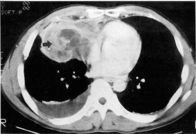
Fig. 2. Postcontrast CT scan shows a large, low- attenuated solid mass with ill-marginated high attenu- ation(arrow) and bilateral pleural effusion. 、
A B
이학수 오1: 피| 모세포종의 단발성 출혈성 뇌 전이
통, 기침, 때로 객혈을 보일 수 있고 광범위한 혈흉으로 나타난 보고도 있다 (9). 종양은 조직학적으로 광범위한 괴사와 출혈을 동반하며( 6), 소아서 발생한 경우는 성인과 달리 암종성 상피 성 세포가 없다 (1 , 3, 7). 본 증례의 조직학적 소견에서도 암종 성 조직이 결여되고 육종성 폐아체 조직으로만 구성되어 있었 다.
단순 흉부 촬영상 주변부 소결절부터 편측 흉강을 채우는 경 계가 분명한 단일성 종괴로도 보일 수 있다.CT상 폐 모세포종 은 내부에 소용돌이 모양의 고빌도를 보이는 고형성분을 포함 한 저밀도의 종괴로 나타난다(1, 3). 예후는 잘 알려져 있지 않 으며 보고자에 따라다르나대개 불량하고(3, 4) 예후가양호한 군과의 조직학적인 차이점은 없고 크기가 5cm 이상시 전이를
‘ ~,‘ Iεr一:~.. κ/ 톨
Fig. 3. Histopathology of the tumor specimen from the lung. The tumor consists of uniform small to medium sized oval cells with no distinct nucleoli. The cytoplasm of tumor cells are scanty. The histiologic features are reminiscent of primitive pulmonary blastemal tissue with no bronchial buddings (Hematoxylin-Eosin stain, X 200).
Fig. 4. A. Axial CT scan shows a 6 X 6 cm sized, round mass with sur rounding edema and marked mass effect in the right temporoparietal lobe. The mass has a focal central low-attenuation area and periph eral high-attenuation hemorrhage.
Acute hyperdense hemorrhage in the frontal horn of right lateral ventricle is also noted
B. Postcontrast CT scan shows small contrast. enhancing areas in the anterior and posterior portions of the mass(open arrows).
- 206 -
대한방사선의학회지 1997; 36 : 205- 208
를 흔히 하며 재발하여 깐칸내 사망한다(1, 3, 7, 10). 드물게 24
년까지 생존한 예도 있다(2). 다른 저자들의 보고를 인용한 Iverson등 (5) 과 Karcioglu등 (4) 의 보고에서도 전체 21예 중 11예에서 국소 부위의 재발이나 원격성 전이를 보고하였는데,
이 경우에도 뇌전이가 2예 포함되어 있었다. Manivel등 (8) 이 보고한 11예 중 9예에서 원격성 전이나 재발을 보고하였는데, 5 예에서 국소 재발을 보였고, 원격성 전이는 3예의 뇌전이를 포 함하여 복강, 두개골, 대퇴골 등에서 관찰되었으나, 그 영상 소 견에 대한 기술은 없었다.
악성 종양의 출혈은 두개내 혈종의 약 10%이며, 뇌전이의 3-14%서 출혈이 일어난다. 출혈을 잘 일으키는 종양은 흑색 종, 융모막 암종, 신 세포암종, 기관지 기원성 암종, 갑상선 암종 등으로 알려져 있다 (11, 12, 13). 종양내 출혈과 다른 원인에 의 한 출혈의 감별은 CT상 어려우며, 비전형적인 위치, 다수의 출 혈 장소, 비교적 초기 의 조영 증강이 도움이 된다고 알려져 있 다(13). 본 예는 내부에 매우 심한 괴사가 출혈과 동반되어 있 었으나, 출혈 부위 이외에 국소적으로 조영증강이 되는 부분이 있어 다른 원인에 의한 출혈과 감별에 도움이 되었다. 원발성 및 전이된 폐 모세포종종양의 내부에 저밀도의 조영증강이 되 지 않는 부분은 심한 괴사를 가지고 있는 부분으로 생각되며,
저자들은 매우 드문 폐 모세포종이 뇌에 전이하여 생긴 출혈과 다른 원인에 의한 출혈의 감별이 매우 어려웠던 1예를 경험하 고의의를검토하였다.
중1- -;-J S긍 등4
딩
1. Senac MO Jr, Wood BP, Isaacs H, WelIer M. Pumonary
m
blastoma: a rare childhood malignancy. Radi%gy 1991; 179 743-746
2. Gibbons JRP, McKeown F, Field TW. pulmonary blastoma with hilar lymph node metastasis: survival for 24 years. Cancer 1981
; 47: 152-155
3. Ohtomo K, Araki T, Yashiro N, Iio M. Pulmonary blastoma in children. Radi%gy 1983; 147: 101-104
4. Karcioglu ZA, Someren AO. Pulmonary blastoma: a case report and review of the literature. Am J Clin Patho/ 1974; 61:
287-295
5. Iverson RE, Straehley CJ. Pulmonary blastoma: longterm sur- vival of juvenile patient. Chest 1973; 63 ‘ 436-440
6. McCann MP, Fu YS, Kay S. Pulmonary blastoma. Cancer 1976;
38: 789-797
7‘ Beuermeister DE, Jennings ER, Beland AH, Judson HA. Pul- monary blastoma, a form of carcinosarcoma. Am J C/in Patho/
1974; 46: 322-329
8. Manivel JC, Priest JR, Watterson J, et al. Pleuropulmonary Blastoma: the so-calIed pulmonary blastoma of children. Cancer 1988;62: 1516-1526
9. Vassilopoulos PP, Vrettou V, Smerniotis V, Zoetopoulos G. Pul- monary blastoma presenting with massive hemothorax. Chest 1992; 102 : 649-650
10. RayChaudhuri M, Winstanley DP. Pulmonary blastoma with diverse metastasis. J Path 1969; 98: 81-82
11. Pechova-Peterova V, Kalvach P. CT findings in cerebral metastases. Neuroradi%gy 1986; 28 ’ 254-258
12. Atlas SW, Grossman RI, Gomori JM, et al. Hemorrhagic intra cranial malignant neoplasms: spin-echo MR imaging. Radi%gy 1987; 164:71-77
13. Mandubur YI. Intracranial hemorrhage caused by metastatic tumors. Neur% gy 1977; 27 ‘ 650-655
이학수 오1: 피| 모세포종의 딘빌성 출혈성 뇌 전이
J Korean Radiol Soc 1997; 36 : 205- 208
Solitary Hemorrhagic Brain Metastasis from Pulmonary Blastoma : A Case Repod
Hak-Soo Lee, M.D., Seung-Ro Lee, M.D., Oh-Keun Bae, M.D., Doung-Woo Park, M.D.
Yo-Won Choi, M.D., Seok-Chol Jeon, M.D., Choong-Ki Park, M.D.
Chang-Kok Hahm, M.D., Jung-Dal Lee, M.D.
1 Department of Diagnostic Radiology, College of Medicine, Hanyang Universiη 2Department of Pathology, College of Medicine, Haηyang University
Pulmonary blastoma is a rare primary lung malignancy consisting of mesenchymal and epithelial components resembling the fetal lung. We report a case of pulmonary blastoma with solitary hemorrhagic brain metastasis in a 32-year-old man. This metastatic lesion was composed mainly of hemorrhagic high density and central necrotic low density areas; on CT it showed partial contrast enhancement and was thus impossible to distinguish from other hemorrhagic lesions.
Index Words: Brain neoplasms, CT
Brain neoplasms, secondary Lung neoplasms, metastasis
Address reprint requests to: Hak-Soo Lee, M.D., Department of Diagnostic Radiology, College of Medicine, Hanyang University
# 17, Haengdang-dong, Sungdong-ku, SeouL 133-792 Korea. Tel. 82-2-290-9164 Fax. 82-2-291-9866
- 208 -