http://dx.doi.org/10.4174/astr.2016.90.1.36 Annals of Surgical Treatment and Research
Outcomes for patients with HCV after liver transplantation in Korea: a multicenter study
Jong Man Kim*, Kwang-Woong Lee1, Gi-Won Song2,*, Bo-Hyun Jung2, Hae Won Lee3, Nam-Joon Yi1, Choon Hyuck David Kwon, Shin Hwang2, Kyung-Suk Suh1, Jae-Won Joh, Suk-Koo Lee, Sung-Gyu Lee2
Department of Surgery, Samsung Medical Center, Sungkyunkwan University School of Medicine, Seoul, 1Department of Surgery, Seoul National University Hospital, Seoul National University College of Medicine, Seoul, 2Division of Hepatobiliary Surgery and Liver Transplantation, Department of Surgery, Asan Medical Center, University of Ulsan College of Medicine, Seoul, 3Department of Surgery, Seoul Metropolitan Government-Seoul National University Boramae Medical Center, Seoul, Korea
INTRODUCTION
An estimated 185 million individuals are chronically infected with HCV worldwide [1]. Of all HCVinfected individuals, 20%–30% develop liver cirrhosis and 1%–4% of all patients with liver cirrhosis develop hepatocellular carcinomas (HCC) [2].
HCV infection is the most common indication for liver trans
plantation (LT) in Western countries. In Korea, 1%–2% of the population is infected with HCV, and 15%–20% of these infected
individuals have chronic liver diseases related to HCV infection [3,4]. As the prevalence has increased, HCVrelated cirrhosis and HCVrelated HCC will gradually become more common indications for LT in Korea [5].
Although LT offers the optimal treatment for HCVrelated endstage liver disease and HCC, graft reinfection with HCV is not acute, but rather immediate and universal in all patients who are HCV RNApositive at transplantation [6]. HCV RNA levels increase when immunosuppression is the highest during Purpose: HCV-related liver disease is the most common indication for liver transplantation (LT) in Western countries, whereas HCV LT is rare in Korea. We conducted a survey of HCV RNA-positive patients who underwent LT and investigated the prognostic factors for patient survival and the effects of immunosuppression.
Methods: We retrospectively reviewed the multicenter records of 192 HCV RNA-positive patients who underwent LT.
Results: The 1-, 3-, and 5-year overall survival rates were 78.8%, 75.3%, and 73.1%, respectively. Excluding the cases of hospital mortality (n = 23), 169 patients were evaluated for patient survival. Most patients were genotype 1 (n = 111, 65.7%) or genotype 2 (n = 42, 24.9%). The proportion of living donors for LT (n = 135, 79.9%) was higher than that of deceased donors (deceased donor liver transplantation [DDLT], n = 34, 20.1%). The median donor and recipient ages were 32 years and 56 years, respectively. Twenty-eight patients (16.6%) died during the observation period. Seventy-five patients underwent universal prophylaxis and 15 received preemptive therapy. HCV recurrence was detected in 97 patients.
Recipients who were older than 60, received DDLT, used cyclosporine, or suffered acute rejection had lower rates of survival.
Conclusion: Patent survival rates of HCV patients after LT in Korea were comparable with other countries.
[Ann Surg Treat Res 2016;90(1):36-42]
Key Words: Hepatitis C virus, Tacrolimus, Acute rejection, Survival, Antiviral treatments
Reviewed January February March April May June July August September October November December
Received June 19, 2015, Revised August 20, 2015, Accepted September 10, 2015
Corresponding Author: Kwang-Woong Lee
Department of Surgery, Seoul National University Hospital, Seoul National University College of Medicine, 101 Daehak-ro, Jongno-gu, Seoul 03080, Korea
Tel: +82-2-2072-2511, Fax: +82-2-766-3975 E-mail: kwleegs@gmail.com
*Jong Man Kim and Gi-Won Song contributed equally to this study as co-first authors.
Copyright ⓒ 2016, the Korean Surgical Society
cc Annals of Surgical Treatment and Research is an Open Access Journal. All articles are distributed under the terms of the Creative Commons Attribution Non- Commercial License (http://creativecommons.org/licenses/by-nc/4.0/) which permits unrestricted non-commercial use, distribution, and reproduction in any medium, provided the original work is properly cited.
the first few months after transplantation. The progression of fibrosis in LT patients is accelerated compared to that in nontransplanted patients because the virus is more aggressive after LT than it is in immunocompetent subjects.
Genetic variation in interleukin28B (IL28B) predicts hepatitis C treatmentinduced viral clearance. Single nucleotide poly
morphisms in IL28B have varied distributions among ethnic groups. East Asian populations such as those in Korea, Japan, and China have the highest frequencies of single nucleotide polymorphisms in alleles associated with HCV clearance [7].
The cumulative number of HCVrelated cirrhosis and HCV
related HCC cases in Korea is very small; therefore, we collected data of LT recipients with HCV from three major centers.
We conducted a survey of HCV RNApositive patients who underwent LT and investigated the prognostic factors for patient survival.
METHODS
Patients
This was a multicenter study involving three LT centers in Korea: Samsung Medical Center (SMC), Asan Medical Center (AMC), and Seoul National University Hospital (SNUH). We did not consent from patients for usage of their clinical records in this study because of retro spective study. Each center’s Institutional Review Board (IRB) approved this study (SMC IRB No. 201407031, AMC IRB No. S201513410003, SNUH IRB No. 1407139597) because written informed consents were not given by patients. We retro spectively evaluated patients undergoing their first LT between 1994 and 2012. Data from all consecutive HCV RNA positive cases were reviewed during this period. Each insti tution utilized a survey with study questionnaire items. The information and/or records of patients deidentified prior to analysis.
HCV RNA quantitation was assessed using the quantitative
branched DNA amplification assay. HCV genotyping was performed following the standard method using reverse hybridization assays after amplification with a polymerase chain reaction assay, based on Simmonds’ classification.
Among the 255 cases with HCVrelated cirrhosis who underwent LT during the study period, 63 cases were excluded due to retransplantation (n = 13), missing results of HCV
RNA (n = 23), and HCVRNA negativity (n = 27). Among the remaining 192 included patients, we identified the causes for graft failure and mortality. We investigated the risk factors associated with patient survival, but exclude hospital mortality (n = 23) for the identification of risk factors which were related with mortality in stable liver transplant recipients with more than three months after transplantation.
Evaluated variables
The following variables were obtained from the medical record review in response to the survey. Collected recipient factors included patient age, gender, pre and posttransplant antiviral treatment, HCV genotype, model for endstage liver disease (MELD) score, the coexistence of HCC, the coexis
tence of HBV or human immunodeficiency virus (HIV), anti
viral treatments received after transplantation, the type of calcineurin inhibitor received, the use of mycophenolate mofetil, steroid withdrawal, biopsyproven acute rejection, HCV recurrence, the response to antiviral treatments, and the outcomes. Additionally, donor age, ischemic time, and the type of partial liver graft were added as variables. Finally, we recorded information on patient survival and calculated time to death. However, we did not incorporate into the analysis any other incomplete variables that may be associated with patient survival, such as IL28 gene polymorphisms, histological findings, biliary complications, and infectious episodes.
The diagnosis of acute rejection followed internationally accepted histologic criteria (Banff guidelines) based on liver biopsies [8]. HCV recurrence was diagnosed based on histology, biochemistry, and/or the detection of HCV RNA in the serum.
Statistical analysis
Continuous data are expressed as the median and range and were compared using the MannWhitney U test. Categorical variables are reported as numbers (proportions). Comparisons between groups for categorical data were performed using the chisquare test or Fisher exact test, when appropriate. Patient survival rates were evaluated using the KaplanMeier method and compared using the logrank test. Clinical variables found to have prognostic significance by univariate analysis were entered into a Cox multivariate proportional hazards model to determine factors that independently predict patient survival. The cutoff values for the continuous variables were set according to each receiver operating characteristic (ROC) curve.
Statistical significance was set at a Pvalue of less than 0.05.
Statistical analysis was performed with IBM SPSS Statistics ver.
21.0 (IBM Co., Armonk, NY, USA).
RESULTS
Patient survival and outcomes
The 1, 3, and 5year cumulative patient survival rates were 78.8%, 75.3%, and 73.1%, respectively (Fig. 1). The causes of graft failure and mortality are summarized in Table 1. Thirty patients (15.6%) developed graft failure during the observation period and 13 patients underwent retransplantation. Fifty patients (26.0%) died during the observation period. Most cases of mortality (38 of 50, 76%) occurred less than one year after transplantation. The patient survival curve showed an
abrupt decrease, but then stable survival after two years after transplantation and onward. Recurrent HCV infection and hepatic failure (n = 17) and chronic rejection (n = 10) were the causes of graft failure. Chronic rejection was the main cause of graft failure in patients less than one year after transplantation.
Infection (n = 21) and recurrent HCV infection with hepatic failure (n = 15) were the leading causes of recipient death.
Infection was the main cause of hospital mortality in patients one year after transplantation.
Baseline characteristics
Among the 192 patients that were identified, we investigated
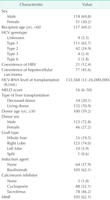
the 169 patients, who represented all patients except those who died in the hospital (n = 23). The characteristics of the 169 HCV RNApositive LT recipients compared in this study are summarized in Table 2. There were 118 men and 51 women, with a median age of 56 years (range, 34–71 years). The median followup period was 38 months (range, 1–157 months), with a wide spectrum of followup duration due to death or shorter observation period from LT. The median MELD score and median HCV RNA levels were 16 (range, 6–50) and 133,568 IU/
mL (range, 12–26,000,000 IU/mL), respectively. One hundred eleven patients (65.7%) had HCV genotype 1 and 42 patients (24.9%) had HCV genotype 2. The number of patients with co- existing HBV infection, HIV infection, and HCC was 21 (12.4%), Table 1. The causes of graft failure and mortality
Graft failure and mortality ≤1 year >1 year (n = 12) (n = 18) (n = 38)
Graft failure
Chronic rejection 8 2
Graft dysfunction 2 0
HCC recurrence 1 0
HCV recurrence 4 6
Hepatic failure 3 4
Mortality
Chronic rejection 2 (1 / 1)a) 1
Graft dysfunction 2 (2 / 0)a) 0
HCC recurrence 3 (0 / 3)a) 1
HCV recurrence 4 (0 / 4)a) 2
Hepatic failure 6 (5 / 1)a) 3
Infection 17 (10 / 7)a) 4
Cerebrovascular accident 1 (1 / 0)a) 1
Bronchial hemorrhage 1 (0 / 1)a) 0
Gastrointestinal bleeding 1 (1 / 0)a) 0 Stressinduced cardiomyo
pathy 1 (1 / 0)a) 0
HCC, hepatocellular carcinoma.
a)Total mortality (hospital mortality / no hospital mortality).
Table 2. Baseline characteristics
Characteristic Value
Sex
Male 118 (69.8)
Female 51 (30.2)
Recipient age (yr), <60 117 (69.2) HCV genotype
Unknown 9 (5.3)
Type 1 111 (65.7)
Type 2 42 (24.9)
Type 3 4 (2.4)
Type 6 3 (1.8)
Coexistence of HBV 21 (12.4)
Coexistence of hepatocellular
carcinoma 77 (45.6)
HCVRNA level at transplantation
(IU/mL) 133,568 (12–26,000,000)
MELD score 16 (6–50)
Type of liver transplantation
Deceased donor 34 (20.1)
Living donor 135 (70.9)
Donor age (yr), ≥30 100 (59.2)
Donor sex
Male 123 (72.8)
Female 46 (27.2)
Graft type
Whole liver 33 (19.5)
Right Lobe 125 (74.0)
Left lobe 10 (5.9)
Split 1 (0.6)
Induction agent
None 64 (37.9)
Basiliximab 105 (62.1)
Calcineurin inhibitor
None 3 (1.8)
Cyclosporin 88 (52.1)
Tacrolimus 78 (46.2)
MMF 105 (62.1)
Values are presented as number (%) or median (range).
MELD, model for endstage liver disease; MMF, mycophenolate mofetil.
Fig. 1. Patient survival rates. The 1, 3, and 5year patient survival rates are 78.8%, 75.3%, and 73.1%, respectively.
0 Cumulativepatientsurvivalrate(%) 0
Time from liver transplantation to death (mo) 120 1.0
0.8 0.6 0.4 0.2
12 24 36 48 60 72 84 96 108
1 (0.6%), and 77 (45.6%), respectively. There were 135 living donor liver transplantations (LDLTs) (79.9%) and 34 deceased donor liver transplantations (DDLTs) (20.1%). The median age of the donors was 32 years (range, 16-70 years), and the graft type in the living donors was the right liver in 125 patients (74.0%).
The median cold ischemic time and median warm ischemic time were 81 and 39 minutes, respectively.
Prognostic factors for patient survival
Recipient and donor factors were analyzed for their associa
tion with overall mortality. The results of the univariate and multivariate analyses are shown in Table 3. The univariate an
alysis revealed that recipient age ≥ 60 years (P = 0.018), HCV
RNA levels at pretransplant (P = 0.023), DDLT (P = 0.020), donor age ≥ 30 years (P = 0.019), the use of cyclosporine (P = 0.025), and biopsyproven acute rejection (P < 0.001) were significant predictors of poor outcome in HCV RNApositive recipients. The duration of steroid use did not affect patient survival. The ROC curve did not reveal a significant cutoff value for HCVRNA levels in terms of patient survival. The multivariate analysis showed that recipient age ≥ 60 years (P = 0.046), DDLT (P = 0.040), the use of cyclosporine (P = 0.029), and biopsyproven acute rejection (P = 0.001) were independent prognostic factors for mortality. The Kaplan–Meier survival curves stratified by these factors are presented in Fig. 2.
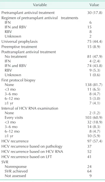
Antiviral treatments in pre- and posttransplant
A summary of the antiviral treatments is shown in Table 4.
Of the 169 recipients, 129 did not receive antiviral treatment in the pretransplant period and 30 underwent antiviral treatment. After LT, 75 patients received universal prophylaxis and 15 patients underwent preemptive treatment due to HCV reactivation. Most patients did not undergo a protocol biopsy, and HCVRNA levels were monitored at every visit. HCV re
currence was detected in 97 patients (57.4%). Among the 97 patients with HCV recurrence, 48 patients were treated with antiviral therapy. The survival rates were higher in patients with sustained viral response (SVR) than in patients without SVR, but there was no statistically significant difference in patient survival between the two groups (P = 0.062) (Fig. 3).
DISCUSSION
Literature from the United Network for Organ Sharing (UNOS) database reported a 5-year patient survival rate of 76%, and a study from the European Liver Transplant Registry (ELTR) reported a 5-year patient survival rate of 65% [9,10]. Recently, nationwide survey in Japan of LDLT reported a 5year patient survival rate of 72% [11]. Our study here is the largest case series of LT for HCV RNApositive recipients in Korea. A total of 192 recipients from three large institutions were reviewed and found to have a 5-year patient survival rate of 73.1%.
Based on these studies, the outcomes of the present study are similar to that of the ELTR, UNOS, and Japanese survey.
Comparisons of the survival rates of HCV recipients between studies should be interpreted with caution because our study excluded patients with operative mortality, hospital mortality, and retransplantation. The proportion of IL28B in Korea is higher than in European countries [7], however survival rates of Korea were similar to other countries despite advanced surgical techniques and perioperative managements in LT.
We selected liver transplant recipients with HCVRNA positive in the pretransplant period because there have been no reports of posttransplant HCV recurrence in HCV RNAnegative Table 3. Risk factors for patient survival
Variable Odds
ratio
95%
Confidence
Interval Pvalue Univariate
Recipient sex, female 0.880 0.387–2.001 0.761 Recipient age (yr), ≥60 2.410 1.133–5.128 0.018 Genotype
Type 1
Type 2 1.392
2.100 0.185–10.486 0.265–16.614 0.748
0.482 Pretransplant antiviral treat
ment 2.048 0.897–4.680 0.089
HCV RNA level 1.000 1.000–1.000 0.023
Coexistence of HBV 0.500 0.119–2.110 0.346 Coexistence of hepatocell
ular carcinoma 0.828 0.390–1.755 0.622
MELD score 0.990 0.941–1.042 0.703
Deceased donor liver trans
plantation 2.475 1.119–5.495 0.020
Donor age (yr), ≥30 3.214 1.216–8.493 0.019 Donor gender, female 0.889 0.376–2.103 0.789 Cold ischemic time 1.000 0.997–1.002 0.858 Warm ischemic time 1.005 0.994–1.016 0.404 Induction agent (Basilixi
mab) 0.643 0.302–1.369 0.252
Use of cyclosporin 2.475 1.089–5.618 0.025
MMF 0.879 0.411–1.881 0.740
Universal prophylaxis 1.421 0.668–3.024 0.362 Preemptive treatment 0.663 0.154–2.862 0.582
HCV recurrence 1.113 0.529–2.344 0.778
Biopsyproven acute rejec
tion 4.013 1.909–8.436 <0.001
Multivariate
Recipient age (yr), ≥60 2.277 1.014–5.113 0.046 Deceased donor liver trans
plantation 2.398 1.041–5.525 0.040
Use of cyclosporin 5.870 1.276–11.909 0.029 Biopsyproven acute rejec
tion 4.338 1.884–9.990 0.001
MELD, model for endstage liver disease; MMF, mycophenolate mofetil.
recipients. Antiviral therapies based on Pegylated interferon
alpha (PEGIFNa) and ribavirin (RBV) have been used to treat HCV in decompensated patients on the transplant waiting list until they are HCV RNAnegative [12]. However, this therapy is limited due to poor tolerance, poor efficacy, and serious adverse events seen in those waiting for LT [13].
Posttransplant viral load is an important marker of disease severity, while pretransplant viral load predicts more severe HCV recurrence after transplantation [14]. Negative HCV viral load at the time of transplantation does not preclude HCV recurrence in the liver graft. A peak posttransplant HCV viral load >107 IU/mL was an independent predictor of graft loss and mortality [15]. Present study showed that prognostic factors, including recipient age > 60 years, DDLT, the use of cyclosporin, and biopsyproven acute rejection were closely associated with patient mortality. High viral load was associated with mortality in univariate analysis, but a cutoff value for the HCV RNA level was not drawn. Universal prophylaxis should be initiated soon after LT because the viral load is at its lowest
level and fibrosis in the graft is absent [16]. However, antiviral therapy may be less effective in the early posttransplant period secondary to strong immunosuppression, and tolerance is low because of the high risk of poor hematological tolerance, acute rejection, and sepsis [17,18].
Posttransplant patients with HCV recurrence have signifi
cantly diminished survival compared to posttransplant patients with no recurrence. The progression of recurrent HCV is variable and the key risk factors remain unclear. Many factors have been reported to play a role prior to LT (genotype 1, viral load, and female gender) or after LT (time of cold or warm ischemia, blood transfusions, steatosis in the liver graft, age of the donor, the use of antilymphocytes, and coinfection with HIV) [13,19].
The early detection of HCV recurrence is crucial because HCV
infected patients appear to respond better to early antiviral therapy [19]. The current clinical standard for early detection is for protocol liver biopsies to be performed every 1–2 years after LT, as HCVinfected recipients are at increased risk of HCV
mediated graft cirrhosis [20]. Antiviral treatment is delayed Fig. 2. Patient survival according to recipient age (A), donor type (B), calcineurin inhibitor (C), and BPAR (D). LDLT, living do
nor liver transplantation; DDLT, deceased donor liver transplantation; Tac, Tacrolimus; CsA, cyclosporin; BPAR, biopsyproven acute rejection.
0 Cumulativepatientsurvivalrate(%) 0
Time from liver transplantation to death (mo) 120 1.0
0.8 0.6 0.4 0.2
12 24 36 48 60 72 84 96 108
A
Recipient age <60
Recipient age >60
P = 0.018 Recipient age <60
Recipient age >60
Recipient age <60-censored Recipient age >60-censored
0 Cumulativepatientsurvivalrate(%) 0
Time from liver transplantation to death (mo) 120 1.0
0.8 0.6 0.4 0.2
12 24 36 48 60 72 84 96 108
B
LDLT
P = 0.020 DDLT
LDLT
DDLT-censored LDLT-censored
DDLT
0 Cumulativepatientsurvivalrate(%) 0
Time from liver transplantation to death (mo) 120 1.0
0.8 0.6 0.4 0.2
12 24 36 48 60 72 84 96 108
C
Tac
CsA
P = 0.025 Cyclosporin
Tacrolimus
Cyclosporin-censored Tacrolimus-censored
0 Cumulativepatientsurvivalrate(%) 0
Time from liver transplantation to death (mo) 120 1.0
0.8 0.6 0.4 0.2
12 24 36 48 60 72 84 96 108
D
No BPAR
P < 0.001 No BPAR
BPAR
No BPAR-censored BPAR-censored
BPAR
until there is histological evidence of recurrent hepatitis in many transplantation centers. However, none of the centers in the present study performed these protocol biopsies.
The successful treatment of recurrent HCV, which is demon
strated by sustained HCV clearance or an SVR, is associated with reduced liverrelated mortality and improved overall sur
vival. The combination of PEGIFNa and RBV is the current standard of care [13,21]. Our study also revealed this effect in the SVR group in recurrent HCV patients, but this did not reach statistical significance. However, the PHOENIX trial of PEGIFNa and RBV given preemptively after the transplant for HCV found no clear benefits when considered in the context of side effects [18]. In a very small study, donor or recipient IL28B genotypes were shown to predict SVR with PEGIFNa and
RBV therapy, and IL28B status was related to SVR after LT [22].
However, this effect of the IL28B genotype was not identified in the present study, and our study did not reveal an association between IL28B and patient survival.
The findings of this retrospective, multicenter study are limited by several factors inherent to the study type, including variability in documentation, differences in the selection criteria and data collection, and missing data. To minimize vari
ability, we sent a standardized collection form containing 56 questions to the transplant centers. The answers were either multiplechoice or involved providing a name or a specific value. However, the quality of the pretransplant interviews from which the baseline data were derived, and the quality of the posttransplant followup data across the three centers may have varied. Furthermore, subjects had varying followup durations. We did not have data on the onset of biopsyproven acute rejection or the date of graft failure. To address these limitations, a welldesigned prospective study is needed.
In conclusion, this retrospective analysis of the largest three LT centers for HCV RNApositive recipients in Korea revealed 1-, 3-, and 5-year survival rates of 78.8%, 75.3%, and 73.1%, respectively. The prognostic factors for patient survival except hospital mortality revealed that recipient age > 60 years old, DDLT, the use of cyclosporin, and biopsyproven acute rejection are closely associated with patient mortality. Present study revealed that patient survival rates in HCV patients after LT in Korea were comparable other countries.
CONFLICTS OF INTEREST
No potential conflict of interest relevant to this article was reported.
Table 4. Antiviral treatment in pre and posttransplant
Variable Value
Pretransplant antiviral treatment 30 (17.8) Regimen of pretransplant antiviral treatments
IFN
IFN and RBV RBV Unknown
6 15 8 2
Universal prophylaxis 75 (44.4)
Preemptive treatment 15 (8.9)
Posttransplant antiviral treatment No treatment
IFN
IFN and RBV RBV Unknown
81 (47.9) 4 (2.4) 74 (43.8)
9 (5.3) 1 (0.6) First protocol biopsy
None <3 mo 3–6 mo 6–12 mo ≥1 yr
138 (81.7) 11 (6.5)
8 (4.7) 5 (3.0) 7 (4.1) Interval of HCV RNA examination
None Every visits <3 mo 3–6 mo 6–12 mo ≥1 yr
2 (1.2) 103 (60.9)
32 (18.9) 14 (8.3)
8 (4.7) 10 (5.9)
HCV recurrence 97 (57.4)
HCV recurrence based on pathology 37
HCV recurrence based on HCV RNA 52
HCV recurrence based on LFT 41
SVR
Nonresponse SVR achieved Not assessed
24 64 9 Values are presented as number (%) or number.
RBV, ribavirin; LFT, liver function test; SVR, sustained viral response.
Fig. 3. Patient survival according to sustained viral response (SVR). Patient survival in patients with SVR was higher than in patients without SVR, but there was no statistically significant difference in patient survival between the two groups.
0 Cumulativepatientsurvivalrate(%) 0
Time from liver transplantation to death (mo) 120 1.0
0.8 0.6 0.4 0.2
12 24 36 48 60 72 84 96 108 SVR
P = 0.062 Nonresponder
SVR achieved
Nonresponder-censored SVR achieved-censored
No SVR
1. Mohd Hanafiah K, Groeger J, Flaxman AD, Wiersma ST. Global epidemiology of hepatitis C virus infection: new esti mates of agespecific antibody to HCV sero
prevalence. Hepatology 2013;57:133342.
2. European Association for the Study of the Liver. EASL Clinical Practice Guidelines:
management of hepatitis C virus infec
tion. J Hepatol 2011;55:24564.
3. Shin HR, Hwang SY, Nam CM. The prevalence of hepatitis C virus infection in Korea: pooled analysis. J Korean Med Sci 2005;20:9858.
4. Kim SJ. Viral hepatitis surveillance sys
tem and statue of C hepatitis sentinel surveillance in Korea. Publ Health Wkly Rep 2012;5:2149.
5. Lee HW, Lee KW, Kim BW, Song GW, Han YS, Kwon CH, et al. Liver Transplantation for Hepatitis C VirusRelated Liver Disease in Korea. J Korean Soc Transplant 2012;
26:26976.
6. Gane EJ, Naoumov NV, Qian KP, Mondelli MU, Maertens G, Portmann BC, et al. A longitudinal analysis of hepa titis C virus replication following liver transplan
tation. Gastroenterology 1996;110:16777.
7. Balagopal A, Thomas DL, Thio CL. IL28B and the control of hepatitis C virus infec
tion. Gastroenterology 2010;139:186576.
8. Banff Working Group on Liver Allograft Pathology. Importance of liver biopsy findings in immunosuppression manage
ment: biopsy monitoring and working criteria for patients with operational tole
rance. Liver Transpl 2012;18:115470.
9. Singal AK, Guturu P, Hmoud B, Kuo YF,
Salameh H, Wiesner RH. Evolving fre
quency and outcomes of liver transplan
tation based on etiology of liver disease.
Transplantation 2013;95:75560.
10. Adam R, Karam V, Delvart V, O'Grady J, Mirza D, Klempnauer J, et al. Evolution of indications and results of liver trans
plantation in Europe. A report from the European Liver Transplant Registry (ELTR). J Hepatol 2012;57:67588.
11. Akamatsu N, Sugawara Y, Kokudo N, Eguchi S, Fujiwara T, Ohdan H, et al.
Outcomes of living donor liver trans
plantation for hepatitis C viruspositive recipients in Japan: results of a nation
wide survey. Transpl Int 2014;27:76774.
12. Everson GT, Trotter J, Forman L, Kugelmas M, Halprin A, Fey B, et al. Treatment of advanced hepatitis C with a low accelera
ting dosage regimen of antiviral therapy.
Hepatology 2005;42:25562.
13. Howell J, Angus P, Gow P. Hepatitis C re
currence: the Achilles heel of liver trans
plantation. Transpl Infect Dis 2014;16:1
16.
14. Charlton M, Seaberg E, Wiesner R, Everhart J, Zetterman R, Lake J, et al. Pre
dic tors of patient and graft survival fol
lowing liver transplantation for hepa titis C. Hepatology 1998;28:82330.
15. Shackel NA, Jamias J, Rahman W, Prakoso E, Strasser SI, Koorey DJ, et al. Early high peak hepatitis C viral load levels inde
pendently predict hepatitis Crelated liver failure postliver transplantation. Liver Transpl 2009;15:70918.
16. Roche B, Samuel D. Hepatitis C virus
treatment pre and postliver transplan
tation. Liver Int 2012;32 Suppl 1:1208.
17. Chalasani N, Manzarbeitia C, Ferenci P, Vogel W, Fontana RJ, Voigt M, et al.
Peginterferon alfa2a for hepatitis C after liver transplantation: two randomized, controlled trials. Hepatology 2005;41:289
98.
18. Bzowej N, Nelson DR, Terrault NA, Everson GT, Teng LL, Prabhakar A, et al.
PHOENIX: A randomized controlled trial of peginterferon alfa2a plus ribavirin as a prophylactic treatment after liver trans
plantation for hepatitis C virus. Liver Transpl 2011;17:52838.
19. Guillouche P, Feray C. Systematic review:
antiviral therapy of recurrent hepatitis C after liver transplantation. Aliment Phar
macol Ther 2011;33:16374.
20. Berenguer M, Rayon JM, Prieto M, Aguilera V, Nicolas D, Ortiz V, et al. Are posttransplantation protocol liver biop
sies useful in the long term? Liver Transpl 2001;7:7906.
21. Berenguer M, Palau A, Aguilera V, Rayon JM, Juan FS, Prieto M. Clinical bene fits of antiviral therapy in patients with recu
rrent hepatitis C following liver transplan
tation. Am J Transplant 2008;8:67987.
22. Fukuhara T, Taketomi A, Motomura T, Okano S, Ninomiya A, Abe T, et al. Va
riants in IL28B in liver recipients and do
nors correlate with response to peginter
feron and ribavirin therapy for recurrent hepatitis C. Gastroenterology 2010;139:
157785, 1585.e13.