Large cell neuroendocrine carcinoma (LCNEC) of the lung is a newly recognized clinicopathologic entity and defined as a poorly differentiated and high-grade neuroendocrine tumor that stands mor- phologically and biologically between atypicallcar- cinoid and small cell lung carcinomal. Histopatho- logically, most LCNECs revealed a marked de- crease in or a loss of organoid architecture and could be mistaken for poorly differentiated adeno- carcinoma or squamous cell carcinoma2,3. Cuta- neous metastasis of pulmonary cancer is common as shown in its rate up to 1.5 to 2.6% and may be presented as a first sign of cancer4, 5. Furthermore, the neuroendocrine features of cutaneous metastasis of LCNEC of the lung may be similar to Merkel cell carcinoma and other neuroendocrine carcinomas and therefore, it should be differentiated from them by histological and immunohistochemical studies. Thus, we herein report a case of cutaneous metastasis of LCNEC of the lung, previously misdi-
agnosed as squamous cell carcinoma.
CASE REPORT
A case of a 72-year-old man presented with a 2×
1cm sized slowly enlarging subcutaneous nodule on his back of 1-month duration. Three months before admittance to our clinic, he was diagnosed as squamous cell carcinoma of the lung (T2N1M0, stage Ib) and operated with right lower lobectomy and with a wedge resection and ligation of right upper lobe bullae. There were no metastasis on the bronchus, pleura and peripheral lymph nodes on operational biopsy findings. With a provisional clinical diagnosis of metastatic carcinoma, we biopsied the skin lesion. The specimen showed a dermal tumor in which tumor cells were arranged in variously sized nests, broad and irregular strands, or solid sheets with prominent stromal lymphoid infiltration and focal necrosis (Fig. 1, 2). At higher magnification, the tumor cells usually were large and polygonal with an abundant, finely granular and eosinophilic cytoplasm, coarse or salt and pepper chromatins, and frequent nucleoli and high mitotic rate. Further observation revealed rosette-like structure in variously sized tumor cell nests (Fig. 3). Tumor cells were stained diffusely
Metastatic Large Cell Neuroendocrine Carcinoma of the Lung Mimicking a Merkel Cell Carcinoma
Won Soon Chung, M.D., Dong Hyun Kim , M.D., Jong Seo Lee, M.D., Soo Chan Kim*, M.D.
Department of Dermatology, College of Medicine Pochon CHA University, Department of Dermatology, Yonsei University College of Medicine*, Seoul, Korea.
Large cell neuroendocrine carcinoma (LCNEC) of the lung is a newly recognized entity of pulmonary neuroendocrine carcinoma. Histologically, it is very difficult to differentiate LCNEC from other pulmonary carcinomas and the prognosis is significantly poor. The cutaneous metas- tasis of LCNEC of the lung shares some features with a Merkel cell carcinoma of the skin in light microscopy and yet it is negatively stained with cytokeratin 20. We report a case of cu- taneous metastasis of LCNEC of the lung, previously misdiagnosed as squamous cell carcinoma.
Our patient showed a poor response to the chemotherapy and also revealed a brain metastasis on follow-up brain CT scan. (Ann Dermatol 14(2) 121-123, 2002).
Key Words : Large cell neuroendocrine carcinoma, Cutaneous metastasis
Received July 24, 2001.
Accepted for publication January 5, 2002.
Reprint request to : Jong Seo Lee, M.D., Pundang CHA Hospital Yatap-dong, Pundang-gu, Sungnam Ky- onggi-do 463-070 Korea
Tel. (031)780-5242, Fax. (031)780-5247
121
with neuron-specific enolase (NSE) and chromo- granin A (Fig. 4A, B) and not with cytokeratin (CK) 20. After confirmation of these histological findings, our patient was rediagnosed as pulmonary LCNEC because of the metastatic cancer on his back. The patient was re-hospitalized to perform follow-up radiological examinations and to take chemotherapy. There was no evidence of metastasis on follow-up whole body bone scan and chest CT but brain CT scan revealed the metastatic lesion on right cerebellum.
DISCUSSION
Large cell neuroendocrine carcinoma (LCNEC) is very difficult to differentiate in light microscopy, especially in cases that are decreased in or loss of organoid architecture. In one study, 18 of 22 cases di- 122 WS Chung, et al.
Annals of Dermatology Vol. 14, No. 2, April 2002
Fig. 1. Tumor cells are arranged in variously sized nests, broad and irregular strands, or solid sheets with prominent stromal lymphoid infiltration and focal necrosis (H × E (40).
Fig. 2. Tumor cells form variously sized nests (Hema- toxylin & eosin stain (100).
Fig. 3. Tumor cells usually are large and polygonal with an abundant, finely granular and eosinophilic cy- toplasm, coarse or salt and pepper chromatins, and fre- quent nucleoli and high mitotic rate. Some rosette-like structures(arrow) are founded (H × E (400).
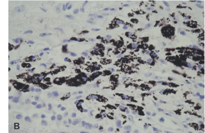
Fig. 4. Tumor cells are stained diffusely with NSE and chromogranin A (A: NSE, B: chromogranin A × 400).
agnosed as LCNEC were misdiagnosed previously and 9 of these 18 cases were squamous cell carcino- ma like our case7. Cutaneous metastatic neuroen- docrine carcinoma must be differentiated from Merkel cell carcinoma and metastatic carcinoma from other visceral disease. Rosette-like structure, suggested by Jiang SX et al.7as the best marker for recognition of neuroendocrine differentiation is described as the small and regular, oval or round lumina, deeply eosinophilic luminal surfaces, and the absence or rare accumulation of nonmucous- secreted material but frequent apoptotic debris in the lumina. Some histologic features of our case re- sembled the Merkel cell carcinoma. However the cell size and the rosette-like structures were different and rather close to the features of pulmonary neu- roendocrine carcinoma. To confirm these neu- roendocrine features, NSE, chromogranin A were used as immunohistochemical staining and to dif- ferentiate from a Merkel cell carcinoma CK 20 was used8. Although some features were similar to the Merkel cell carcinoma, the result elucidated that the tumor contained significant neuroen- docrine natures but it could be differentiated from the Merkel cell carcinoma by its negative staining for CK 20. Cutaneous metastasis from visceral organ was usually from hematogenous spreading but in our case, direct extension of tumor cell was doubted because the location of skin lesion was near that of previous diagnostic fine needle aspiration biopsy site. We also found similar immunohistochemical features in the previous surgically resected lung cancer sections. It represents that previous squa- mous cell carcinoma was a misdiagnosis and re- sected lung cancer was infact pulmonary neuroen- docrine carcinoma. In various pulmonary neu- roendocrine carcinoma, these histological findings corresponded with the histologic criteria for LC- NEC proposed by Travis et al1. Clinically, the
prognosis of LCNEC is significantly worse than that for stage-comparable non-small cell lung cancer and our patient had a poor response to chemotherapy and a brain metastasis on follow-up brain CT scan.
REFERENCES
1. Travis WD, Linnoila RI, Tsokos MG, et al.: Neu- roendocrine tumors of the lung with proposed crite- ria for large-cell neuroendocrine carcinoma. An ul- trastructural, immunohistochemical, and flow cyto- metric study of 35 cases. Am J Surg Pathol 15:529- 53, 1991.
2. Hammond ME, Sause WT: Large cell neuroen- docrine tumors of the lung: clinical significance and histologic definition. Cancer 56;1624-9,1985.
3. Jung KJ, Lee KS, Han JH et al.: Large cell neuroen- docrine carcinoma of the lung: Clinical, CT, and Pathologic findings in 11 patients. J Thoracic Imag- ing 16:156-162, 2001.
4. Schwartz RA: Cutaneous metastatic disease. J Am Acad Dermatol 33:161-82,1995.
5. Argila DD, Bureo JC, Marquez FL, et al.: Small-cell carcinoma of the lung presenting as a cutaneous metastasis of the lip mimicking a Merkel cell carci- noma. Clin Dermatol 24:170-72, 1999.
6. Forster BB, Muller NL, Miller RR, et al.: Neuroen- docrine carcinomas of the lung: clinical, radiologic and pathologic correlation. Radiology 170:441- 5,1989.
7. Jiang SX, Kameya T, Shoji M, et al.: Large cell neuroendocrine carcinoma of the lung: a histologi- cal and immunohistochemical study of 22 cases.
Am J Surg Pathol 22:526-37,1998.
8. Scott MP, Helm KF: Cytokeratin 20: a marker for diagnosing Merkel cell carcinoma. Am J Der- matopathol 21:16-20,1999.
Metastatic Large Cell Neuroendocrine Carcinoma of the Lung Mimicking a Merkel Cell Carcinoma 123