Copyright ⓒ 2019 by Korean Society for Surgery of the Hand, Korean Society for Microsurgery, and Korean Society for Surgery of the Peripheral Nerve. All Rights reserved.
This is an Open Access article distributed under the terms of the Creative Commons Attribution Non-Commercial License (http://creativecommons.org/licenses/by-nc/4.0/) which permits unrestricted non-commercial use, distribution, and reproduction in any medium, provided the original work is properly cited.
INTRODUCTION
Metacarpal bone fracture is a commonly encountered upper limb trauma, comprising one fifth of upper extrem- ity fractures and one half of hand fractures1,2. Treatment for metacarpal bone fractures can be either surgical or nonsurgical, and the former may further be divided into
open reduction and closed reduction using percutaneous pinning3.
In many cases, metacarpal fractures can be treated non- operatively. However, fractures with severe angulation4,5, rotation6, shortening7, unstable fractures such as long oblique fractures, fractures with bone loss, and multiple fractures3 require surgical management. Due to the lack Hand and
Microsurgery
중수골 단독 골절에 대한 최소 관혈적 정복술
정연진
1ㆍ오세영
2ㆍ최지선
2ㆍ임진수
2ㆍ심형섭
21가톨릭대학교 의과대학 은평성모병원 성형외과학교실, 2가톨릭대학교 의과대학 성빈센트병원 성형외과학교실
Mini-Open Reduction of Isolated Metacarpal Bone Fracture
Yeon Jin Jeong
1, Se Young Oh
2, Ji Seon Choi
2, Jin Soo Lim
2, Hyung-Sup Shim
21Department of Plastic and Reconstructive Surgery, Eunpyeong St. Mary’s Hospital, College of Medicine, The Catholic University of Korea, Seoul, Korea
2Department of Plastic and Reconstructive Surgery, St. Vincent’s Hospital, College of Medicine, The Catholic University of Korea, Suwon, Korea
Purpose: Metacarpal bone fracture is a commonly encountered. The authors applied a minimally invasive open reduction technique that comprises only a stab incision to treat metacarpal bone fractures, thereby minimizing complications that accompany traditional open reduction methods while retaining the advantages of closed reduction techniques.
Methods: A 5-year retrospective study was carried out of all patients who underwent surgical treatment performed by two separate hand surgeons. Total 37 patients were operated. Fourteen patients of conventional open reduction group and 23 patients of minimal invasive group were included in the study.
Results: Mini-open reduction group had shorter operative time, comparable radiological reduction result, lower subjec- tive pain, comparable mean active range of motion of the metacarpophalangeal joint, similar complication rate and supe- rior outcome scar quality than conventional open reduction group.
Conclusion: Mini-open reduction method may be an alternative to conventional open reduction in treating metacarpal fractures.
Key Words: Metacarpal fracture, Open reduction, Internal fixation, Closed reduction, Kirschner wire
Received July 8, 2019, Revised August 4, 2019, Accepted September 4, 2019 Corresponding author: Hyung-Sup Shim
Department of Plastic and Reconstructive Surgery, St. Vincent’s Hospital, College of Medicine, The Catholic University of Korea, 93 Jungbu- daero, Paldal-gu, Suwon 16247, Korea
TEL: +82-31-249-7206, FAX: +82-31-241-0005, E-mail: sharpshim@catholic.ac.kr, ORCID: https://orcid.org/0000-0001-5156-2239
Original Article
of high-level evidence from prospective cohorts or ran- domized controlled trials, however, debate still exists on whether closed or open reduction technique produces op- timal results5,8,9.
The open reduction technique may easily be applied to stable fractures, and is also favored when dealing with unstable fractures with poor maintenance of reduction.
Open reduction is usually indicated for transverse shaft fractures that either are significantly displaced or have residual angulation of more than 10 degrees in the second and third metacarpals, 20 to 30 degrees in the ring meta- carpal, and 30 to 40 degrees in the small finger metacar- pal and indicated for most spiral and oblique fractures, particularly if there is evidence of a rotational deformity on physical examination, because fracture reduction is difficult to maintain by closed techniques and the ana- tomical positions of these bones may hinder complete reduction.
Authors applied a mini-open reduction technique that comprises only a stab incision to treat metacarpal bone fractures, thereby minimizing complications that accom- pany traditional open reduction methods while retain- ing the advantages of closed reduction techniques, and analyzed the results. This study was approved by the Institutional Review Board of the Catholic University of Korea. All data were analyzed anonymously and accord- ing to the principles in the Declaration of Helsinki (1975, revised in 2008).
MATERIALS AND METHODS
1. Patients
Patients with isolated second to fifth metacarpal bone fractures that underwent surgical treatment at our institute from January 2010 through December 2015 were includ- ed and data were retrospectively reviewed in the study.
Patients with other concomitant trauma, or those exhibit- ing a limited range of motion from previous traumatic event were excluded. A total of 37 patients were treated as subjects, with 14 patients (37.8% of total) under go- ing open reduction and internal fixation (ORIF) using
titanium plate and screws (open reduction group, group I), and 23 patients (62.2% of total) undergoing mini-open reduction (mini-open reduction group, group II).
All patients were admitted to the hospital through out- patient clinic or emergency room, and surgery was done under general anesthesia within two weeks from the date of the injury. Depending on the degree of fracture and de- gree of associated injury, the admission date was varied from pod 2 to 7. Pain control started with nonsteroidal anti-inflammatory drugs and applied pethidine intrave- nous (IV) when control was difficult and patients were treated with IV antibiotics until discharge. The patient maintained a short arm splint for 4-6 weeks.
Results in both groups were analyzed based on opera- tion time, and the total sum score (ten being the highest score) given by two separate hand surgeons on a scale of one to five after reviewing post-reduction radiographic images. Also, other variables including the degree of recovered range of motion of the involved metacarpopha- langeal (MCP) joint six months after the surgery, subjec- tive pain reported by the patient using the visual analogue scale (VAS) postoperative two weeks, number of cases that required secondary procedures to release postsurgi- cal adhesions, and postoperative complications were also evaluated. The surgical scar sites were also assessed after 6 months using the Vancouver scar scale (VSS).
We received informed consent from the patients to sub- mit the imanges containing any part of the patient’s body to the paper.
2. Surgical technique: open reduction and internal fixation (group I)
Under general anesthesia, a three to five centimeter- long dorsal incision was made depending on the location and the degree of the fracture. The periosteum of the metacarpal bone was exposed with careful dissection to avoid any injury to the extensor digitorum communis tendons and adjacent superficial veins and nerves. The periosteum was elevated for subsequent reduction and plate fixation, with the aid of bone holding forceps to maintain reduction if deemed necessary. Depending on
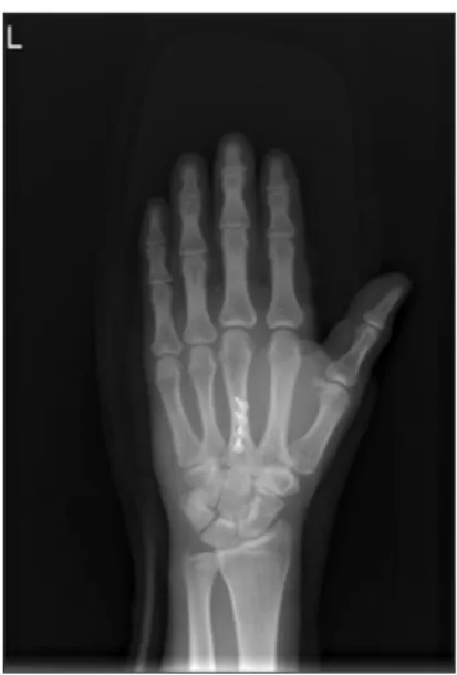
the severity of the fracture, a 4 to 6-hole plate along with 8 to 12mm screws were used for bicortical plate fixation (Fig. 1). The periosteum was then closed using Vicryl 4-0 sutures, and anti-adhesion agent was applied before skin closure with 5-0 Vicryl and 5-0 Nylon sutures. Sutures were removed 10 to 14 days after the surgery, during a short arm splint was applied.
3. Surgical technique: mini-open reduction (group II)
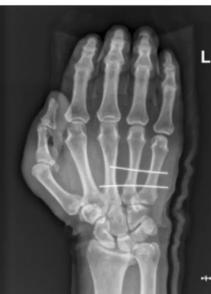
Under fluoroscopic guidance, a 3 mm sized longitudi- nal incision was made on the dorsum above the fracture site visualized, while taking care not to injure the exten- sor digitorum communis tendons. Authors reached the fracture site with gentle dissection using the Freer eleva- tor (Fig. 2). The fractured segment was then reduced to appropriate position through simultaneous distal traction of the involved finger and direct palpation using the el- evator (Fig. 3), while any intervening tissue was pulled aside to facilitate the procedure. A 0.9-1.1 mm sized Kirschner wire (K-wire) was inserted from the proximal side of the fractured metacarpal bone toward the opposite side of the bone to avoid injury to the MCP joint. The K- wire was advanced to include the whole fractured area, while maintaining the reduction and alignment with the help of the Freer elevator (Fig. 4). In patients with unfa- vorable fractures which reduction cannot be maintained with intramedullary pinning only, transverse pining method was utilized (Fig. 5). An anti-adhesion agent was applied through the stab incision site followed by skin closure, and the tip(s) of the K-wire was buried under the deep skin to minimize unwarranted interference on the
Fig. 2. Reached the fracture site with gentle dissection using the Freer elevator.
Fig. 1. Depending on the severity of the fracture, a 4 to 6-hole plate along with 8 to 12 mm screws were used for bicortical plate fixation.
Fig. 3. The fractured segment was then reduced to appropriate position through simultaneous distal traction of the involved finger and direct palpation using the elevator.
range of motion.
In both groups, after the operation, the surgeons confirmed full range of motion of the MCP, proximal interphalangeal, and distal interphalangeal joint of the involved digit. Subsequent scar management and postop- erative massage using silicone ointment and sheet were carried out. In the mini-open reduction group, the K- wire(s) was removed 4 weeks after the surgery.
RESULTS
Statistical analyses were conducted with an indepen- dent sample t-test, and p<0.05 was considered significant.
Statistical analyses was conducted using SAS software ver. 9.3 (SAS Institute, Cary, NC, USA).
Patient demographic data are presented in Table 1. The mean age in group I and group II was 41.2 and 43.7 re- spectively, with no significant difference. Male patients were dominant in both groups. The number of cases of unfavorable fractures was 9 in group I (total 14) and 14 in group II (total 23), with no significant difference. The mean follow-up duration was 15.4 months in group I, and 14.8 months in group II. There is no significant differ- ence in patients demographics between group I and group II.
The mean operative time was 41 minutes in group I and 24 minutes in group II, which was significantly shorter than the former (p=0.02). There was no significant dif- ference between two groups in the mean radiological reduction result, with group I scoring 8.2±1.1 and group II scoring 8.5±0.9. The mean VAS score was 4.3±0.7 and 2.7±0.8 in each group, showing a significantly higher re- sult in group I (p=0.02). The mean active range of motion of the MCP joint at six months was 88.4o in group I and 88.0o in group II, with no significant difference. In group I, there was one case that underwent an additional adhe- siolysis at five months after the surgery, whereas group II required no further procedures; however, there was no significant difference between two groups.
Fig. 4. After comfirmed reduction and alignment, authors further advanced the initially inserted K-wire to reach the opposite side of the metacarpal base.
Fig. 5. In patients with unfavorable fractures, K-wires are inserted transverse direction while maintaining the reduction with Freer elevator.
Table 1. Demographics and summary of the patients enrolled in the study
Group I (n=14) Group II (n=23) Age (yr) 41.2 (range, 25-57) 43.7 (range, 27-60) Sex
Female/male (%) 4 (28.6)/10 (71.4) 7 (30.4)/16 (69.6) Fracture type
Favorable/
unfavorable (%) 5 (35.7)/9 (64.3) 9 (39.1)/14 (60.9) Follow-up (mo) 15.4 (range, 13-21) 14.8 (range, 14-19) Values are presented as number only or number (%).
Group I: open reduction group, group II: mini-open reduction group.
Both groups had no major postoperative complications that required surgical intervention. There was one case of stitch abscess and one case of minimal wound disruption in group I, and two patients developed superficial infec- tion at the wire insertion site in group II. All such minor complications were resolved with conservative care. Scar quality assessment using the VSS resulted in 5.7±1.1 in group I and 3.9±0.9) in group II, indicating superior out- come in the latter (p=0.03) (Table 2).
DISCUSSION
There exists yet no absolute guideline on operative techniques for treating metacarpal fractures. Before choosing a certain surgical technique, many variables such as the location of the fracture (intra-articular or ex- tra-articular), pattern of the fracture (transverse, oblique, spiral or comminuted), or presence of any bony displace- ment must all be taken into consideration, as well as the surgeon’s preference and skill3.
In addition to closed reduction, there are various open reduction fixation techniques depending on the type and position of the fracture. K-wires may be used in nearly any fracture pattern. Pinning technique is relatively easy, requires minimal dissection and minimizing soft tissue trauma. It can be used single or multiple and may be used in combination of other fixation technique. Composite wiring is used in a combination of K-wires. It provides additional stability and fracture compression and superior strength, stiffness, and approximation compared with crossed K-wires alone. Cerclage and interosseous wiring can be applicable for oblique and spiral metacarpal shaft
fractures. These wires are rigid enough to permit early motion. But these techniques need additional dissection and are contraindication when there is bone loss, commi- nution, or osteopenia. Intramedullary fixation can be used alone and successfully employed for transverse fractures.
It is easy to perform and allow active motion. Also, it has advantage of no exposed pins and secondary removal is unnecessary. But, rotational control may be difficult and in case of infection or re-fracture, hardware removal may be very difficult. External fixation can be indicated for highly comminuted open shaft fracture with or without bone loss and comminuted articular fracture and fractures with injury of loss of surrounding soft tissue. It also has disadvantage that prominence of the device make diffi- cult for activities of daily life and there are highly risk of pin-site infection, may impede tendon gliding and cause of adhesion.
Debate still exists on whether closed reduction using K-wires or open reduction using plate and screws pro- duces optimal results. Ozer et al.10 compared intramed- ullary nailing using K-wire with open reduction using plate and screws, and concluded that the former resulted in increased loss of reduction, more injury to MCP joint, and a higher rate of secondary procedures necessary to remove K-wires11. On the other hand, Greeven et al.12 reported that patients treated with open reduction showed a higher tendency to require secondary procedures such as tenolysis compared with those treated otherwise. In all three studies, there was no significant difference between patient groups regarding final functional outcome. Facca et al.13 held closed reduction to be superior to open reduc- tion, as he reported that patients with fifth metacarpal Table 2. Postoperative result of the both groups
Group I Group II
Mean operation time (min) 41.0±6.8 24.0±4.9
Mean immediate postoperative reduction score 8.2±1.1 8.5±0.9
Mean six months postoperative ROM (deg) 88.4±0.9 88.0±1.1
Mean postoperative pain (VAS) 4.3±0.7 2.7±0.8
Number of postoperative adhesion cases requiring secondary procedure 1 0
Vancouver scar scale 5.7±1.1 3.9±0.9
Values are presented as mean±standard deviation or number only. Group I: open reduction group, group II: mini-open reduction group.
ROM: range of motion, VAS: visual analogue scale.
neck fractures who were treated with locking plates ex- hibited paradoxical poor movement of MCP joint despite of early mobilization, compared with those treated with K-wire fixation.
Authors were able to directly manipulate the fracture site with surgical instrument through minimal skin inci- sion, which enabled authors to constantly maintain the reduced position while inserting wires. In cases of un- stable fractures, such method can facilitate primary bone healing better than simple closed reduction by allowing secure bone-to-bone contact. Fractures can be generally categorized into stable and unstable groups depending on whether reduction is well-maintained or not. Most sur- geons opt for ORIF when treating unstable fractures, such as transversely displaced fractures, long spiral fractures, short oblique fractures, and displaced condyle fractures with more than 25% of articular surface involvement14. In unstable fractures, one of the main complications is meta- carpal shortening due to poor reduction maintenance, and also angulation may develop; such outcomes are not uncommon when unstable fractures are managed sim- ply with closed reduction and K-wire fixation. Authors were able to minimize such complications by inserting a Freer elevator or a bone hook through a stab incision and using the instrument to securely maintain reduction while pinning the fracture site. While maintaining secure bone-to-bone contact, authors inserted the wire into the metacarpal bone from cortex to cortex under fluoroscopy guidance, thereby eliminating unwarranted interference with the joint movement and thus enabling limited but early mobilization.
Both groups showed comparable clinical outcomes regarding post-reduction x-ray evaluation and range of motion recovery. There was no significant difference in range of motion six months after the surgery between two groups, as all the patients regained full motion re- covery. Authors also evaluated the degree of range of motion two months after the surgery to compare recovery speed, which also did not show significant difference be- tween two groups. One of the notable advantages of this proposed technique is the reduction of operation time.
Although not included in this study, authors feel that
operating time was well comparably reduced to that of closed reduction and K-wire fixation, which is the most frequently applied technique in treating stable fractures.
Also, this method showed superior outcome regarding postoperative pain, as soft tissue injury following dis- section is minimal. Furthermore, there was one case in the ORIF group that developed adhesion which required surgical release, whereas no adhesion was noted in the mini-open reduction group. Although this does not have any statistical significance, it can be assumed that such mini-open reduction should carry much lower risk of de- veloping postsurgical adhesion compared with the open reduction technique, as the former requires much less incision and dissection than the latter. As for complica- tions, both groups had acceptable outcomes as only a few patients developed minor acute wound-related problems.
Another advantage is the reduction of postoperative scar, which was nearly unnoticeable compared with that of conventional dorsal incision in the open reduction group.
Patient satisfaction regarding scar quality was significant- ly higher, making it an aesthetically tolerable technique that may be applied to socially active patients, as well as young female patients.
Although not included in this study, authors assume that this technique may prevent many complications that arise from open reduction method, such as contracture, extension lag, tendon rupture due to fixating material, or plate prominence15,16, which are all consequences of wide dissection required for open reduction method, while maintaining the fixation until bony union, especially in the case of unfavorable fracture. Also, it may minimize secondary procedures compared with open reduction method, as 25% of patients treated with open reduction are reported to undergo removal procedures due to plate- related complications15. Furthermore, for patients who feel uncomfortable having titanium plates remained in their body after ORIF, or for those that need to undergo specific imaging studies such as magnetic resonance imaging in the future, mini-open reduction may be an at- tractive alternative. This technique is relatively easy to perform and does not require extensive surgical expertise, although care must be taken not to injure the extensor
digitorum tendons and associated structures, as the visual field obtained through the stab incision can be seem- ingly limited. Also, the surgeon can simply extend the stab incision intraoperatively to switch to ORIF in case complete reduction seems unlikely due to soft tissue im- pingement in the fracture site, or in case multiple, severe fractures not previously detected in preoperative images are diagnosed during the surgery. Finally, it puts signifi- cantly less financial burden on patients compared with open reduction using plate and screws.
Admittedly, the technique presented in this study has several potential drawbacks. Our technique cannot re- place the traditional open reduction and plate fixation in case of multiple, comminuted fracture that requires rigid fixation. This type of fracture is likely to require plan- ning of traditional open reduction techniques or external fixation. Also, it still accompanies low risk of inflicting injury to vessels or nerves adjacent to the incision site, especially in case of an untrained surgeon. All such draw- backs must therefore be taken into account before execu- tion.
The restricted number of cases, along with the fact that the study was conducted by a single surgeon, places limitations to this study. A more reliable result may be achieved by both increasing the number of subjects and minimizing the surgeon’s bias. Also, authors excluded first metacarpal bone fractures from the study, since first metacarpal bones have unique anatomical structure and location different from those of other metacarpal bones, and thus would be difficult to assess its range of motion in the similar context to the rest of the metacarpal bones.
Finally, authors mainly focused on comparing with ORIF in treating unstable fractures; however, additional studies that compare with closed reduction and K-wire fixation, yet another widely performed technique in treating meta- carpal fractures, need to be conducted as well.
To our knowledge, no randomized or non-randomized studies comparing ORIF with K-wire fixation have been reported, and this is the first comparative study for K- wire fixation with ORIF in metacarpal fractures. By retaining the ability of ORIF to achieve complete reduc- tion and yet minimizing downsides such as soft tissue
adhesion and scarring, authors were able to produce sat- isfactory outcomes, and thus propose this method as an alternative to ORIF in treating most metacarpal fractures excluding intra-articular fractures.
CONCLUSION
We have obtained satisfactory results through the mini- open reduction technique. Therefore mini-open reduction method may be an alternative to conventional open re- duction in treating isolated metacarpal fractures.
CONFLICTS OF INTEREST
The authors have nothing to disclose.
REFERENCES
1. Chung KC, Spilson SV. The frequency and epidemiology of hand and forearm fractures in the United States. J Hand Surg Am. 2001;26:908-15.
2. Feehan LM, Sheps SB. Incidence and demographics of hand fractures in British Columbia, Canada: a population- based study. J Hand Surg Am. 2006;31:1068-74.
3. Ben-Amotz O, Sammer DM. Practical management of metacarpal fractures. Plast Reconstr Surg. 2015;136:370e- 9e.
4. Henry MH. Fractures of the proximal phalanx and meta- carpals in the hand: preferred methods of stabilization. J Am Acad Orthop Surg. 2008;16:586-95.
5. Diaz-Garcia R, Waljee JF. Current management of meta- carpal fractures. Hand Clin. 2013;29:507-18.
6. Freeland AE, Lindley SG. Malunions of the finger meta- carpals and phalanges. Hand Clin. 2006;22:341-55.
7. Low CK, Wong HC, Low YP, Wong HP. A cadaver study of the effects of dorsal angulation and shortening of the metacarpal shaft on the extension and flexion force ratios of the index and little fingers. J Hand Surg Br.
1995;20:609-13.
8. Friedrich JB, Vedder NB. An evidence-based approach to metacarpal fractures. Plast Reconstr Surg. 2010;126:2205- 9.
9. Bloom JM, Hammert WC. Evidence-based medicine:
metacarpal fractures. Plast Reconstr Surg. 2014;133:1252- 60.
10. Ozer K, Gillani S, Williams A, Peterson SL, Morgan S.
Comparison of intramedullary nailing versus plate-screw fixation of extra-articular metacarpal fractures. J Hand Surg Am. 2008;33:1724-31.
11. Mumtaz MU, Farooq MA, Rasool AA, Kawoosa AA, Badoo AR, Dhar SA. Unstable metacarpal and phalangeal fractures: treatment by internal fixation using AO mini- fragment plates and screws. Ulus Travma Acil Cerrahi Derg. 2010;16:334-8.
12. Greeven AP, Bezstarosti S, Krijnen P, Schipper IB. Open reduction and internal fixation versus percutaneous trans- verse Kirschner wire fixation for single, closed second to fifth metacarpal shaft fractures: a systematic review. Eur J
Trauma Emerg Surg. 2016;42:169-75.
13. Facca S, Ramdhian R, Pelissier A, Diaconu M, Liverneaux P. Fifth metacarpal neck fracture fixation: Locking plate versus K-wire? Orthop Traumatol Surg Res. 2010;96:506- 12.
14. Dabezies EJ, Schutte JP. Fixation of metacarpal and pha- langeal fractures with miniature plates and screws. J Hand Surg Am. 1986;11:283-8.
15. Bosscha K, Snellen JP. Internal fixation of metacarpal and phalangeal fractures with AO minifragment screws and plates: a prospective study. Injury. 1993;24:166-8.
16. Page SM, Stern PJ. Complications and range of motion following plate fixation of metacarpal and phalangeal fractures. J Hand Surg Am. 1998;23:827-32.
중수골 단독 골절에 대한 최소 관혈적 정복술
정연진
1ㆍ오세영
2ㆍ최지선
2ㆍ임진수
2ㆍ심형섭
21가톨릭대학교 의과대학 은평성모병원 성형외과학교실, 2가톨릭대학교 의과대학 성빈센트병원 성형외과학교실
목적: 중수골 골절은 흔히 발생하는 골절 중 하나이다. 저자는 중수골 골절의 치료로 최소한의 절개를 통한 최소 침 습적 관혈적 정복술을 제시하고 있다. 이를 통해서 전통적인 관혈적 정복술이 가져올 수 있는 각종 부작용을 최소 화하면서, 동시에 비관혈적 정복술의 장점을 취할 수 있다.
방법: 2명의 수부 외과의에 의해 수술적 치료를 받은 모든 환자를 대상으로 5년간의 후향적 연구를 진행하였다. 총 37명의 환자를 대상으로 시행되었고, 이 중 14명은 전통적인 관혈적 정복술을 시행 받았고, 23명은 최소 침습적 관혈적 정복술을 시행 받았다.
결과: 최소 관혈적 정복술을 시행 받은 군은 전통적인 관혈적 정복술을 시행 받은 군과 비교하여, 부작용의 발생 빈 도는 비슷하였으나 수술 시간이 더 짧았으며, 영상의학적으로 더 나은 정복 결과를 보였으며, 환자가 호소하는 주 관적인 통증의 정도가 더 작았고 중수지 수족 관절의 술 후 능동적 운동 가능 범위가 더 넓었고 흉의 발생 정도는 더 경미한 결과를 보였다.
결론: 최소 관혈적 정복술 방식이 중수골 골절의 치료에 있어서 전통적인 관혈적 정복술을 대체할 수 있는 좋은 방 법이 될 수 있다.
색인단어: 중수골 골절, 관혈적 정복술, 내고정술, 비관혈적 정복술, K-강선
접수일 2019년 7월 8일 수정일 2019년 8월 4일 게재확정일 2019년 9월 4일 교신저자 심형섭
16247, 경기도 수원시 팔달구 중부대로 93, 가톨릭대학교 의과대학 성빈센트병원 성형외과학교실 TEL 031-249-7206 FAX 031-241-0005 E-mail sharpshim@catholic.ac.kr
ORCID https://orcid.org/0000-0001-5156-2239