ABSTRACT
Weiss-Kruszka syndrome (WSKA), caused by heterozygous loss-of-function variants in ZNF462 gene, is a recently described and extremely rare genetic disorder. The main phenotypes include characteristic craniofacial features, ptosis, dysgenesis of the corpus callosum, and neurodevelopmental impairment. We report the first Korean boy with molecularly confirmed WSKA presenting with an atypical manifestation. A 16-year-old boy with a history of bilateral ptosis surgery presented with short stature (−3.49 standard deviation score) and delayed puberty. The patient showed characteristic craniofacial features including an inverted triangular-shaped head, exaggerated Cupid's bow, arched eyebrows, down-slanting palpebral fissures, and poorly expressive face. He had a mild degree of intellectual disability and mild hypotonia. Endocrine studies in the patient demonstrated complete growth hormone deficiency (GHD) associated with empty sella syndrome (ESS), based on a magnetic resonance imaging study for the brain that showed a flattened pituitary gland and cerebrospinal fluid space herniated into the sella turcica. To identify the genetic cause, we performed whole exome sequencing (WES). Through WES, a novel de novo heterozygous nonsense variant, c.4185del; p.(Met1396Ter) in ZNF462 was identified. This is the first case of WSKA accompanied by primary ESS associated with GHD. More clinical and functional studies are needed to elucidate this association.
Keywords: ZNF462; Weiss-Kruszka Syndrome; Empty Sella; Growth Hormone Deficiency
INTRODUCTION
Weiss-Kruszka syndrome (WSKA; MIM#618,619) is a rare autosomal dominant disorder associated with zinc-finger protein 462 (ZNF462) gene variants or the deletion of the 9q31.2 chromosome region involving ZNF462. It is characterized by mild global developmental delay, ptosis, dysmorphic craniofacial abnormalities including metopic ridging or synostosis and triangular shape forehead with/without autistic features.1,2 The ZNF462 gene encodes a zinc-finger protein of unknown function3 and it was reported that its heterozygous loss-
Case Report
Received: Feb 9, 2021 Accepted: Apr 11, 2021 Address for Correspondence:
Sujin Kim, MD
Department of Pediatrics, Inha University Hospital, Inha University College of Medicine, Northwest Gyeonggi Regional Center for Rare Disease, 27 Inhang-ro, Jung-gu, Incheon 22332, Republic of Korea.
E-mail: [email protected] Ji Eun Lee, MD, PhD
Department of Pediatrics, Inha University Hospital, Inha University College of Medicine, Northwest Gyeonggi Regional Center for Rare Disease, 27 Inhang-ro, Jung-gu, Incheon 22332, Republic of Korea.
E-mail: [email protected]
© 2021 The Korean Academy of Medical Sciences.
This is an Open Access article distributed under the terms of the Creative Commons Attribution Non-Commercial License (https://
creativecommons.org/licenses/by-nc/4.0/) which permits unrestricted non-commercial use, distribution, and reproduction in any medium, provided the original work is properly cited.
ORCID iDs Jisun Park
https://orcid.org/0000-0003-3793-6577 Dong Jun Ha
https://orcid.org/0000-0002-4331-6801 Go Hun Seo
https://orcid.org/0000-0003-1518-1791 Seri Maeng
https://orcid.org/0000-0001-6850-5548 Sung Mo Kang
https://orcid.org/0000-0001-7722-596X
Jisun Park ,1,2 Dong Jun Ha ,1 Go Hun Seo ,3 Seri Maeng ,2,4 Sung Mo Kang ,2,5 Sujin Kim ,1,2 and Ji Eun Lee 1,2
1Department of Pediatrics, Inha University Hospital, Inha University College of Medicine, Incheon, Korea
2Northwest Gyeonggi Regional Center for Rare Disease, Inha University Hospital, Incheon, Korea
33 Billion Inc., Seoul, Korea
4Department of Psychiatry, Inha University Hospital, Inha University College of Medicine, Incheon, Korea
5 Department of Ophthalmology, Inha University Hospital, Inha University College of Medicine, Incheon, Korea
Empty Sella Syndrome Associated with Growth Hormone Deficiency:
the First Case Report of Weiss- Kruszka Syndrome
Human Genetics & Genomics
Sujin Kim
https://orcid.org/0000-0003-0893-0512 Ji Eun Lee
https://orcid.org/0000-0002-7386-0015 Funding
This work was supported by Inha University Research Grant, 2021.
Disclosure
The authors have no potential conflicts of interest to disclose.
Author Contributions
Conceptualization: Park J, Kim S, Lee JE. Data curation: Ha DJ. Formal analysis: Seo GH.
Writing - original draft: Park J. Writing - review
& editing: Maeng S, Kang SM, Kim S, Lee JE.
of-function variants presented with a pattern of overlapping phenotype in eight individuals from six families in 2017.4 To date, only 25 affected individuals from 22 families have been described.1,2 In most cases (95%), WSKA occurs due to de novo variants in ZNF462, and only 5% of individuals diagnosed with WSKA have an affected parent.1 Brain lesions related to WSKA are identified as ventriculomegaly and midline structural abnormalities of brain such as corpus callosum hypoplasia in about 25% of patients.2 However, there has been no report of structural malformation of the pituitary gland related to midline brain structures and endocrine dysfunction in individuals with WSKA. Here, we present the case of a Korean boy with
molecularly confirmed WSKA and previously unreported clinical manifestations of primary empty sella syndrome (ESS), which was associated with growth hormone deficiency (GHD).
CASE DESCRIPTION
A 16-year-old boy was referred to our hospital for the evaluation of growth retardation and delayed puberty. He was born at full term to healthy and non-consanguineous Korean parents, with a birth weight and length of 3.0 kg (−0.30 standard deviation score [SDS]) and 50 cm (0.06 SDS), respectively. The heights of his father and mother were 174 cm (−0.08 SDS) and 170 cm (1.74 SDS). At the first visit, his height was 152.8 cm (−3.49 SDS) and weight was 42.7 kg (−2.82 SDS). There were no postnatal problems, such as microphallus and cryptorchidism, except bilateral ptosis. He underwent surgery for the correction of ptosis at the age of 4 years. He had no previous history of trauma or chronic illness. He complained of mild hypotonia when he exercised or did hard work. He felt frequent tiredness and muscle weakness in ordinary life. According to his mother, he worked alone at 15 months- old and his language development was also delayed when he was a young child. According to the results of his cognitive and intellectual function in the department of psychiatry at 18 years old, the full scale intelligence quotient was 75 according to the results of Korean Wechsler Adult Intelligence Scale-IV administered at the age of 18. However, the social age for estimating social adaptation function was social quotient 84, and considering the stable subtest performance level, his intellectual ability was estimated to be at the lower average level (80–89). In the comprehensive attention test, a decrease in the function of interference in selective attention and divided attention was observed.
From school age, he gradually showed a decrease in height velocity, and there has been no pubertal development in adolescence. He had two younger brothers who grew up normally without any growth and developmental issues and had no family history of genetic disorders.
The blood pressure was 109/73 mmHg. His pubertal stage was assessed as Tanner 2 for the external genitalia development (testes volume 4 mL). He had dysmorphic craniofacial features, including an inverted triangular-shaped forehead, exaggerated Cupid's bow, arched eyebrows, down-slanting palpebral fissures, pterygium in the left eye with corneal opacity, mild ptosis and poorly expressive face (Fig. 1). He presented with mild hypotonia without other neurological symptoms and signs such as headache or visual disturbance. Laboratory tests for chemistry and thyroid hormone showed normal levels. The serum insulin-like growth factor (IGF)-1 and IGF binding protein-3 levels were 155.3 ng/mL (normal range: 360.0–885.0 ng/mL) and 1,698 ng/mL (normal range: 1,574–4,260 ng/mL), respectively. The bone age was over 2 years delayed. Two different provocative growth hormone (GH) testing, glucagon stimulation test and L-dopa stimulation test, revealed complete GHD as the result of low peak GH levels (0.34 and 1.99 ng/mL, respectively). Gonadotropin-releasing hormone stimulation testing showed early stage of puberty (peak luteinizing hormone 8.93 mlU/mL and peak follicle stimulating
hormone 12.10 mIU/mL). Testosterone level was 0.23 ng/mL. Magnetic resonance imaging (MRI) of the brain showed a flattened pituitary gland and cerebrospinal fluid (CSF) space herniated into the sella turcica, which indicated ESS (Fig. 2). The other pituitary hormones showed normal levels. To identify the underlying genetic cause, we performed karyotype and chromosomal microarray analysis, which showed unremarkable results. Whole exome sequencing (WES) was performed and a heterozygous novel nonsense variant, c.4185del;
p.(Met1396Ter), in ZNF462 was identified, which was confirmed by Sanger sequencing (Fig.
3). This variant resulted from the deletion of nucleotides “C” in exon 3 of ZNF462, making stop codon in the protein-coding sequence, which was expected to produce truncated protein.
This variant in ZNF462 has not been reported in a large population database as well as in the Genome Aggregation Database (https://gnomad.broadinstitute.org/). The patient's phenotype matched the typical phenotypes of previously reported individuals with WSKA.5 Segregation analysis using Sanger sequencing showed no ZNF462 variant in his mother. Although his father and two younger brothers could not undergo genetic tests for detecting the ZNF462 variant, there were no clinically affected family members. Therefore, this variant was categorized as pathogenic according to the standards and guidelines of the American College of Medical Genetics and Genomics.6 There were no additional congenital anomalies such as congenital heart defects, optic nerve hypoplasia, papilledema and hearing impairment. Recombinant
A B C D
Fig. 1. Patient's craniofacial manifestation. (A, B) The patient has arched eyebrows, exaggerated Cupid's bow, and short upturned nose with bulbous nasal tip.
(C) Inverted triangular-shaped forehead was shown. (D) Inverted triangular-shaped head shape was shown in back. The figures are published with the consent of the patient and his parents.
A B C
Fig. 2. MRI studies of the brain showing ESS. (A) Sagittal view of brain MRI shows a flattened pituitary gland and CSF space herniated into the sellar turcica. The posterior pituitary gland appears normal. (B) Coronal view of brain MRI shows the pituitary fossa which is largely empty of tissue, replaced by CSF. (C) Axial view of brain MRI shows ESS.
MRI = magnetic resonance imaging, ESS = empty sella syndrome, CSF = cerebrospinal fluid.
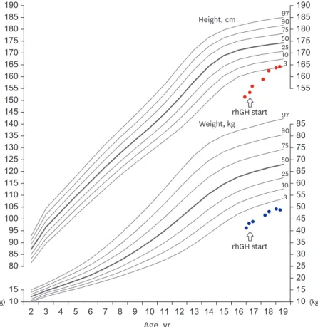
human growth hormone (rhGH) replacement therapy was started at the dose of 0.23 mg/kg/
week administered as daily subcutaneous injections and the dose of rhGH has been gradually increased up to 0.3 mg/kg/week until 18 years old. An improvement in height velocity (8 cm/
year) was observed and his current height is 168.2cm (−1.15 SDS). Gradual pubertal progress (Tanner stage 2 for the external genitalia development, testes volume 6 mL at 18 years old) was also observed (Fig. 4). Now, he is 18 years 11 months-old and his bone age was estimated at 15 years. He continues rhGH replacement therapy and we consulted a urologist to evaluate and manage sexual maturity and fertility.
Ethics statement
The study was approved by the Institutional Review Board of Inha University Hospital (No.
2020-08-014). The images are published with the consent of the patient and his legal guardian.
Independent informed consent for the publication of this case report was also obtained.
DISCUSSION
ZNF462 located in the 9q31.2 chromosome encodes a C2H2 type of zinc-finger transcription factor, which is believed to play an important role in embryonic development, especially in brain development, and it controls early patterning of the central nervous system.2,7-9 According to the literature, the phenotype associated with variations in ZNF462 is
10 190 185 180 175 170 165 160 155
190 185 180 175 170 165 160 155
85 80 75 70 65 60 55 50 150
145 140 135 130 125 120 115 110 105 100 95 90 85 80
45 40 35 30 25 20 15 10 15
2 3 4 5 6 7 8 9 10 11 12 13 14 15 16 17 18 19 Age, yr
rhGH start rhGH start
Height, cm 9097
75 5025 103
97 90 75 50 25 10 3 Weight, kg
(kg) (kg)
Fig. 3. Growth chart of the patient under rhGH replacement therapy. Upper side shows height chart and lower side shows weight chart. The dots indicate the height and weight measurement. The arrow marks the beginning of rhGH treatment start. The patient's height velocity after the start of rhGH was increased 8 cm/year.
characterized by craniofacial anomalies, corpus callosum dysgenesis, ptosis, and developmental delay.4,7 Since the first reported case of a ZNF462-related syndrome in 2003,1,2,8 only 25 individuals from 22 families have been identified to have pathogenic variants in ZNF462, including chromosomal rearrangements disrupting ZNF462.1,2,7 The most common clinical features of WSKA are ptosis and developmental delay, described in 83% and more than 75% of affected individuals, respectively.1,2 Patients with WSKA show various types of developmental delay, such as global delay, motor delay, speech delay, or a combination of these. In addition, down-slanting palpebral fissures (54%), an exaggerated Cupid's bow (54%), arched eyebrows (50%), epicanthal folds (46%), and a short, upturned nose with a bulbous tip (46%) have been described in a large proportion of patients.1,2
However, metopic ridging or craniosynostosis involving the metopic or lambdoid suture is less common (38%).1,2,8 About 25% of patients with WSKA show corpus callosum abnormalities on brain imaging (Table 1).1,2 Our patient had typical craniofacial features, which were previously reported in individuals with WSKA, including bilateral ptosis, mild intellectual disability without autism, and mild hypotonia. Interestingly, after the diagnosis of WSKA based on the results of WES, “reverse phenotyping” was performed to confirm the diagnosis.
As previously reported, the extensive use of facial analysis technologies can help increase the number of patients diagnosed with this syndrome.1,2 Additionally, brain MRI of this patient indicated ESS, which is associated with hypothalamic-pituitary dysfunction, such as complete GHD; this clinical finding was unique and has not been reported. The index patient in our study carried a novel pathogenic heterozygous nonsense variant, c.4185del; p.(Met1396Ter), in exon 3 of the ZNF462 gene. Most of the reported pathogenic variants in ZNF462 are loss-of- function variants in exon 3, which accounts for 54% of the coding region of ZNF462.8 Another heterozygous variant in exon 3 of the ZNF462 gene, c.4165C>T; p.(Gln1389Ter) (NM_021224.5), has already been reported; it is located in close proximity to our patient's variant position.8 A patient with a Gln1389-to-Ter substitution showed grossly similar craniofacial features with no mention of growth failure and he did not undergo brain MRI.8
Primary ESS is observed less frequently in children than in adults, and it is frequently associated with hypothalamic-pituitary dysfunction such as GHD, hypogonadism, and multiple pituitary hormone deficiency.10-12 The exact etiology of primary ESS is not known, but several
ZNF462 gene c.4185del; (p.Met1396Ter)
Patient
Mother
Fig. 4. Sanger sequencing result of the patient with a novel heterozygous nonsense variant at position 4185 (c.4185del) in ZNF462 gene. The arrow points deletion of ‘c,’ which generates stop codon. The patient's mother did not have the ZNF462 pathogenic variant.
rhGH = recombinant human growth hormone.
etiopathogenetic hypotheses have been proposed, including congenital deformities of the sellar diaphragm and pituitary and/or upper sellar factors.10,13 The following syndromes have been reported to be associated with primary ESS: Turner syndrome, Moyamoya disease, Bartter's syndrome, nevoid basal cell carcinoma syndrome, Hunter syndrome, Prader-Willi syndrome, Alstrom syndrome, Meniere's disease, and Erdheim-Chester disease.13 To date, a few patients with WSKA who underwent brain imaging (64%, 16/25) have shown corpus callosum dysgenesis (30%, 5/16).2 Since the causative gene for this syndrome has recently been identified, its clinical manifestation and molecular basis need to be established simultaneously.
However, considering that the pituitary gland, pons, cerebellar vermis and corpus callosum are Table 1. Phenotypes of patients with reported variants of ZNF462
Patients Sex Age,
yr Variant type Inheritance DD Ptosis Down- slanting palpebral
fissures
Arched eyebrows Short
upturned nose
Cupid's
bow Epicanthal folds Cranio-
synostosis/
metopic ridging
Hypotonia Midline brain abnormal-
ities Index P M 16 c.4185del; p.(Met1396Ter) Mother
negative, father unknown
+ + + + + + + + + +
P1 M 1 c.2590C>T p.(Arg864
*
) Maternal (mosaic) + + − + − + + − + −P2 M 10 c.2542del p.(Cys848Valfs
*
66) De novo + + + + + + + − − −P3 M 6 c.831_834del
p.(Arg277Serfs
*
26) De novo + − − − + − − − + −P4 M 2 c.6214_6215del
p.(His2072Tyrfs
*
8) De novo + + − − − + + + −P5 F 14 c.763C>T p.(Arg255
*
) De novo + + − + − − + + −P6 F 0 c.7057-2A>G De novo + + + + + + + − + +
P7 M 13 c.6794dup p.(Tyr2265
*
) De novo + − − + − + − + −P8 M 2 c.882dup p.(Ser295Glnfs
*
64) De novo + + + − − − − − − +P9 M 15 c.4165C>T p.(Gln1389
*
) De novo + + + + − + − − +P10 M 8 c.1234_1235insAA; p.(Ser412
*
) De novo + + − − − − − − −P11 F 2 c.6214_6215del
p.(His2072Tyrfs
*
8) De novo − + − − − − − +P12 M 0 c.2049dup p.(Pro684Serfs
*
14) De novo + + + + + + + − + −P13 M 8 c.6631del p.(Arg2211Glyfs
*
59) De novo − + + − − + − − −P14 F 8 c.2695G>T; p.(Glu899
*
) negative, Motherfather unknown
+ − − + + − − − + −
P15 F 2 c.3787C>T p.(Arg1263
*
) Paternal − + + + + + − + − +P16 F 4 c.3787C>T p.(Arg1263
*
) Paternal − + + − − + + + − −P17 M 34 c.3787C>T p.(Arg1263
*
) Maternal − + − − − − − + −P18 M 2 c.2979_2980delinsA
p.(Val994Trpfs
*
147) De novo + + + + + − + + − −P19 M 2 c.4263del p.(Glu1422Serfs
*
6) De novo + + + − + − + + + −P20 F 5 Chr9:g.(108940763–110561397)
del(hg19) De novo − − + + + + + − + +
P21 F 15 Chr9:g(108464368–110362345)
del (hg19) De novo + + − − − + − − −
P22 M 9 c.5145delC p.(Tyr1716Thrfs
*
28) De novo + + − − − − − − + −P23 F 5 t(2;9)(p24;q32); disrupting
ZNF462 and ASXL2 De novo + + + + + + − − + +
P24 M 24 t(9;13)(q31.2; q22.1) disrupting
ZNF462 and KLF12 De novo + + + − − − + − + +
P25 F 3 c.3306dup;
p.(Gln1103Thrfs
*
10) De novo + + + − − − + − +Inheritance types were maternal 8% (2/25), paternal 12% (3/25), unknown 8% (2/25), de novo 72% (18/25). Clinical characteristics were below; DD in 76%
(19/25), 84% (21/25) with ptosis, 56% (14/25) with down-slanting palpebral fissures, 52% (13/25) with arched eyebrows, short upturned nose in 48% (12/25), Cupid's bow in 52% (13/25), metopic ridging in 40% (10/25), hypotonia in 52% (13/25) and midline brain abnormalities in 28% (7/25, 10 had normal brain MRI findings and 8 were not tested) including our patient. Blank means no mention about the clinical features and/or no test results have been reported.
P = patient, DD = developmental delay, MRI = magnetic resonance imaging.
included in brain midline structures, we infer ESS might be a type of deformity in brain midline structures.14 As ZNF462 significantly contributes to the regulation of brain morphogenesis,15,16 further functional studies on ZNF462 variants are required.
This is the first case of WSKA accompanied by primary ESS associated with GHD. This case contributes to the diagnosis of WSKA and the identification of its clinical features. More clinical and functional studies are needed to elucidate this association.
ACKNOWLEDGMENTS
We thank the patient and his family for their participation in this study.
REFERENCES
1. González-Tarancón R, Salvador-Rupérez E, Miramar Gallart MD, Barroso E, Díez García-Prieto I, Pérez Delgado R, et al. A novel mutation in the ZNF462 gene c.3306dup; p.(Gln1103Thrfs*10) is associated to Weiss-Kruszka syndrome. A case report. Acta Clin Belg 2020;1-4.
PUBMED | CROSSREF
2. Kruszka P. Weiss-Kruszka syndrome. In: Adam MP, Ardinger HH, Pagon RA, Wallace SE, Bean LJH, Mirzaa G, et al., editors. GeneReviews®. Seattle, WA: University of Washington; 1993.
3. Nagase T, Nakayama M, Nakajima D, Kikuno R, Ohara O. Prediction of the coding sequences of unidentified human genes. XX. The complete sequences of 100 new cDNA clones from brain which code for large proteins in vitro. DNA Res 2001;8(2):85-95.
PUBMED | CROSSREF
4. Weiss K, Wigby K, Fannemel M, Henderson LB, Beck N, Ghali N, et al. Haploinsufficiency of ZNF462 is associated with craniofacial anomalies, corpus callosum dysgenesis, ptosis, and developmental delay. Eur J Hum Genet 2017;25(8):946-51.
PUBMED | CROSSREF
5. Uliana V, Percesepe A. Reverse phenotyping comes of age. Mol Genet Metab 2016;118(4):230-1.
PUBMED | CROSSREF
6. Richards S, Aziz N, Bale S, Bick D, Das S, Gastier-Foster J, et al. Standards and guidelines for the
interpretation of sequence variants: a joint consensus recommendation of the American College of Medical Genetics and Genomics and the Association for Molecular Pathology. Genet Med 2015;17(5):405-24.
PUBMED | CROSSREF
7. Cosemans N, Vandenhove L, Maljaars J, Van Esch H, Devriendt K, Baldwin A, et al. ZNF462 and KLF12 are disrupted by a de novo translocation in a patient with syndromic intellectual disability and autism spectrum disorder. Eur J Med Genet 2018;61(7):376-83.
PUBMED | CROSSREF
8. Kruszka P, Hu T, Hong S, Signer R, Cogné B, Isidor B, et al. Phenotype delineation of ZNF462 related syndrome. Am J Med Genet A 2019;179(10):2075-82.
PUBMED | CROSSREF
9. Nowick K, Gernat T, Almaas E, Stubbs L. Differences in human and chimpanzee gene expression patterns define an evolving network of transcription factors in brain. Proc Natl Acad Sci U S A 2009;106(52):22358-63.
PUBMED | CROSSREF
10. De Marinis L, Bonadonna S, Bianchi A, Maira G, Giustina A. Primary empty sella. J Clin Endocrinol Metab 2005;90(9):5471-7.
PUBMED | CROSSREF
11. Lee SS, Han AL, Ahn MB, Kim SH, Cho WK, Cho KS, et al. Growth without growth hormone in combined pituitary hormone deficiency caused by pituitary stalk interruption syndrome. Ann Pediatr Endocrinol Metab 2017;22(1):55-9.
PUBMED | CROSSREF
12. Son HW, Lee JE, Oh SH, Keum C, Chung WY. Effects of long-term growth hormone therapy in a girl with Floating-Harbor syndrome. Ann Pediatr Endocrinol Metab 2020;25(2):126-31.
PUBMED | CROSSREF
13. Chiloiro S, Giampietro A, Bianchi A, Tartaglione T, Capobianco A, Anile C, et al. Diagnosis of endocrine disease: primary empty sella: a comprehensive review. Eur J Endocrinol 2017;177(6):R275-85.
PUBMED | CROSSREF
14. Hayakawa K, Konishi Y, Matsuda T, Kuriyama M, Konishi K, Yamashita K, et al. Development and aging of brain midline structures: assessment with MR imaging. Radiology 1989;172(1):171-7.
PUBMED | CROSSREF
15. Al-Naama N, Mackeh R, Kino T. C2H2-type zinc finger proteins in brain development,
neurodevelopmental, and other neuropsychiatric disorders: systematic literature-based analysis. Front Neurol 2020;11:32.
PUBMED | CROSSREF
16. Krishna SS, Majumdar I, Grishin NV. Structural classification of zinc fingers: survey and summary. Nucleic Acids Res 2003;31(2):532-50.
PUBMED | CROSSREF