This is an Open Access article distributed under the terms of the Creative Commons Attribution Non-Commercial License (http://creativecommons.org/licenses/
The reduction methods of operator’s radiation dose for portable dental X-ray machines
Objectives: This study was aimed to investigate the methods to reduce operator’s radiation dose when taking intraoral radiographs with portable dental X-ray machines.
Materials and Methods: Two kinds of portable dental X-ray machines (DX3000, Dexcowin and Rextar, Posdion) were used. Operator’s radiation dose was measured with an 1,800 cc ionization chamber (RadCal Corp.) at the hand level of X-ray tubehead and at the operator’s chest and waist levels with and without the backscatter shield. The operator’s radiation dose at the hand level was measured with and without lead gloves and with long and short cones. Results: The backscatter shield reduced operator’s radiation dose at the hand level of X-ray tubehead to 23 - 32%, the lead gloves to 26 - 31%, and long cone to 48 - 52%. And the backscatter shield reduced operator’s radiation dose at the operator’s chest and waist levels to 0.1 - 37%. Conclusions:
When portable dental X-ray systems are used, it is recommended to select X-ray machine attached with a backscatter shield and a long cone and to wear the lead gloves. (Restor Dent Endod 2012;37(3):160-164)
Key words: Dental digital radiography; Portable dental X-ray; Radiation dosage;
Radiation protection
Introduction
Since portable dental X-ray machine was developed to be used in military field, it has been used for forensics at disaster areas and for patients who cannot access care in dental offices, i.e. those who are home-bound or handicapped.1-6 Recently, portable dental X-rays have been used for identification of the dead bodies by Indonesia’s tsunami damage or by hurricane in Louisiana, USA.7 In Korea, portable dental X-ray machines are sometimes used for implant surgery and endodontic treatment and even in general diagnosis, since it is convenient. Some American states including Michigan, Ohio, and Washington allow the limited use of portable dental X-ray machines and define the conditions of use for these machines.8-10 But, Korea doesn’t have rules or guidelines on radiation protection for the portable dental X-ray machine.
The operator leakage and scattered radiation from portable dental radiography were greater than those from fixed dental radiography because it is handheld.11 According to National Institute of Food and Drug Safety Evaluation (NIFDS) regulation in Korea, when portable dental X-ray machine is used at places other than the operating room, the emergency room and the intensive care room, it is imperative to wear the lead apron and to use radiation protective partition.12 This inspecting standard is mostly about patient radiation protection, not for the operator who directly handles the
Jeong-Yeon Cho, Won- Jeong Han*
Department of Oral & Maxillofacial Radiology, Dankook University College of Dentistry, Cheonan, Korea
Received July 14, 2012;
Last Revised July 31, 2012;
Accepted July 31, 2012.
Cho JY; Han WJ, Department of Oral and Maxillofacial Radiology, Dankook University Colledge of Dentistry, Cheonan, Korea
*Correspondence to
Won-Jeong Han, DDS, MSD, PhD.
Associate Professor, Department of Oral & Maxillofacial Radiology, Dankook University College of Dentistry, 119 Dandae-ro, Dongnam-gu, Cheonan, Korea 330- 714
TEL, +82-41-550-1922; FAX, +82- 41-556-7127; E-mail, wjhan@
dankook.ac.kr
ISSN 2234-7658 (print) / ISSN 2234-7666 (online) http://dx.doi.org/10.5395/rde.2012.37.3.160
※ This research was supported by a grant (11172KFDA528) from Korea Food and Drug Administration in 2011.
equipment. The operator was prohibited from holding of the tube housing or cone with his or her hand to reduce the chance of occupational exposure. Moreover the operator should stand 6 feet from the X-ray tube during the exposure. Dentists or assistants using portable dental X-ray unit usually pay little attention on radiation protection due to their little understanding of operator exposure.
Therefore this study was done to evaluate the efficiency of various remedies for reduction of the operator’ radiation dose in dental radiography by portable dental X-ray machines.
Materials and Methods
1. Materials
Dose measurements were conducted using a Human skull DXTRR III (Dentsply Rinn, Elgin, IL, USA) (Figure 1) and were recorded using an ionization chamber (Medical X-ray system test equipments Model 9015RM with 1,800 chamber, RadCal Corp., Monrovia, CA, USA) (Figure 2).
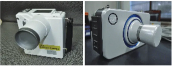
Two kinds of portable dental X-ray machines were included in this study. One (DX3000, Dexcowin, Seoul, Korea) was operated at 60 kVp, 1 mA and could be equipped with a removable external lead shield in place at the end of the cone. The other (Rextar, Posdion, Seoul, Korea) could be attached with two types of cones, 6 cm and 14 cm in length and operated at 70 kVp, 2 mA (Figure 3).
2. Methods
1) Operator’s radiation dose measurement with the use of the backscatter shield
(1) At the operator’s hands level
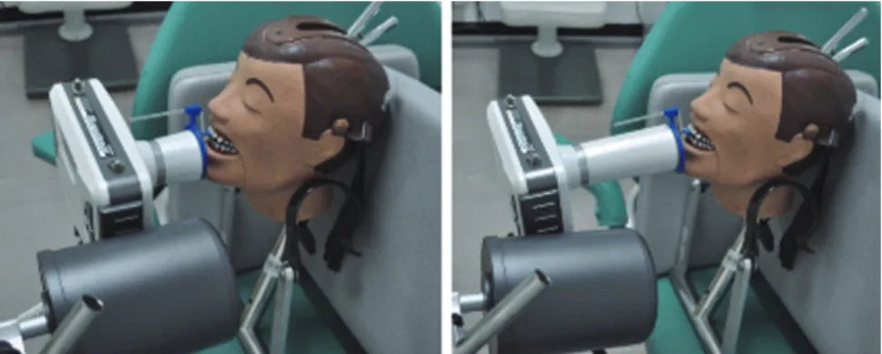
Dose measurement was carried out at the operator’s hand level during the exposure of lower anterior and posterior teeth, with and without a lead backscatter shield attached at the end of cone. The setting for exposure was 60 kVp, 1 mA and 0.4 seconds for the lower anterior teeth, 0.8 seconds for the lower posterior teeth using digital sensor (Figures 4 and 5).
Figure 1. Human skull phantom DXTRR III.
Figure 2. An 1,800 cc ionization chamber for low-level radiation measurement.
Figure 3. Two kinds of portable hand-held X-ray machines (Left-DX3000; Right-Rextar).
Figure 4. Operator’s radiation dose was measured by an ionization chamber at the operator’s hand level using the portable dental X-ray machine without a lead shield.
(2) At the operator’s chest and waist levels
The scattered radiation was measured at the operator’s chest and waist levels at a horizontal distance of 20 cm away from the X-ray machine during the exposure of lower posterior teeth, with and without a lead backscatter shield attached at the end of cone. A vertical distance of 130
cm from the floor was used as the chest height and 70 cm from the floor as the waist height (Figure 6).
2) Operator’s radiation dose measurement with the use of the lead gloves
The doses at the hand level were measured in cases of wearing the lead gloves and not wearing them when human skull DXTTR III was exposed to portable X-ray unit.
The lead sleeves which have lead equivalence 0.23 mm Pb at 60 kVp were used instead of the lead gloves due to size and form of ionization chamber (Figure 7).
3) Operator’s radiation dose measurements according to the cone length of tubehead
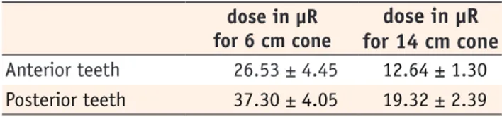
Rextar X-ray machine (Posdion, Seoul, Korea) could attach two kinds of cone, 6 cm and 14 cm in length. Operator’s radiation dose at the hand level were measured with both of them. Each setting time for anterior and posterior teeth exposure was 0.1 and 0.2 second in a 6 cm length cone, respectively. In a 14 cm cone, exposure time was 0.2 and 0.43 second, respectively (Figure 8).
Figure 5. Portable dental X-ray machine with a lead shield.
Figure 6. Scattered radiation dose was measured by an ionization chamber at the operator’s waist level.
Figure 7. Radiation doses were measured at the hand level of operators using an ionization chamber covered with a lead sleeve.
Figure 8. Operator’s radiation dose at hand level of operator was measured using short and long cones.
Results
1. Operator’s radiation dose with the use of the backscatter shield
When a lower periapical radiograph was taken using a portable dental X-ray machine, the backscatter shield to the end of cone reduced operator’s radiation dose to 32%
(posterior teeth) and 23% (anterior teeth) at the hand level, 0.1% at the chest level and 37% at the waist level (Table 1).
2. Operator’s radiation dose with the use of the lead gloves
When the lower anterior and posterior periapical radiography was taken using a portable dental X-ray machine, the lead gloves decreased operator’s radiation dose at the hand level to 26% and 31% respectively (Table 2).
3. Operator’s radiation dose according to the cone length of tubehead
When the lower anterior and posterior periapical radiography was taken using portable dental X-ray machine, the long cone (14 cm length cone) reduced operator’s radiation dose at the hand level to 48% and 52% respectively (Table 3).
Discussion
The portable dental X-ray machine has the advantage of free movement outside the X-ray room. For this reason, the use of this equipment is on the increase in the dental clinics. Though its convenience and utility are accepted in the disaster area, the weak point is that the operator has the equipment in his/her hands. The operator has the possibility of direct exposure to leakage radiation from the tube head and scattered radiation from the patient.
Some authors reported that operator exposure due to leakage and scattered radiation using the hand-held dental X-ray system are well below established occupation exposure limits and insignificant compared with established radiation safety guidelines of 50 mSv per year.7,13-15 Even so, operator exposure to radiation should be reduced to keep the ALARA (as low as reasonably achievable) principle of dose optimization. Methods of dose reduction that can be used in portable dental radiography were offered. The focus in this study is not about the risk of the operators but the effect of dose reduction methods like backscatter shield, lead gloves and long cone. In further study, it is recommended to do dose measurements using TLD dosimetry to estimate operator’s biologic effect.
Operator’s radiation dose is mitigated by the use of shielding devices to reduce leakage exposure and to minimize backscatter from the patient.16 NOMAD (Arbiex, Orem, UT, USA) has an external lead-acrylic backscatter Table 1. Operator’s radiation dose during the exposure of
lower teeth with and without the backscatter shield dose in µR
without shield
dose in µR with shield Hand level (anterior) 70.46 ± 0.35 22.79 ± 0.61 Hand level (posterior) 128.53 ± 18.90 29.17 ± 5.59 chest level 42.87 ± 4.88 0.04 ± 0.11 waist level 61.72 ± 3.92 22.79 ± 4.42 chest level, 130 cm height from floor; waist level, 70 cm height from floor.
*dose in µR means the leakage and scattered radiation dose and standard deviation measured by an 1,800 cc ionizing chamber.
Table 2. Operator’s radiation dose at the hand level during the exposure of lower teeth with and without the lead gloves
dose in µR without shield
dose in µR with shield Anterior teeth 82.96 ± 1.33 21.37 ± 0.65 Posterior teeth 144.19 ± 15.85 44.70 ± 5.50 anterior teeth, lower anterior periapical radiography;
posterior teeth, lower posterior periapical radiography.
*dose in µR means the average leakage and scattered radiation dose and standard deviation measured by an 1,800 cc ionizing chamber.
Table 3. Operator’s radiation dose at the hand level during the exposure of the lower teeth using 6 cm and 14 cm length cone
dose in µR for 6 cm cone
dose in µR for 14 cm cone Anterior teeth 26.53 ± 4.45 12.64 ± 1.30 Posterior teeth 37.30 ± 4.05 19.32 ± 2.39 anterior teeth, lower anterior periapical radiography;
posterior teeth, lower posterior periapical radiography.
*dose in µR means the average leakage and scattered radiation dose and standard deviation measured by 1,800 cc ionizing chamber.
shield permanently attached at the end of the tube, with which the low operator exposure is described by the manufacturer.17 The use of the NOMAD presented no significant scattered radiation risk to any member of the operators.15,18 When three portable X-ray machines were tested for the occupational dosimetry, the dose at the operator’s hand with protective shielding was the lowest.18 In this study, we found that the dose at the operator’s hand level of the machine provided with the circular lead shields was reduced to 23 - 32%, in comparison to the case with the shields.
Most of portable dental X-ray machines, recently available in the Korea market, have no radio-protective shielding on the cone. Also, there is no legal standard for the additional reduction in radiation exposure to the operator.According to the results, we intend to provide a guideline for safe use of portable dental X-ray machines so that operators are able to use the appropriate shielding device while taking radiographs using a portable dental X-ray unit.
Conclusions
Use of a backscatter shield reduced the operator’s radiation dose at hand level of X-ray tubehead to 23 - 32%, the lead gloves to 26 - 31%, and long cone of 14 cm to 48 - 52%. When portable dental X-ray systems are used, it is recommended to select the X-ray machine attached with a backscatter shield and a longer cone, and to wear the lead gloves.
References
1. Coy JD. Use of lightweight X-ray machine and processor during Riverine medical readiness training exercise on the Amazon River. Mil Med 1991;156:623-628.
2. Van Dis ML, Miles DA, Parks ET, Razmus TF. Information yield from a hand-held dental x-ray unit. Oral Surg Oral Med Oral Pathol 1993;76:381-385.
3. Coy J. Hand-held dental X-ray (HDX) with medical collimator: use in casualty radiology. Mil Med 1996;
161:428-431.
4. Coy J, Vandre RH, Davidson WR. Use of the hand- held dental X-ray machine during joint operation, NATO exercise Display Determination-92. Mil Med 1997;162:575-577.
5. Varghese S, Kimmel A, Radmer T, Bradley TG, Bahcall J.
In vitro evaluation of the XR-15 portable x-ray unit for forensic odontology. J Forensic Odontostomatol 2004;
22:5-8.
6. Charlton DG. Portable dental equipment: dental units and x-ray equipment. Gen Dent 2009;57:336-341.
7. Hermsen KP, Jaeger SS, Jaeger MA. Radiation safety for the NOMAD™ portable X-ray system in a temporary
morgue setting. J Forensic Sci 2008;53:917-921.
8. Department of Licensing and regulatory affairs: Ionizing radiation rules, Part 9. Dental X-ray installations, R325.5396. Hand-held portable dental x-ray systems. Available from: http://www.michigan.gov/
lara/0,4601,7-154-35299_28142_3579-46448--,00.
html (updated 2012 July 30).
9. Ohio department of Health: Dental radiation- generating equipment. Available from: http://www.
odh.ohio.gov/~/media/ODH/ASSETS/Files/rules/
final/37011-66/37011-66-06.ashx (updated 2012 July 30).
10. Washington state legislature: Radiation safety and diagnostic image quality standards for dental facilities, chapter 246-225A-085 Hand-held X-ray system.
Available from: http://apps.leg.wa.gov/wac/default.
aspx?cite=246-225A&full=true#246-225A-085 (updated 2012 July 30).
11. Kim EK. Leakage and scattered radiation from hand- held dental x-ray unit. Korean J Oral Maxillofac Radiol 2007;37:65-68.
12. National Institute of Food and Drug Safety Evaluation.
Regulation for the safety management of diagnostic x-ray equipments. Available from: http://www.nifds.
go.kr/nifds/01_about/about08.jsp?mode=view&article_
no=4813&pager.offset=0&board_no=2 (updated 2012 July 30).
13. Danforth RA, Herschaft EE, Leonowich JA. Operator exposure to scatter radiation from a portable hand-held dental radiation emitting device (Aribex™ NOMAD™) while making 915 intraoral dental radiographs. J Forensic Sci 2009;54:415-421.
14. Goren AD, Bonvento M, Biernacki J, Colosi DC.
Radiation exposure with the NOMAD™ portable X-ray system. Dentomaxillofac Radiol 2008;37:109-112.
15. Gray JE, Bailey ED, Ludlow JB. Dental staff doses with handheld dental intraoral x-ray units. Health Phys 2012;
102:137-142.
16. White SC, Pharoah MJ. Oral radiology; principles and interpretation. 6th ed. St. Louis: Mosby-Year Book Inc.;
2009. p148-149.
17. Aribex, Inc. Aribex NOMAD™ dental portable x-ray system for intraoral radiographic imaging. User manual.
Orem UT: Aribex, Inc., 2006. Available from: http://
aribex.com/portable-x-ray-machine/dental-x-ray- machine/nomad-x-ray/nomad-safety (update 2012 Jan 30).
18. Pittayapat P, Oliveira-Santos C, Thevissen P, Michielsen K, Bergans N, Willems G, Debruyckere D, Jacobs R. Image quality assessment and medical physics evaluation of different portable dental X-ray units.
Forensic Sci Int 2010;201:112-117.