Journal of Korean Spine Surg.
Vol. 13, No. 4, pp 225~233, 2006
Address reprint requests to Yong-Soo Choi, M.D.
Department of Orthopaedics, Kwangju Christian Hospital, 264, Yanglim-dong, Nam-gu, Gwangju, 503-715, Korea
Tel: 82-62-650-5062, Fax: 82-62-650-5066, E-mail: [email protected]
� 연구비지원: 본 연구는 한국과학기술 평가원(STEPI)의 특정연구개발과제 연구비(M1-0312-00-0010)의 지원을 받아 시행되었음.
성숙 생쥐 뇌로부터 다기능 신경줄기세포주의 확립
김기수∙최용수∙서승용∙김병학∙김영선
#∙이민철
#광주기독병원 정형외과, 전남대학교 의과대학 병리학교실#
Establishment of a Multipotent Neural Stem Cell Line from the Adult Mouse Cerebrum
Ki-Soo Kim, M.D., Yong-Soo Choi, M.D., Seung-Yong Seo, M.D., Byung-Hak Kim, M.D.,Young-Seon Kim, M.D.#, Min-Cheol Lee, M.D.#
Department of Orthopaedics, Kwangju Christian Hospital, Department of Pathology, Chonnam National University Medical School#, Gwangju, Korea
– Abstract –
Study DDesign: Establishment of a multipotent neural stem cell line from the adult mouse cerebrum.
Objectives: To establish a daughter cell line, B2A1, from B2 cells through the limiting dilution method, and to determine if the cells have the characteristics of neural stem cell (NSCs) using immunocytochemistry and RT-PCR methods.
Summary aand LLiterature RReview: In the development of NSCs, differentiated organ or tissue-derived multipotent stem cells have attracted considerable interest because of the lack of ethical issues. Previously, a glial precursor cell line (B2 cells) was generated from the primary cultures of oligodendrocytes/ astrocytes in an adult BALB/c mouse brain. These cells exhibited the cell-type specific markers for immature neuroectodermal cells, astrocytes, and oligodendrocytes in serum-contained media.
Materials aand MMethods: The primary cultures of oligodendrocytes/astrocytes were established from the whole brains of 12 to 16-week-old BALB/c mice from either gender. After 6 months with 25 serial passages, the culture consisted of a morphologi- cally homogeneous cell population, which was designated as B2 cells. A subclone, B2A1, was isolated from B2 cells through two consecutive limiting dilutions.
Results: More than 90% of B2A1 cells showed immunopositivity for nestin, a specific marker for NSC. The cells also showed immunopositivity for the neuronal, astrocytic and oligodendroglial markers. These cells expressed the genotypic mRNA mes- sages for both neural progenitor cells and differentiated neuronoglial cells. These positive immunocytochemical reactions and mRNA messages for neuronoglial cells varied according to the extrinsic growth factors used. However, the treatment of extrinsic growth factors did not produce any significant differences in the nestin-immunopositive cells.
Conclusions: B2A1 cells have the immunocytochemical and cytogenetic properties of NSCs, and the capacity to differentiate into neuronoglial cells.
Key WWords: Neural stem cell, B2A1 cell, Adult mouse cerebrum
서 론
외상성 척수 손상은 손상된 중추신경이 재생 능력이 매우 낮거나 전혀 재생되지 않아 심각한 신경장애와 합 병증을 야기한다. 줄기세포 이식술은 손상된 중추신경 계의 해부학적 및 기능적 회복을 위해서 손상에 의한 사멸된 세포를 대체할 수 있는 치료법으로 연구 되고 있다.
줄기세포는 자가 재생산, 분화, 다기능성을 갖는 세포 로서 여러 장기들, 특히 조혈기계1)와 중추신경계2,3)에서 잘 알려져 있다. 최근에 인간 질병의 치료적인 면에서 보면 수정된 난자나 초기 분열세포에서 기원하는 전능 줄기세포나 포배기의 내세포종에서 기원하는 배아줄기 세포 대신에, 성숙된 장기나 조직에서 기원하는 성체 줄 기세포의 이용에 관심이 모아지고 있다4,5)
. 성체 줄기세
포는 성인에게서도 얻을 수 있으므로 윤리적 문제를 야 기하지 않으며, 배아줄기세포에 비하여 세포의 분화력 이 이미 제한되어 있어 특정한 형질을 지닌 세포를 얻기 가 오히려 용이하다는 장점이 있다.신경줄기세포의 존재는 이미 포유류의 대뇌 피질에서 보고된 바 있으며, 그 세포들은 세 가지 타입의 중추신 경계세포 즉 신경원, 성상세포, 희돌기교세포로 분화 될 수 있다6,7)
.
이 전구세포들은 일반적으로 뇌실하부(Sub-ventricular zone)에 위치하며, 성숙생쥐나 인간의 뇌에서
도 확인되었다8,9).
성장인자, cytokine, adhesion molecule 같은 여러 가지 인자들이 신경줄기세포의 분화와 자가 재생산을 조절한다고 보고되어 있다10,11).
최근 골수세포나 아기가 출생할 때 탯줄에 존재하는 제대혈이 신경이나 근육 및 지방세포로 분화할 수 있다 는 사실이 알려지면서 성체 줄기세포를 이용해 다양한 질병을 치료할 가능성도 밝혀지고 있다.
이전의 연구에서, B2 신경교 세포주는 성숙 생쥐의 대 뇌로부터 분리한 희돌기 교세포 분획을 1년 이상 계대 배양 함으로써 얻어졌다12)
.
이 세포주는 희돌기 교세포(O4, galactocerebroside), 성상세포(GFAP), 신경외배엽
세포(vimentin) 표지자에 면역조직화학적으로 양성반응 을 나타냈으며 이 세포들은 cAMP의 일종인 dibutyrylcAMP를 첨가하여 배양하였다. 저자들은 본 연구에서
성숙한 실험동물의 뇌 조직으로부터 다능성 줄기세포 를 생성하기위해 B2 세포로부터 복제 세포주를 확립하 였으며, 이를 B2A1 세포라고 명명하였고, 이 세포가 신 경줄기세포주의 특성을 가지는지 조사하였다.연구대상 및 방법
1. B2 세포로부터 B2A1 세포의 발생
대뇌 세포 중 희돌기 교세포 분획의 일차 배양은 앞서 기술한 방법대로 12~16주 연령의 성숙 BALB/c 생쥐의 대뇌로부터 얻어졌다
12,13). 대뇌에서 희돌기 교세포 분획의 일차 배양법은 다음과 같다. 분리된 세포들은 물리적 인 방법으로 잘게 부수고 분해효소를 처리하여 Percoll 을 이용한 밀도구배 원심 분리법을 이용하여 대뇌로부 터 enriched-oligodendrocyte /astrocyte가 얻어졌다. 이 세 포들은 10% 우태아혈청(FBS), 100 U/ml penicillin과 100
㎍/ml streptomycin이 첨가된 Dulbecco’s Modified Eagle Medium (DMEM)배양액을 사용하였고 5% CO2/ 95
% air, 37�C의 배양기에서 배양되었으며 배양액은 4 일마다 교체하였다.
배양 첫 주 동안 세포의 60~80% 가 희돌기 교세 포에 특이한 표지자인 galactocerebroside (GalC)에 양성을 보였다. 2~3주 후 배양 결과 비희돌기교세포 의 과증식이 관찰되었으며 이들은 glial fibrillary acidic
protein (GFAP)을 발현하는 편평 성상세포 (20~40%), Ricinus communis agglutinin-1 (RCA-1)을 발현하는 소교세포(1~5%), 그리고 형태학적으로 다른 교세포들과는 구별되는 작은 양극 또는 삼극세포(30~40%)로 구성되 어 있었다. 이 새로운 세포들을 채집하여 0.1% trypsin
/1ml EDTA 로 처리 후 배양 배지 상에 106cells/ml로 75cm2T형 플라스크에 배양하였다. 계대배양은 일주일에
한 차례씩 하였다.
이 연속적인 배양 과정에서 혼합된 성상세포와 소교세 포는 점점 수가 줄어들게 되었다. 25번의 계대배양이 있 은 6개월 후에 배양세포는 형태학적으로 균질의 세포집 단을 형성하게 되었고 이를 B2세포라 명하였다. 혈청첨 가 배지에서 B2세포는 배양 배지 상에서 어느 성장인자 도 필요 없이 충분히 성장을 했다. 그것들은 10% 마혈청
(HS)을 포함하는 배양 배지에서 25번의 계대배양 과정을거친 6개월 동안 더 유지되었다. 생쥐에서 추출한 세포 임을 확인하기 위하여 G-banding법
14)을 이용한 핵형분석 을 한 결과 B2세포의 총 염색체 수는 40개로 BALB/C 생 쥐의 정상 체세포의 염색체 수와 동일하였다.
B2A1 세포는 B2세포로부터 두 번의 연속적인 최대희
석법(limiting dilution)을 통해 분리하였다
15). 단일 세포를얻기 위해 B2세포를 0.1% trypsin/0.02% EDTA에서 37�
C에서 15분 동안 배양하였고, 10% FBS를 포함하는 배
지에 배양하고, 96 well microtiter plate 상에 한 well당 하
나의 세포를 분주하였다. 배양액은 일주일에 2번씩 바
꾸었고 잘 분리된 세포 군체를 선택하여 3~4주 후에 이 런 과정을 반복하였다. 첫 번째 희석배양상 3개의 well 에서 세포증식을 확인하였고 이를 B2A라고 명명하였 다. 두 번째 희석 실험에서 28개의 well에 단일세포를 배 양한 결과 하나의 well에서 증식, 분리된 세포를 B2A1 으로 명명하였다. 이 세포들은 10% HS를 포함하는
DMEM에서 유지되었다.
2. 면역세포화학 검사
면역세포 화학적 염색을 위하여 B2A1세포는 poly-L-
lysine (PLL)를 처리한 9 mm Aclar coverslip에 10
4cells/coverslip의 세포를 배양하였다. 세포들은 또한 10%
HS가 첨가된 DM4 media
16)에서 5일 동안 배양하였고,DM4 media는 10 μg/ml insulin, 10 μg/ml trans ferrin, 0.3 nM triiodothyronine, 30 nM sodium selenite, 그리고 50 nM hydro cortisone이 첨가된 DMEM으로 구성되었다.
Insulin (Novo Laboratories, Willow dale, ON, Canada)을
제외한 모든 약품들은 Sigma (St. Louis, MO)사로부터 얻었다.신경세포 표현형을 확인하기 위해 세포 배양물은 세 포형 특이 표지자 즉, 신경 줄기세포의 nestin, 신경원의 βtubulin isotype과 low molecular weight neuro-filament
protein, 성상세포의 GFAP, 희돌기 교세포의 galactocere- broside (GalC), 소교세포의 Ricinus communis agglutinin lectin (RCA-1) 항체를 이용한 면역조직화학염색을 시행
하였다. Mouse anti-βtubulin isotypeⅢ 단일크론 항체와biotinylated RCA-1은 Sigma사, GFAP와 myelin basic pro- tein (MPB)에 대한 Rabbit antibody는 DAKO사로부터 구
입하였으며, Mouse anti GalC monoclonal antibody는 Dr.S. Miller가 제공했다
17).
일차항체와 반응 후에 biotinylated secondary antibody 와 avidin-biotin complex (ABC, Vector, Burlingame, CA)반 응을 거쳐 3-amino-9-ethyl carbazole (AEC, Sigma) chro-
mogen으로 발색시켰다. 신경세포들의 면역화학적 특징
인 항체를 이용하였다 (Table. 1).3. 성장인자로 처리한 B2A1세포의 면역세포 화학적
& RT-PCR 분석
B2A1
세포에 작용하는 다양한 성장인자들과 약물에대한 신경세포 분화능력에 대해 연구하기 위해 세포들 은 104
cells/coverslip 의 비율로 PPL-coated coverslip에 준
비하였다. 그리고 DM4에서 triiodothyronine과 hydrocor-tisone supplement와 함께 다음의 성장인자를 각각 첨가
하여 72시간 동안 배양하였다: 10% HS, 10 mg/mlinsulin, 10 mg/ml transferrin, 30 nM selenite혼합액 (ITS,Sigma), 1 mM dbc-AMP (Sigma), 10 μM retinoic acid (RA)(Sigma), 100 ng/ml recombinant human basic fibrob- last growth factor (bFGF)(GIBCO BRL, Gaithersburg, MD), 100 ng/ml recombinant human platelet-derived growth factor (PDGF) AA (Upstate Biotechnology, Lake Placid, NY), 10 ng/ml leukocyte inhibitory factor (LIF, Sigma) or 100 nM phorbol 12-myristate 13-acetate (PMA, Sigma). Nestin, βtubulin Ⅲ, GFAP, GalC에 대한 면역세
포화학적인 염색도 앞서 기술한 배양 절차를 따랐다.RT-PCR
분석을 위해 B2A1 세포를 6 well plate에 5×10
5cells/well로 분주한 후 위의 cytokine들 중 하나를 처
리한 DM4배양액에서 5일간 배양하였다. RT-PCR은oligonucleotide primer (Table. 2)를 가진 각각의 well에서
추출한 세포에서 시행하였다18,19).
신경줄기세포는 표지 자로서 nestin, Notch1, PDGFRα의 발현으로 결정하였다.B2A1세포의 신경원의 특성을 확인하기 위해 cDNA는 NF-L로 35회 PCR cycle을 거친다. 추가로 다른 중추신
경계 세포 표지자들 즉 성상세포에는 GFAP, 희돌기 교 세포에는 MBP를 검사하였다.표준 표지자는 Glyceraldehyde-3-phosphate dehydroge-
nase (G3PDH)를 이용하였다. 전체의 RNA는 각각의 검
Table 1. Cell type specific immunocytochemical markers
Antigen Specificity Clone name Subtype Source
Nestin Neural stem cell IgG1 Chemicon
NF-L Neuron MAb IgG1 Neo Marker
βtubulin III Neuron SDL.3D10 MAb IgG1 Sigma
GFAP Astrocyte Rabbit DAKO
GalC Oligodendrocyte Mouse Dr. S. Miller
MBP Oligodendrocyte Rabbit DAKO
RCA-1 Microglia Lectin Sigma
NF-L: low molecular weight neurofilament protein, GFAP: glial fibrillary acidic protein, GalC: galactocerebroside, MBP: myelin basic protein, RCA-1: Ricinus communis agglutinin-1 lectin
체들로부터 TRIzol reagent (GIBCO-BRL, Gaithersberg,
MD)를 사용하여 추출하였다. 각각의 검체로부터 보체 DNA (cDNA) 가닥들이, 35~40회의 PCR 증폭주기(30초
동안 94�C, 60초 동안 55�C에서 서서히 달구고 90초동 안 72�C에서 extension)를 거친 MMLV reverse transcrip-tase (GIBCO-BRL) 400 unit을 사용하여 oligo dT primer
들로부터 얻어진 2 μg의 total RNA로부터 준비되었다.각각의 PCR product의 10 μl가 1.5% agarose gel 전기 영 동법에 의해 분석하였고 신뢰되는 band는 선택적 효소 분해에 의해 결정하였다.
결 과
1. 면역조직 화학법에 의한 B2A1 세포의 신경줄기세 포의 표현형질
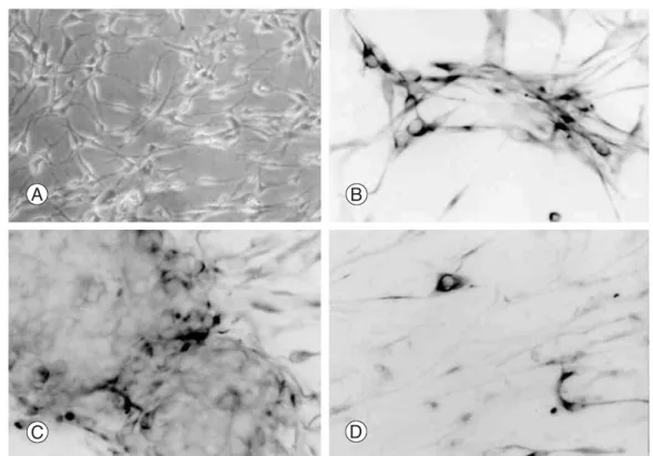
혈청을 함유한 배지에서 B2A1세포는 7~8 μm 크기를 가 진 양극 또는 삼극 세포로 보였고, 개별적으로 성장하거 나 작은 덩어리로 응집하는 경향을 보였다(Fig. 1A). 그 세 포는 nestin에 91.8%에서 면역반응성을 나타냈다(Fig. 1B).
또한 NF-L (3.1%)(Fig. 1C), βtubulin Ⅲ(2.7%) 양성의 신경원세포가 관찰되었으며, GFAP (성상세포 표지자) 를 포함하는 다른 중추신경계 표지자에서도 세포의
6.9%에서 면역양성을 보였으며(Fig. 1D), GalC와 MBP (희돌기 교세포 표지자)는 각각 5.6%와 4.7%를 나타냈
다. 소교세포 표지자인 RCA-1은 면역 음성반응을 나타 냈다(Table. 3).2. 성장인자 처리에 의한 B2A1 세포의 면역 세포화 학적 특징
혈청과 다양한 성장인자로 처리된 DM4 무혈청 배양 액에서 배양된 B2A1 세포에 대한 Nestin, βtubulin Ⅲ,
GFAP, GalC에 양성인 세포들은 Fig. 2와 같다. 10% HS, ITS, c-AMP, bFGF, PDGF, LIF 또는 PMA를 함유하는 배
양 배지에서 배양되면, B2A1 세포의 88~93%에서 nestin 에 면역반응성을 보였다(Fig. 2A). nestin에 양성인 세포 의 비율은 RA를 포함하는 배양 배지에서 약간 감소하였 다(70.5%).PDGF,
βtubulin Ⅲ로 처리된 배양에서 B2A1 세포의3.8%가 발현하였고, 반면 10% HS, cAMP, RA, bFGF로
처리된 배지에서는 더 적게(1.8~2.7%) 발현을 했다(P<0.05). 단지 몇몇 세포(0.5%이하)만이 ITS, LIF, PMA
로 처리된 배양 배지에서 βtubulin Ⅲ에 양성을 보였다(Fig. 2B).
cAMP나 PDGF를 함유하는 배양 배지에서 배양하면
Table 3. Immunocytochemical expression of B2A1 cells
Antigen B2A1 cells
Nestin 91.8%
NF-L 03.1%
βtubulin III 02.7%
GFAP 06.9%
GalC 05.6%
MBP 04.7%
RCA-1 -
%: proportion of positive immunoreactivity in B2A1 cells -: negative immunoreactivity
Table 2. Sequence of PCR primers
Gene Sequence (Sense and Antisense) Product size (bp)
Nestin 5’-AGT GTG AAG GCA AAG ATA GC-3’ 317
5’-TCT GTC AGG ATT GGG ATG GG-3’
Notch1 5’-TCA AGG CCC GGA GGA AGA AGT C-3’ 976
5’-TCA GGG GAT GGG GTG AGG AAG-3’
PDGFRα 5’-CTG TAA CTG GCA GGC TCG GAG-3’ 331
5’-GTT GTC TGC AGT ACA AGT TGG CG-3’
NF-L 5’-TCC TAC TAC ACC AGC CAT GT-3’ 284
5’-TCC CCA GCA CCT TCA ACT TT-3’
GFAP 5’-GCA GAG ATG ATG GAG CTC AAT GAC C-3’ 266
5’-GTT TCA TCC TGG AGC TTC TGC CTC A-3’
MBP 5’-ACA CGG GCA TCC TTG ACT CCA TCG G-3’ 510
5’-TCC GGA ACC AGG TGG GTT TTC AGC G-3’
G3PDH 5’-CCA TGT TCG TCA TGG GTG TGA ACC A-3’ 251
5’-GCC AGT AGA GGC AGG GAT GAT GTT C-3’
Fig. 1. Cytologic characteristics of B2A1 cells disclosed bi- or tripolar cells (A) phase contrast microscopy (B) and show immunopositivities for nestin (C) βtubulin III, (D) and GFAP .
Fig. 2. Multipotential differentiation of B2A1 cells by treatment of growth factors. Most of all cells were immunopositive for nestin in any culture condition (A). Immunopositive cells for βtubulin III significantly increased in PDGF-contained media (B), while GFAP (C) and galactocerebroside (D) positive cells markedly increased in c-AMP or PDGF-contained media.
GFAP는 B2A1세포의 14.7~16.4%에서 발현되었고, 10%
HS, ITS, RA, bFGF, LIF
를 함 유 하 는 배 지 에 서 는4.1~6.9%에서 발현되었다(P<0.01). GFAP에 대한 면역
양성반응은 PMA를 함유하는 배지에서는 B2A1세포가2.2%에서 보였다(Fig. 2C).
10% HS, cAMP, PDGF를 함유하는 배지에서 배양되
면 B2A1 세포의 5.6~6.8%가 GalC에 양성인 세포들이었 고, 4.2~2.9%는 RA, ITS, bFGF, PMA 함유하는 배지에서 양성이었다(P<0.05)(Fig. 2D).3. Cytokine 처리에 의한 B2A1 세포의 유전형질적 발현
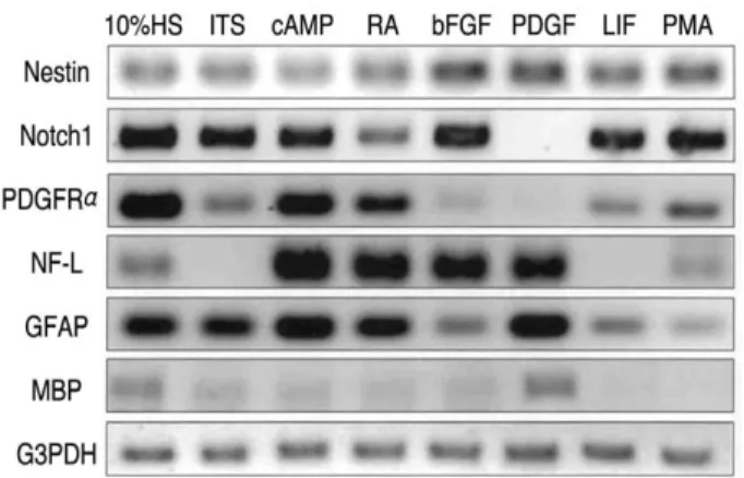
B2A1세포가 초기 분열하는 신경원, 성상세포, 희돌기
교세포 뿐만 아니라 신경줄기세포의 발현 표지자인지 알아보기 위해, 다양한 cytokine으로 처리하여 배양된 세포에서 RT-PCR을 시행했다. 발현된 정도는 housekeeping 유전자 G3PDH의 발현을 대조군으로 삼았으며,
그 결과는 중추신경계의 줄기세포 혹은 초기 전구세포 표지자(nestin, Notch1, PDGFα), 신경원 표지자인 NF-L, 성상세포 표지자인 GFAP, 희돌기 교세포 표지자인MBP가 동시에 발현되었다(Fig. 3). 발현의 정도가 처리
된 혈청 혹은 성장인자에 대해 다양하게 보였지만, 유전 자형의 표지자는 상대적으로 면역세포화학적 표현형질 과 관련되어 있었다.고 찰
척수 손상에서 줄기세포의 적용은 미분화된 줄기세포 가 손상된 신경을 복원하는데 필요한 세포로 분화되어 축삭의 재생 및 목표 방향으로 성장 유도, 신경교성 반 흔 또는 낭종의 복원 등 아직까지 확인되지 않은 기전까 지의 복원과정에 기여할 것으로 기대하고 있다.
줄기세포를 연구하기 위해서는 조직이나 기관으로부 터 줄기세포를 순수하게 분리하고 많은 세포를 얻어야 한다. 특정한 세포의 종류를 풍부하게 하는 방법 중의 하나는 형광 활성화된 세포 분류법(FACS)과 자기적으 로 활성화된 세포 분류법(MACS)이다20)
.
이러한 방법들 은 세포표면에 있는 단백에 항체가 결합하는 능력에 따 라 결정되고, 주로 조혈계 줄기세포를 분리하는 데에 사 용된다. 줄기세포를 분리시키는 다른 방법은 장기간에 걸친 배양이다21,22). 간단히 말하면, 중요한 조직으로부터
세포집단이 분리되어 배양하며 성숙되어 줄기세포가 증식하고 줄기세포의 특징을 유지할 수 있게 된다. 이러 한 방법은 장간막 줄기세포와 신경줄기세포의 분리에 적용되어져 왔다.이 연구에서 B2A1이라 명시된 신경줄기 세포주는 성 숙생쥐의 대뇌로부터 추출한 세포에서 장기간의 두 단 계의 배양과정에서 생겨났다. 첫 단계는 희돌기 교세포 가 풍부한 분획의 일차 배양과 1년 이상의 기간에 걸친 계대배양으로부터 B2세포를 생성했다. B2 세포는 10%
HS
함유 배지에서 미성숙 신경외배엽 세포23,24)의 주요 중간 필라멘트 단백인 vimentin에 면역양성을 나타내는 양극 혹은 삼극형 세포의 모양을 보였다. 혈청이 없는 배지에서 1mM dbcAMP의 존재하에 5일간 배양하여,vimentin, GFAP (성상세포의 표지자), O4와 GalC (희돌
기 교세포의 표지자)에 면역양성을 보였다. B2세포는 신경원성, 소교세포성, 내피 세포에 대해서는 어느 세포 특이적인 표지자도 발현하지 못했다. 이러한 면역세포 화학적인 특징은 B2세포가 미성숙 신경외배엽 세포와 신경교 전구세포로 구성되어 있다는 것을 의미한다.두 번째 단계는 희석법을 사용하여 B2세포의 응집체 로부터 분리된 단일 세포 배양으로부터 B2A1세포를 생 성했다. 고도로 정제된 양극 또는 삼극형 작은 세포들이 배양으로부터 얻어졌다. 혈청이 제거된 배지 DM416)에 서 배양된 세포는 혈청과 성장인자들에 의해 처리되어 면역세포 화학과 RT-PCR에 의해 검증되었다. B2A1세 포는 다음과 같은 신경줄기세포의 여러 가지 특징이 확 인되었다.
먼저 대부분의 세포(91.8%)가 중추신경계의 신경줄기 세포에 특이적으로 발현하는 중간필라멘트 단백인
nestin에 대해 면역양성이었다
25,26,27).둘째로 B2A1세포 또 한 신경원, 성상세포, 희돌기 교세포 표지자에 대해 양Fig. 3. Gene expression of cell type specific markers studied by RT-PCR in B2A1 cells. The cells expressed both neural progenitor cell markers (nestin, Notch1, PDGFα) and differentiated cell markers for neurons (NF-L), astro- cytes (GFAP), and oligodendrocytes (MBP). The expression levels were variable related to addition of growth factors.
성 면역반응을 보였다. 이러한 면역세포 화학적인 특징 들은 B2A1세포가 중추신경계에서 세포분화에 있어서 다능성을 가진다는 것을 암시한다. 셋째로, B2A1세포는
ITS, cAMP, RA, bFGF, PDGF, LIF, PMA 같은 성장인자
들 투여 하에 배양된 신경 세포 표지자를 유지했다. 신 경원성 분화는 PDGF의 투여로 증가했다. 성상세포 분 화는 cAMP와 PDGF의 투여 하에 유의하게 증가했고, 희 돌기 교세포는 PDGF투여 하에 증가했다. 넷째로 RT-PCR결과 B2A1세포에서의 유전형의 발현은 nestin, Notch1, PDGFRα에 대한 mRNA를 발현했다. 현재 분자
유전학 연구에서는 많은 분자들은 배아기 신경 발달 초 기 단계에서 발현된다고 한다. 이것들은 Brain1 (Brn1),Numb, NeuroD 그리고 Notch1, Hes1, Presenilin, Sox2의 effector같은 전사 요소를 포함한다
28,29,30,31,32). Notch1은 쥐
에서 뇌실하 구역내의 원시세포에서 발현을 했고, 포유 류의 피질 신경발생 동안 원시세포 증식과 신경분화의 두 가지 면에서 생물학적 역할을 의미한다28). PDGFα발
현은 주로 초기 중추신경계 발달 동안 희돌기 교세포 전 구세포에서 보인다33).
최근의 연구에서 PDGFα는 쥐의 배아기 8.5일에 생후 소뇌에 발달하고 있는 과립세포, 성숙 쥐의 소뇌에 있는 Purkinje 세포, 그리고 후근신경 절 세포를 발달시키는 과정에서 뿐만 아니라 신경판의 신경상피 세포에서 관찰되었다34).
이러한 보고는 PDGF α가 미분화세포, 즉 신경원이나 교세포로 분화할 수 있 는 세포에 대한 또 다른 표지자라는 것을 의미한다. 이 연구에서 B2A1 세포가 PDGF에서 배양될 때, 분화된 신 경원, 성상세포, 희돌기 교세포의 증가된 분획이 있었 다. B2A1 세포의 세포학적인 분화는 PDGFα의 존재하 에 일어날 수 있다. 다섯째, NF-L, GFAP, MBP같은 다른 분화된 신경세포 mRNA가 B2A1세포에서 동정되었다.이 연구에서는 RT-PCR과 면역세포 화학적인 측면에 의 한 mRNA 발현사이에 연관성이 있었다.
지금까지 쥐에서의 다능성 신경 줄기세포의 발생에 관한 몇 가지 연구가 있었다. 중추신경계에서 기원한 불 멸의 신경 전구세포주는 retroviral vector를 사용하여
NGF를 분비하는 세포를 만들기 위해서 발생되었다
35).
다 른 신 경 세 포 주 는 성 숙 생 쥐 의 대 뇌 로 부 터 flow
cytomery에 의해 얻었고, 신경, 비신경 세포를 발생시키
는 능력에 대해 연구했다36). 다른 다능성 신경 전구세포 (2Y6f1)는 성숙 p53-/- 쥐의 소뇌로부터 만들었다
37).
본 연구의 B2A1 세포는 지금까지 보고 되었던 종양유 전자를 이용하거나, 유전자를 조작하여 확인된 신경 줄 기세포와 달리 정상적인 생리 조건하에 성숙 생쥐의 대 뇌로부터 장기간의 일차 배양으로부터 신경줄기세포를 얻었다는 것은 커다란 의미가 있어 신경 줄기세포 생물 학을 연구하는 데에 유용할 것으로 사료된다. 임상적 의
의는 향후 중추신경계 내의 손상된 축삭의 해부학적 및 기능적 회복을 위해 성인 다기능 줄기 세포주를 적용할 수 있는 근거를 제시하는 바이며, 인간 신경계 발달을 유도하는 인자의 확인 또는 약제의 개발 등에 응용될 수 있으리라 사료된다.
결 론
B2A1세포는 성숙대뇌로부터 수립된 신경줄기세포주
로서, 이들 세포는 각종 성자인자들에 의하여 신경원.별세포 및 희돌기 교세포로 분화될 수 있음을 확인할 수 있었다.
참고문헌
01) Bartlett PF, Brooker GJ, Faux CH et al: Regulation of neural stem cell differentiation in the forebrain. Immun Cell Biol 1998;76:414-418.
02) Brustle O, Spiro AC, Karram K, Choudhary K, Okabe S, McKay RD: In vitro-generated neural precursors par- ticipate in mammalian brain development. Proc Natl Acad Sci USA 1997;94:14809-14814.
03) Flax JD, Aurora S, Yang C et al: Engraftable human neural stem cells respond to developmental cues, replace neurons, and express foreign genes. Nat Biotechnol 1998;16:1033-1039.
04) Thomoson JA, Itskovitz-Eldor J, Shapiro SS et al:
Embryonic stem cell lines derived from human blastocysts.
Science 1998;282:1145-1147.
05) Shamblott MJ, Axelman J, Wang S et al: Derivation of pluripotent stem cells from cultured human primordial germ cells. Proc Natl Acad Sci USA 1998;95:13726- 13731.
06) Davis AA, Temple S: A self-renewing multipotential stem cell in embryonic rat cerebral cortex. Nature 1994
;372:263-266.
07) Gage FH: Mammalian neural stem cells. Science 2000;287:1433-1438.
08) Kuhn HG, Dickinson-Anson H, Gage FH: Neurogenesis in the dentate gyrus of the adult rat: age-related decrease of neuronal progenitor proliferation. J Neurosci 1996
;16:2027-2033.
09) Kukekov VG, Laywell ED, Suslov O et al: Multipotent stem/progenitor cells with similar properties arise from
two neurogenic regions of adult human brain. Exp Neurol 1999;156:333-344.
10) Anderson DJ: Stem cells and pattern formation in the nervous system: the possible versus the actual. Neuron 2001;30:19-35.
11) Benraiss A, Chmielnicki E, Lerner K, Roh D, Goldman SA: Adenoviral brain-derived neurotrophic factor induces both neostriatal and olfactory neuronal recruitment from endogenous progenitor cells in the adult forebrain. J Neu- rosci 2001;21:6718-6731.
12) Satoh J, Tabira T, Kim SU: Rapidly proliferating glial cells isolated from adult mouse brain have a differentia- tive capacity in response to cyclic AMP. Neurosci Res 1994;20:175-184.
13) Satoh J, Kim SU, Kastrukoff LF, Takei F: Expression and induction of intercellular adhesion molecules (ICAMs) and major histocompatibility complex (MHC) antigens on cultured murine oligodendrocytes and astro- cytes. J Neurosci Res 1991;29:1-12.
14) Rey JA, Bello MJ, Decampos JM, Kusak ME, Moreno S: Cytogenetic analysis in human neurinomas. Cancer Genet Cytogenet 1987;28:187-188.
15) Satoh J, Gallyas F Jr, Endoh M, Yamamura T, Kun- ishita T, Tabira T: Coexistence of cholinergic, cate- cholaminergic, serotonergic, and glutamatergic neuro- transmitter markers in mouse clonal hybrid neurons derived from the septal region. J Neurosci Res 1992
;32:127-137.
16) Kim SU, Stern J, Kim MW, Pleasure DE: Culture of purified rat astrocytes in serum-free medium supplement- ed with mitogen. Brain Res 1983;274:79-86.
17) Ranscht B, Clapshaw PA, Price J, Noble M, Seifert W:
Development of oligodendrocytes and Schwann cells stud- ied with a monoclonal antibody against galactocerebro- side. Proc Natl Acad Sci USA 1982;79:2709-2713.
18) Nagai A, Suzuki Y, Baek SYet al: Generation and Char- acterization of Human Hybrid Neurons Produced between Embryonic CNS Neurons and Neuroblastoma Cells. Neu- robiol Dis 2002;11:184-198.
19) Cai J, Wu Y, Mirua T et al: Properties of a fetal multipo- tent neural stem cell (NEP cell). Dev Biol 2002;251:221- 240.
20) Goodell MA, Brose K, Paradis G, Conner AS, Mulli- gan RC: Isolation and functional properties of murine hematopoietic stem cells that are replicating in vivo. J Exp Med 1996;183:1797-1806.
21) Bjornson CR, Rietze RL, Reynolds BA, Magli MC, Vescovi AL: Turning brain into blood: a hematopoietic fate adopted by adult neural stem cells in vivo. Science 1999;283:534-537.
22) Pittenger MF, Mackay AM, Beck SC et al: Multilineage potential of adult human mesenchymal stem cells. Science 1999;284:143-147.
23) Dahl D, Rueger DC, Bignami A, Weber K, Osborn M:
Vimentin, the 57 000 molecular weight protein of fibrob- last filaments, is the major cytoskeletal component in immature glia. Eur J Cell Biol 1981;24:191-196.
24) Bignami A, RajuT, Dahl D: Localization of vimentin, the nonspecific intermediate filament protein, in embryonal glia and in early differentiating neurons. In vivo and in vitro immunofluorescence study of the rat embryo with vimentin and neurofilament antisera. Dev Biol 1982;91:286-295.
25) Lendahl U, Zimmerman LB, McKay RD: CNS stem cells express a new class of intermediate filament protein.
Cell 1990;60:585-595.
26) Gates MA, Thomas LB, Howard EM et al: Cell and molecular analysis of the developing and adult mouse sub- ventricular zone of the cerebral hemispheres. J Comp Neurol 1995;361:249-266.
27) Dahlstrand J, Lardelli M, Lendahl U: Nestin mRNA expression correlates with the central nervous system progenitor cell state in many, but not all, regions of devel- oping central nervous system. Brain Res Dev Brain Res 1995;84:109-129.
28) Zhong W, Jiang MM, Weinmaster G, Jan LY, Jan YN:
Differential expression of mammalian Numb, Numblike and Notch1 suggests distinct roles during mouse cortical neurogenesis. Development 1997;124:1887-1897.
29) Josephson R, Muller T, Pickel J et al: POU transcrip- tion factors control expression of CNS stem cell-specific genes. Development 1998;125:3087-3100.
30) Kageyama R, Ohtsuka T: The Notch-Hes pathway in mammalian neural development. Cell Res 1999;9:179-188.
31) Yang X, Handler M, Shen J: Role of presenilin-1 in murine neural development. Ann N Y Acad Sci 2000;920:165-170.
32) Kishi M, Mizuseki K, Sasai N et al: Requirement of Sox2-mediated signaling for differentiation of early Xeno- pus neuroectoderm. Development 2000;127:791-800.
33) Johe KK, Hazel TG, Muller T, Dugich-Djordjevic MM, McKay RD: Single factors direct the differentiation
of stem cells from the fetal and adult central nervous sys- tem. Genes Dev 1996;10:3129-3140.
34) Andrae J, Hansson I, Afink GB, Nister M: Platelet- derived growth factor receptor-alpha in ventricular zone cells and in developing neurons. Mol Cell Neurosci 2001;17:1001-1013.
35) Martinez-Serrano A, Lundberg C, Horellou P et al:
CNS-derived neural progenitor cells for gene transfer of nerve growth factor to the adult rat brain: complete res- cue of axotomized cholinergic neurons after transplanta-
tion into the septum. J Neurosci 1995;15:5668-5680.
36) Rietze RL, Valcanis H, Brooker GF, Thomas T, Boss AK, Bartlett PE: Purification of a pluripotent neural stem cell from the adult mouse brain. Nature 2001;412:736-739.
37) Ohkawara B, Okuno M, Ishii T, Horiuchi M, Tomooka Y: Characterization of a multipotent neural progenitor cell line cloned from an adult p53-/- mouse cerebellum.
Brain Res 2003;959:11-19.
※ 통신저자 : 최 용 수
광주광역시 남구 양림동 264 광주기독병원 정형외과
Tel: 82-62-650-5062 Fax: 82-62-650-5066 E-mail: [email protected] 연구계획: 성숙생쥐 대뇌로부터 다기능 신경줄기세포주의 확립
연구목적: 성숙한 실험동물의 뇌조직으로부터 다능성 줄기세포를 생성하기 위해 B2 세포로부터 복제 세포주를 확 립하였으며, 이를 B2A1 세포라 명명하였고, 이 세포가 신경줄기 세포주의 특성을 가지는지 조사하였다
대상 및 방법: 실험동물은 생후 12~16주된 BALB/c 용 생쥐를 사용하였다. 대뇌를 적출하여 잘게 부수고 단백분해 효소를 처리하여 각각 분리된 세포를 만든후 percoll을 이용한 밀도구배 원심분리법을 이용하여 희돌기교세포/별세 포 분획을 분리하여 일차배양하였다. 미분화된 소세포를 분리하여 6개월 이상에 걸쳐 25회 계대배양하면서 2차례에 걸쳐 최대한 희석시켜 단일 세포를 배양하였다. 단일 세포 배양으로부터 증식된 하나의 세포군(B2A1세포)을 분리하 였고, B2A1세포는 면역세포화학적 염색과 RT-PCR법을 통하여 동정하였다.
결과: B2A1세포는 면역세포화학적으로 90% 이상에서 nestin에 양성으로 염색되어 신경줄기세포임을 확인할 수 있 었고, 그 외에 NF-L, β-tubulin Ⅲ, GFAP, MBP, Ga1C에 양성으로 염색되었다. 그리고 B2A1세포에 각종 성장인자를 투여하여 관찰한 결과 nestin 양성세포는 약 90%로 동일하였다. 또한 이들 성장인자 투여에 인한 RT-PCR결과 B2A1 세포에서는 nestin, PDGFRα, Notch1, NJ-L, GFAP, MBP에 대한 mRNA가 다양하게 발현되었다.
결론 : B2A1세포는 성숙대뇌로부터 수립된 신경줄기세포주로서, 이들 세포는 각종 성장인자들에 의하여 신경원, 별 세포 및 희돌기교세포로 분화될 수 있음을 확인할 수 있었다.
색인단어: 신경줄기세포, B2A1세포, 성숙생쥐 대뇌 국 문 초 록