Effi cacy of Postoperative Radiograph for
Evaluating the Prevertebral Soft Tissue Swelling after Anterior Cervical Discectomy and Fusion
Kyung-Jin Song, MD, Byung-Wan Choi, MD*, Hye-Young Kim, MD, Taek-Su Jeon, MD*, Han Chang, MD*
Department of Orthopedic Surgery, Chonbuk National University Hospital, Jeonju,
*Inje University Haeundae Paik Hospital, Busan, Korea
Received August 4, 2011; Accepted November 1, 2011 Correspondence to: Byung-Wan Choi, MD
Department of Orthopedic Surgery, Inje University Haeundae Paik Hospital, 875 Haeun-daero, Haeundae-gu, Busan 612-862, Korea
Tel: +82-51-797-0240, Fax: +82-51-797-0249 E-mail: alla1013@naver.com
Cervical spine surgery by the anterior approach is current- ly used to treat diverse diseases of the cervical spine. How- ever, many complications can occur. In particular, postop- erative airway obstruction is a potentially life-threatening complication.
Cervical spine prevertebral soft tissue swelling (PSTS) has been observed on plain radiographs of the lateral cervical spine, and used as an indirect indicator for
Background: After surgery for degenerative spinal disease by the anterior approach, the degree of soft tissue swelling can be assessed simply using plain radiographs. However, there are little studies according to the surgical methods or extent of surgery, and no study had addressed the clinical meaning of swelling determined by plain radiography. The purpose of this study was to evaluate the clinical signifi cance of prevertebral soft tissue swelling (PSTS) after anterior cervical fusion with plate fi xation for the treatment of degenerative cervical spinal disorders.
Methods: One hundred and thirty-fi ve patients that underwent anterior cervical fusion with plate augmentation for degenerative cervical spondylosis were included in this study. PSTS differences were analyzed with respect to numbers of fusion segments and location of fusion. Cases were divided into two groups based on the amount of PSTS, and incidences of dyspnea, dysphagia, dys- phonia were evaluated.
Results: PSTS increments were signifi cantly greater in patients that had undergone multi-level or high-level fusion. Complications of dyspnea, dysphagia and dysphonia were found more frequently in patients with marked PSTS group.
Conclusions: Increments of PSTS after anterior cervical fusion for degenerative spinal disorders are greater and incidences of complications are higher in patients that undergo multi-level or high-level fusion. Thus, measurement of PSTS using consecutive cervical lateral radiographs after anterior cervical surgery is clinically meaningful procedure.
Keywords: Cervical spine, Prevertebral soft tissue swelling, Anterior cervical discectomy and fusion
identifying degrees of damage by trauma or pathologic conditions, such as a retropharyngeal abscess.1-6) After surgery for degenerative spinal diseases by the anterior approach, the degree of soft tissue swelling were assessed simply by using plain radiographs. However, few studies have been conducted according to the surgical methods or extent of surgery, and no study has addressed the clinical meaning of swelling determined by plain radiography.
In the present study, we evaluated soft tissue swelling observed in plain lateral radiographs aft er anterior cervical fusion surgery to identify the relationships between PSTS and the extent and location of surgery, and to explore cor- relations between PSTS and complications related to soft tissue damage.
Copyright © 2012 by Th e Korean Orthopaedic Association
Th is is an Open Access article distributed under the terms of the Creative Commons Attribution Non-Commercial License (http://creativecommons.org/licenses/by-nc/3.0) which permits unrestricted non-commercial use, distribution, and reproduction in any medium, provided the original work is properly cited.
Clinics in Orthopedic Surgery • pISSN 2005-291X eISSN 2005-4408
METHODS
Materials
One hundred and seventy-six patients underwent anterior interbody fusion with cage and plate augmentation due to degenerative cervical disease between April 2004 and June 2007 at our institution. Th e exclusion criteria applied were: revision surgery, the performance of anterior and posterior combined fusion, and preoperative dysphasia or dysphonia. We enrolled 135 patients, 78 males and 57 fe- males with a mean age of 54.8 years (range, 30 to 86 years).
Th e mean follow-up period was 38.7 months (range, 25 to 84 months). In 48 cases, fusion was performed on a single segment, in 67 cases on two segments, and in 20 cases on three segments. In 86 cases, fusion was conducted below the C5, and in 49 cases above the C5 included.
Operation Method
Surgery was performed under general anesthesia in all patients. First, cancellous bone for bone graft ing was har- vested percutaneously using a trocar (diameter 7 mm; AO Synthes, Bettlach, Switzerland) via a 1 cm mini-incision placed at least 2 cm from the lateral side of the anterior superior iliac spine. A standard Smith-Robinson method was used to expose the cervical spine.7) Th e cervical spine was approached from the left side unless the patient had undergone a prior approach from the left side. Aft er com- plete decompression by removing osteophytes and rem- nant disc materials, endplate cartilage was removed with a high-speed burr and a curette until bleeding occurred.
Lateral radiographs of the cervical spine were checked to determined cage size, plate lordosis, and screw insertion angles. Finally, a polyetheretherketone cage filled with cancellous bone graft was inserted into the intervertebral space and anterior plating was performed. Solis cages (Stryker, Kalamazoo, MI, USA) were used throughout. In addition, the Maxima Anterior Cervical Plate System (U&I Corporation, Uijeongbu, Korea) or the cervical spine lock- ing plate (CSLP) system (Synthes Inc., Paoli, PA, USA) was used for anterior stabilization. Closed suction drain was routinely used and the drain was removed approximately 48 hours after surgery. After operations, a Philadelphia cervical orthosis was applied for 4 weeks and a soft collar was recommended for an additional 2 weeks.
Physical examinations and radiological examina- tions, including plain radiography and MRI, were per- formed preoperatively in all patients. Plain radiography (anterior-posterior [AP], lateral, and fl exion/extension lat- eral views) was repeated at 6 weeks and at 3, 6, 9, 12, and 18 months postoperatively, and then annually.
Methods
Measurement of PSTS
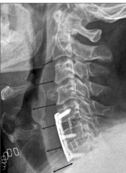
In all cases, plain radiographs of the lateral cervical spine were taken before surgery, immediately aft er surgery, and daily until 5 days after surgery. Plain radiographs of the lateral cervical spine were taken with patients standing with their chins facing forward and their heads placed nat- urally without tension. Films were centered at the shoulder and the radiation was aligned with the 5th cervical spine and located around 1.8 m from patients. In plain radio- graphs of lateral cervical spines, the AP diameters of the PSTS from the 2nd cervical spine (C2) to the 7th cervical spine (C7) were determined by measuring distances from the inferior margin of each vertebral body perpendicularly to the point where the air shadow of the airway began. In the cases of bony spur formation on the vertebra, we mea- sured the distance from the non-spurred anterior margin of the vertebral body. Th e shortest lengths were used as a value. In the areas where the plates were fi xed, the distanc- es from the anterior margins of plates to a shadow were measured (Fig. 1). PSTS measurements from day 0 (the day of surgery) to day 5 aft er surgery were documented to monitor patterns of change. Th e values of soft tissue swell- ing change from the preoperative state to the days after the operation were used for analysis. The mean value of PSTS before surgery was 5.47 mm on C2, 5.16 mm on C3, 7.84 mm on C4, 15.36 mm on C5, 15.03 mm on C6, and 12.64 mm on C7. Two blinded observers independently measured the segment dimensions twice; average values
Fig. 1. Measurement method of prevertebral soft tissue swelling. The prevertebral soft tissue was measured between the anterior surface of each vertebral body and the air shadow of the airway.
were used in the analysis. To verify the reliabilities of mea- sured values, intra- and inter observer consistencies were checked using interclass correlation coeffi cient (ICC). ICC (Cronbach’s α) showed excellent correlation in intraob- server (0.81), interobserver (0.70) correlation.
Analysis according to the number and location of fusion segments
Correlations between operation time, number of fusion segments, and location of fusion areas were analyzed, based on degrees of soft tissue swelling at the C3 on day 2 aft er surgery, where changes in soft tissue swelling were the largest. Cases were divided into one level fusion group and two or more level fusion group for comparison pur- poses. Cases were also divided into two groups of above and below the C5. When the case included the level of C4- 5, we regarded this case as above the C5 level group. 86 cases with a fusion range below the C5 and 49 cases with a fusion range, including the 3rd and 4th cervical spines above the C5 level, were compared.
Analysis according to PSTS and postoperative complica- tions
Based on a mean soft tissue swelling change of 7.3 mm at C3 area on day 2 aft er surgery, where changes in soft tis- sue swelling were at their largest, subjects were divided into 71 cases with a swelling < 7.3 mm (group A) and 64 cases with a swelling > 7.3 mm (group B). Dyspnea was re- garded to have occurred in cases where patients reported subjective symptoms during hospitalization aft er surgery or treatment, such as oxygen administration or bed lift ing that was necessary because of the symptoms. Dysphagia was defined as being present when difficulties in swal- lowing liquid or solids persisted for at least three months aft er surgery.8) Dysphonia was defi ned as the presence of persisting changes in phonation at least three months aft er surgery.9)
The degrees of longitudinal changes of soft tissue swelling were analyzed using a paired sample t-test. Diff er- ences in degrees of swelling relative to the number of fu- sion and the location of fusion were tested using ANOVA and an independent sample t-test. Pearson’s correlation was used to analyze the correlation between the time when dyspnea occurred and the time of soft tissue swelling. De- velopment of dyspnea, dysphagia, and dysphonia between the groups was analyzed using the chi-square test. SPSS ver. 12.0 (SPSS Inc., Chicago, IL, USA) was used for statis- tical analysis, and signifi cance was accepted for p-values of
< 0.05.
RESULTS
Postoperative Progress of Soft Tissue Swelling
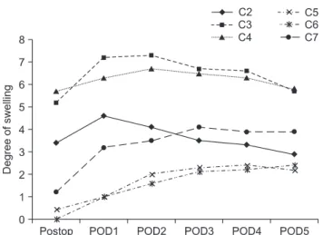
Based on PSTS measured at each segment after surgery, for upper spines (C2, C3, C4), there was significantly more soft tissue swelling during the measurement period compared with the preoperative state (p < 0.01). Th e de- gree of swelling was highest on days 2 and 3 aft er surgery, and then showed a gradually decreasing tendency. For lower cervical spines (C5, C6, C7), soft tissue swellings did not increase significantly immediately after surgery, but showed significant increases from day 2 (p < 0.001).
Unlike upper cervical spines, soft tissue swelling persisted during the measurement period (Fig. 2). Before surgery, the PSTS of C3 was 4.8 mm on average and on day 2, it was 12.1 mm average when it was at its largest. Th us, the average maximum increase due to swelling was 7.3 mm.
Analysis according to the Number and Location of Fusion Segments
Degrees of PSTS increases were not found to be sig- nificantly correlated with operation time (p = 0.33), but significant differences were observed between numbers of operated segments and PSTS increases as 6.38 mm in one level fusion group and 8.2 mm in two or more levels fusion group (p = 0.03). Regarding fusion areas, a mean PSTS increase of 5.64 mm was found in cases with areas below C5 and of 9.08 mm in cases above C5 (p < 0.01).
Fig. 2. The histogram shows the time course of prevertebral soft tissue swelling (mm) in the postoperative period. Postop: operation day, POD1:
day 1 after operation, POD2: day 2 after operation, POD3: day 3 after operation, POD4: day 4 after operation, POD5: day 5 after operation.
Analysis according to PSTS and Postoperative Complications
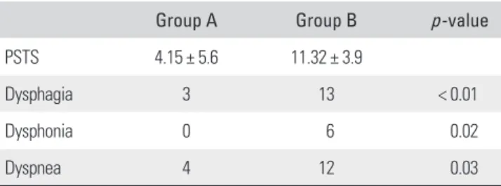
Th e incidences of complications (dyspnea, dysphagia, and dysphonia) that occurred in groups A and B were analyzed and compared. No significant differences were observed between these two groups in terms of age, gender, opera- tive time, or smoking history (Table 1).
In the analysis of relationships between increases in PSTS and the occurrence of complications after surgery, group B showed significantly more occurrences. Com- plication of dysphagia was found in 3 cases in group A and 13 cases in group B (p < 0.01). Dysphonia was found in 0 cases in group A and 6 cases in group B (p = 0.02).
Dyspnea was found in 4 cases in group A and 12 cases in group B (p = 0.03) (Table 2). The time when dyspnea mainly occurred (average of 1.3 days aft er operation) and the time when soft tissue swelling was most severe (average of 2.48 days aft er operation) were not the same and there was no correlation (p = 0.32).
DISCUSSION
Soft tissue swelling aft er anterior surgery for degenerative cervical spinal disease occurs in almost all patients, and this is identifi ed by increases in the width of prevertebral soft tissues on plain radiographs. Many studies have at- tempted to determine whether cervical spine areas have been damaged by measuring the widths of prevertebral soft tissues in trauma patients. Weir1) advised that the normal range of PSTS at C3 was 2.6-4.8 mm, Templeton et al.4) defi ned a PSTS of > 10 mm in the retropharyngeal space as abnormal, Miles et al.5) defined a PSTS size of larger than half of the diameter of the vertebral body in the retropharyngeal space as abnormal, and Pope and Rid- dervold defi ned a PSTS of > 7 mm at C2-3 as abnormal.6) However, few studies have reported the abnormal range of PSTS observed after surgery for degenerative cervical spinal diseases. Sanfi lippo et al.10) advised that most swell-
ings disappear within 6 weeks and that large abnormal swellings should be monitored or treated, but they did not mention the normal range or a treatment method.
Th e general progress of swelling peaks at day 2 or 3 aft er surgery and thereafter decreases.11-13) Sanfilippo et al.10) advised that although soft tissue swelling was observed even at two weeks aft er surgery, it normalized at 6 weeks.
Similarly, in our study, degrees of swelling progressed and peaked on day 2 at the anterior 3rd. cervical spine at an average of 12.1 mm. When quantifying swelling, the diam- eters of prevertebral soft tissues measured in the middle of the area between the lower end plate of the C3 and C4 refl ect the most sensitive swelling.13) It was noted that the lower vertebral levels experienced little change postopera- tively when compared with the upper cervical spine. Th is is most likely because of the more constrained anatomy of the lower cervical spine. Th e potential retropharyngeal space appears to be much greater in the pharynx and hypopharynx than in the more distal trachea/esophageal portion of the neck. Th erefore, greater consistency would be found between the preoperative and postoperative measurements. Furthermore, in our study, differences in results were also compared based on PSTS anterior to the C3 on day 2 when changes in PSTS were at their largest. In this study, we identifi ed that the multiple level fusion and upper level fusion showed signifi cant correlations with de- grees of swelling. In comparing single level and two level fusions, Suk and colleagues11,12) reported that a diff erence in postoperative degrees of swelling was not evident, but advised the possibility that results might diff er when more than two levels are involved. In addition, they advised that when surgery is performed at an area proximal to the 5th cervical spine, soft tissue swelling is severe in the C2 and C3 areas, whereas Andrew and Sidhu13) advised that al- though swelling increased greatly in the upper level fusion, it was not related to the location. We found that if PSTS re- fl ects the degree of damage to soft tissues occurring during surgery, its degree would be higher for multiple segment surgery. Th erefore, degrees of damage to soft tissues occur- Table 1. Demographic Data according to Prevertebral Soft Tissue
Swelling
Variables Group A Group B p -value
Sex (M:F) 37:34 41:23 0.21
Smoking 19 20 0.70
Age 51.56 ± 11.07 53.64 ± 11.90 0.29
Operation time 84.10 ± 23.87 89.02 ± 18.28 0.18 Fusion level 1.63 ± 0.66 1.97 ± 0.67 0.004
Table 2. Analysis according to PSTS and Postoperative Complications
Group A Group B p -value
PSTS 4.15 ± 5.6 11.32 ± 3.9
Dysphagia 3 13 < 0.01
Dysphonia 0 6 0.02
Dyspnea 4 12 0.03
PSTS: prevertebral soft tissue swelling.
ring during surgery in degenerative cervical spinal disease may also be indirectly identifi ed using plain radiographs as with acute cervical spine trauma patients. Although air- way obstruction rarely occurs aft er cervical spine surgery, it is potentially fatal, and has been reported with swell- ing and hematoma.14-16) In our study, re-intubation after surgery was not observed in airway obstruction, dyspnea that required treatment was observed in 13% of the 135 study subjects, and its incidence was higher in those who showed more severe PSTS. However, the time when dys- pnea mainly occurred (between day 0 and day 2) and the time when soft tissue swelling was most severe (days 2 or 3) were not the same, and thus, the degrees of soft tissue swelling cannot be safely used to predict the development of dyspnea.
Dysphagia is a complication that occurs relatively frequently aft er anterior cervical spine surgery. Although no clear mechanism has been suggested, swelling by trac- tion, hematoma, damage to the pharyngeal nerve plexus and to the hypoglossal nerves during surgery in the upper area have been suggested as risk factors.17-20) In addition, damaged soft tissues may adhere to the larynx or esopha- gus during the healing process and induce dysphagia.21,22) Bazaz et al.8) reported that after anterior cervical spine surgery, swelling occurred more frequently in females and those patients that underwent multiple segment surgery.
No significant differences were shown between revision surgery and primary surgery, and between the locations of surgery. Th e present study shows that soft tissue swell- ing was not significantly different in males and females, but that it increased markedly when multiple segments and upper level surgery cases were included. Furthermore,
dysphagia frequently occurred in patients that experienced marked soft tissue swelling.
Dysphonia after surgery in the anterior cervi- cal spine occurs less frequently than dysphagia, and is known to be caused by traction damage to the laryngeal nerves.17,19) However, laryngeal swelling could also be a cause, and both are more likely to occur when damage to soft tissue during surgery is more severe. In our study, dys- phagia and dysphonia persisting for at least three months after surgery occurred in 12% and 4% of our study sub- jects, respectively, and these frequencies were signifi cantly related to PSTS.
The limitations of this study are that corrections were not made for other factors, such as, the degrees of traction during surgery, which might aff ect postoperative soft tissue swelling or the occurrence of complications.
Furthermore, patterns of complications relative to PSTS severity were not quantitatively analyzed. Nevertheless, for the fi rst time, the present study describes the links between PSTS and the complications associated with surgery, and thus, sheds light on the clinical relevance of PSTS.
Increments in PSTS aft er anterior cervical fusion for degenerative spinal disorders are greater and incidences of complications are higher in patients that undergo multi- level or high-level fusion. Th us, the measurement of PSTS using consecutive cervical lateral radiographs after ante- rior cervical surgery is a clinically meaningful procedure.
CONFLICT OF INTEREST
No potential confl ict of interest relevant to this article was reported.
REFERENCES
1. Weir DC. Roentgenographic signs of cervical injury. Clin Orthop Relat Res. 1975;(109):9-17.
2. Chong VF, Fan YF. Radiology of the retropharyngeal space.
Clin Radiol. 2000;55(10):740-8.
3. Boucher C, Dorion D, Fisch C. Retropharyngeal ab- scesses: a clinical and radiologic correlation. J Otolaryngol.
1999;28(3):134-7.
4. Templeton PA, Young JW, Mirvis SE, Buddemeyer EU.
The value of retropharyngeal soft tissue measurements in trauma of the adult cervical spine: cervical spine soft tissue measurements. Skeletal Radiol. 1987;16(2):98-104.
5. Miles KA, Maimaris C, Finlay D, Barnes MR. Th e incidence and prognostic significance of radiological abnormalities in soft tissue injuries to the cervical spine. Skeletal Radiol.
1988;17(7):493-6.
6. Keats TE, ed. Emergency radiology. 2nd ed. St Louis: Year Book Medical Publishers; 1989.
7. Smith GW, Robinson RA. Th e treatment of certain cervical- spine disorders by anterior removal of the intervertebral disc and interbody fusion. J Bone Joint Surg Am. 1958;40(3):607- 24.
8. Bazaz R, Lee MJ, Yoo JU. Incidence of dysphagia aft er ante- rior cervical spine surgery: a prospective study. Spine (Phila Pa 1976). 2002;27(22):2453-8.
9. Schwartz SR, Cohen SM, Dailey SH, et al. Clinical practice guideline: hoarseness (dysphonia). Otolaryngol Head Neck Surg. 2009;141(3 Suppl 2):S1-31.
10. Sanfi lippo JA Jr, Lim MR, Jacoby SM, et al. "Normal" pre- vertebral soft tissue swelling following elective anterior cervical decompression and fusion. J Spinal Disord Tech.
2006;19(6):399-401.
11. Suk KS, Kim KT, Lee SH, Park SW. Prevertebral soft tissue swelling aft er anterior cervical discectomy and fusion with plate fi xation. Int Orthop. 2006;30(4):290-4.
12. Suk KS, Kim KT, Lee JH, Lee SH, Kim JS, Choi IH. Prever- tebral soft tissue swelling aft er anterior cervical discectomy and fusion: comparison between plate fixation and cage insertion. J Korean Orthop Assoc. 2009;44(2):249-55.
13. Andrew SA, Sidhu KS. Airway changes after anterior cervical discectomy and fusion. J Spinal Disord Tech.
2007;20(8):577-81.
14. Epstein NE, Hollingsworth R, Nardi D, Singer J. Can airway complications following multilevel anterior cervical surgery be avoided? J Neurosurg. 2001;94(2 Suppl):185-8.
15. Sagi HC, Beutler W, Carroll E, Connolly PJ. Airway com- plications associated with surgery on the anterior cervical spine. Spine (Phila Pa 1976). 2002;27(9):949-53.
16. Emery SE, Smith MD, Bohlman HH. Upper-airway ob- struction aft er multilevel cervical corpectomy for myelopa-
thy. J Bone Joint Surg Am. 1991;73(4):544-51.
17. Frempong-Boadu A, Houten JK, Osborn B, et al. Swallow- ing and speech dysfunction in patients undergoing anterior cervical discectomy and fusion: a prospective, objective preoperative and postoperative assessment. J Spinal Disord Tech. 2002;15(5):362-8.
18. Martin RE, Neary MA, Diamant NE. Dysphagia following anterior cervical spine surgery. Dysphagia. 1997;12(1):2-8.
19. Winslow CP, Winslow TJ, Wax MK. Dysphonia and dys- phagia following the anterior approach to the cervical spine.
Arch Otolaryngol Head Neck Surg. 2001;127(1):51-5.
20. Riley LH 3rd, Skolasky RL, Albert TJ, Vaccaro AR, Heller JG. Dysphagia aft er anterior cervical decompression and fu- sion: prevalence and risk factors from a longitudinal cohort study. Spine (Phila Pa 1976). 2005;30(22):2564-9.
21. Fogel GR, McDonnell MF. Surgical treatment of dys- phagia after anterior cervical interbody fusion. Spine J.
2005;5(2):140-4.
22. Seidl RO, Todt I, Ernst A. Esophagus atresia aft er cervical spine surgery: case report and literature review. Eur Arch Otorhinolaryngol. 2007;264(3):291-3.