Iron deficiency anemia in infants and toddlers
Eun Young Joo, Keun Young Kim, Dong Hyun Kim, Ji-Eun Lee, Soon Ki Kim
Department of Pediatrics, Inha University College of Medicine, Incheon, Korea
p-ISSN 2287-979X / e-ISSN 2288-0011 https://doi.org/10.5045/br.2016.51.4.268 Blood Res 2016;51:268-73.
Received on July 29, 2016 Revised on October 20, 2016 Accepted on November 18, 2016
Background
In Korea, the prevalence of anemia and iron deficiency anemia (IDA) among older infants and young children remains high. To detect IDA early and to reduce its adverse impact, we assessed the characteristics of infants and young children who had IDA or were at risk of developing IDA, or who exhibited characteristics associated with severe anemia.
Methods
Among the 1,782 IDA-affected children aged 6 months to 18 years who visited the hospi- tal, we retrospectively analyzed the medical records and laboratory data of 1,330 IDA-af- fected children aged 6‒23 months who were diagnosed between 1996 and 2013. We ex- cluded patients with a C-reactive protein level ≥5 mg/dL.
Results
IDA was predominant in boys (2.14:1) during infancy and early childhood. The peak IDA incidence was noted among infants aged 9‒12 months. Only 7% patients exhibited symp- toms of IDA, while 23.6% patients with severe IDA demonstrated classic symptoms/signs of IDA. Low birth weight (LBW) infants with IDA demonstrated low adherence to iron supplementation. In a multivariate analysis, prolonged breastfeeding without iron for- tification (odds ratio [OR] 5.70), and a LBW (OR 6.49) were identified as risk factors of severe anemia.
Conclusion
LBW infants need more attention in order to increase their adherence to iron supple- mentation. For the early detection of IDA, nutritional status of all infants, and iron batteries of high-risk infants (LBW infants, infants with prolonged breastfeeding, picky eaters, and/or infants with the presence of IDA symptoms) should be evaluated at their health screening visits.
Key Words Iron deficiency anemia, Infant, Child, Risk factors, Breastfeeding, Low birth weight
*This work was supported by an Inha University Research grant (53780-01).
Correspondence to Soon Ki Kim, M.D., Ph.D.
Department of Pediatrics, Inha University School of Medicine, 27, Inhang-ro, Jung-gu, Incheon 22332, Korea
E-mail: [email protected]
Ⓒ 2016 Korean Society of Hematology
INTRODUCTION
The prevalence of anemia and iron deficiency anemia (IDA) remains high in late infancy and early childhood de- spite the increased breastfeeding rate, improvements in pub- lic health, and development of iron-fortified foods [1-4].
Hopkins et al. [2], using the anemia definition set forth by the World Health Organization (WHO) (hemoglobin [Hb]
<11 g/dL), showed that the prevalence of anemia was 23%
at 8 months of age, and 18% at 12 months of age. The prevalence of non-anemic iron deficiency may be as high as 30% in toddlers from developed countries [3, 5, 6].
According to the WHO criteria, anemia prevalence was esti-
mated at 15% in Korean preschool-aged children (6–59 mo), and to date, anemia remains an important health concern [7]. Additionally, the demands and expenses for hospital care for IDA-affected children have increased from 2006 to 2014 [8].
IDA in infants remains underdiagnosed as infant blood sampling and obtaining sufficient blood volume for labo- ratory detection of IDA are difficult. Most infants do not under- go blood tests unless reasonable clinical events are present.
Further, the symptoms of IDA (pallor, irritability, poor feeding, fatigue, lethargy, and pica) are non-specific. Although anemia and iron deficiency are usually corrected by the age of 2–3 years, children are adversely affected by IDA.
IDA is associated with impaired neurocognitive function
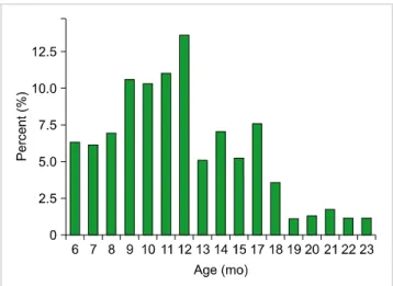
Fig. 1. Distribution of iron deficiency anemia in patients aged 6–23 months.
and exercise intolerance [9-12], and the association exists even after its successful treatment [11]. Therefore, preventing the progression of iron deficiency is especially important during infancy and early childhood when the rapid growth and development rate, especially of the brain [12], increases the vulnerability to IDA-induced impairment.
To detect IDA at an early age and to reduce the adverse impact of iron deficiency, we assessed the clinical character- istics of infants and young children with IDA who visited the Inha University Hospital in the last 17 years. We inves- tigated the distribution of IDA according to age (in months) and gender and analyzed the risk factors of severe IDA.
MATERIALS AND METHODS
Patients and study design
Of the 1,782 IDA patients who visited the pediatric depart- ment of the Inha University Hospital Pediatric Department between January 1997 and December 2013, 1,330 IDA pa- tients aged 6–23 months were included in the study. We retrospectively reviewed the medical records of these patients. Additionally, we conducted a questionnaire-based survey for IDA at pediatric hemato-oncology outpatient clin- ic to collect information regarding the following.: (1) late initiation of weaning food – age at the initiation of weaning food >6 months; (2) use of non-iron-fortified weaning food;
(3) picky eaters, or infants who required more than 1 month for successful weaning; (4) cow’s milk intake during infancy or a high intake of cow’s milk (>700 mL/day) after infancy;
(5) IDA symptoms and signs such as pallor, pica or night crying twice or more per night; and (6) no iron supplementa- tion in low birth weight (LBW) infants.
From venous blood sampling, laboratory tests for Hb, hem- atocrit, mean corpuscular volume (MCV), mean corpuscular hemoglobin (MCH), red cell distribution width (RDW), ferri- tin, and transferrin saturation (iron/total iron binding ca- pacity×100) were performed on venous blood samples.
Laboratory test data were recorded for inpatients on the day of discharge, and for outpatients on their second visit except for when they visited the hospital for health screening, vaccination, or symptoms of anemia. Anemia was defined and classified according to the WHO criteria [13], and the severity of anemia was defined according to patients’ Hb levels (severe anemia, Hb<7.0 g/dL; moderate anemia, Hb 7.0–9.9 g/dL; and mild anemia, Hb 8.0–10.9 g/dL). IDA was defined as anemia with abnormal values for two out of the three iron status parameters. The cut-off values for iron status parameters were as follows: RDW elevation ≥15%, serum ferritin level <12 ng/mL, and/or transferrin saturation (TS) <16%. Toddlers with microcytic anemia (MCV<70 fL), who did not exhibit any abnormal iron batteries, were diagnosed with IDA if an Hb increment of >1 g/dL was observed after 4 weeks of iron treatment [13-15]. We ex- cluded children with a C-reactive protein (CRP) level ≥5 mg/dL to rule out active inflammation or bacterial infection.
Prolonged breastfeeding was defined as exclusive breastfeed-
ing until >6 months of age. LBW was defined as a body weight of <2.5 kg at birth. Follow-up end point was defined as Hb ≥11 g/dL, and/or an Hb increment of >1 g/dL.
Follow-up loss was defined as no visit after an IDA diagnosis or no Hb test results after iron supplementation treatment.
Statistical analysis
Statistical analysis was performed using IBM SPSS statistics software version 19. Descriptive statistics [mean, standard deviation (SD), and proportion] were calculated. Unpaired t-tests were used for group comparisons. Logistic regression and multiple regression analyses were performed to stratify the risk factors of severe anemia. For all statistical analyses, a P<0.05 was considered significant.
RESULTS
IDA was observed predominantly in boys than in girls [906 boys (M), and 424 girls (F); M:F=2.14:1]. The mean CRP level of the study population was 0.62 (±1.01) mg/dL.
The median follow-up duration was 10 days (range, 0–11 mo), and the follow-up loss rate was 47.2% (N=628). The mean age of IDA was 11.9 (±3.9) months. The peak IDA in- cidence was observed among infants aged 9–12 months; the incidence then decreased until the age of 18 months and ex- hibited a plateau thereafter (Fig. 1). The proportions of mild, moderate, and severe anemia were 36.9% (N=491), 59.3%
(N=789), and 3.8% (N=50), respectively. Laboratory-based measurements for all parameters (MCV+ferritin+TS+RDW) were conducted in only 59.2% (N=788) of the patients, while only MCV and RDW levels were measured in 86.5% of the patients (Table 1). In this study, the mean gestational age of LBW infants was 35 (±2.5) weeks. Among the LBW infants, marginally LBW (MLBW) (body weight≤2 kg and <2.5 kg) was noted in 68.4% infants.
The chief complaint at the time of hospital visit was the presence of upper respiratory symptoms (46.6%), followed
Table 2. Chief complaints in infants and young children with iron deficiency anemia at the time of hospital visit.
Chief complaints IDA (N=1,330) Severe IDA (N=50)
N (%) N (%)
Respiratory symptomsa) 635 (46.6) 18 (35.3)
Gastrointestinal symptomsb) 182 (13.4) 6 (11.8)
Vaccination or infant health screening 97 (7.1) 2 (3.9)
Symptoms of anemiac) 96 (7.0) 12 (23.6)
Urinary tract infections 83 (6.1) 3 (5.9)
Fever alone 80 (5.9) 6 (11.8)
Skin rashes 69 (5.1) 1 (2.0)
Seizure 66 (4.9) 1 (2.0)
Miscellaneousd) 35 (2.5) 2 (3.9)
Bleedinge) 11 (0.8) 0 (0.0)
a)Respiratory symptoms: cough, rhinorrhea, and nasal obstruction. b)Gastrointestinal symptoms: vomiting, diarrhea, and abdominal pain.
c)Symptoms of anemia: pallor, night irritability (≥2 arousals and/or cries), and pica. d)Miscellaneous: the presence of a neck mass, cellulitis, hernia, etc. e)Bleeding: epistaxis, melena, and hematuria.
Abbreviation: IDA, iron deficiency anemia.
Table 1. Clinical characteristics of iron deficiency anemia in infants and young children aged <24 months (N=1,330).
Values
Age (mo, mean±SD) 11.9±3.9
Gender (M:F) 2.14:1
CRP (mg/dL, mean±SD) 0.62 (±1.01)
Severity (%)
Mild (N=491) 36.9
Moderate (N=789) 59.3
Severe (N=50) 3.8
Test done (%)
MCV+RDW (N=1,150) 86.5
MCV+ferritin (N=981) 73.8
MCV+TS+ferritin (N=882) 66.3
MCV+RDW+TS+ferritin (N=788) 59.2
Follow-up duration [median (range)] 10 days (0–11 mo)
Follow-up loss (N=628) 47.2%
Abbreviations: M, male; F, female; SD, standard deviation; MCV, mean corpuscular volume; RDW, red cell distribution width; TS, transferrin saturation [iron/ total iron binding capacity×100].
by gastrointestinal symptoms such as vomiting or diarrhea (Table 2). Only 7.0% (N=96) of the patients visited the hospi- tal due to symptoms of IDA, such as pallor, night irritability (≥2 arousals and/or cries), or pica. Even in cases where a subsequent diagnosis of severe anemia was made, only 23.6% patients presented to the hospital with symptoms of IDA, and the remaining 76.4% were diagnosed based solely on laboratory tests, without any accompanying symp- toms. The incidence of bleeding-induced IDA (melena, hem- aturia, epistaxis) was 0.8% (N=11) in our study population.
The most common cause of IDA was prolonged breastfeed- ing without the use of iron-fortified foods or iron supple- mentation (35.6%, N=474). Other causes of IDA included allergic diseases such as food allergy and atopic dermatitis
(9.2%, N=123), LBW (7.1%, N=95), and a failure to thrive (2.8%, N=37), as listed in Table 3.
However, a multivariate analysis showed that a history of low birth weight was the mostly likely indicator of in- creased severe anemia risk (odds ratio [OR], 6.49; 95% con- fidence interval [CI], 3.34–12.60). Prolonged breastfeeding without iron fortification (OR, 5.70; 95% CI, 2.67–12.19) was also associated with an increased risk of severe IDA in infants. Failure to thrive and the presence of allergic diseases did not increase the risk of severe anemia at infancy (Table 4). The rate of breastfeeding among LBW infants was 56.8%. Among the LBW infants, none of the MLBW infants received regular iron supplements after discharge from the neonatal unit, while only 13% of the non-MLBW infants received regular iron supplements.
DISCUSSION
We assessed the clinical characteristics of IDA in infants and young children. Similar to studies conducted in the United States [16], Sweden [17], and in Southeast Asia [18], IDA in infants was more prevalent in boys than in girls (M:F=2.14:1; age<2 yr). According to Domellöf et al. [17], at 9 months of age, male infants have significantly lower Hb level and exhibit a 10-fold higher risk of being diagnosed with IDA than female infants. They further suggested that the reasons for increased IDA risk in the male infants were a higher pre- and post-natal growth rate, an increased fetal erythropoietic activity resulting in a low iron storage state [17, 19], lower iron absorption, larger intestinal iron loss, and more frequent infections in boys than in girls. However, these gender-based differences disappear when iron-fortified foods are administered, and the amount of oral iron require- ment is reported to be 6–10 mg/day [17, 18].
In this study, the highest prevalence of IDA was noted in the infants aged 9–12 months. This is due to an inadequate
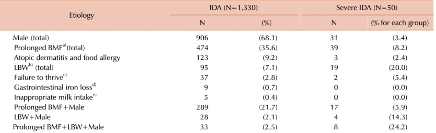
Table 3. The frequency of risk factors in iron deficiency anemia affected infants and young children aged 6 to 23 months (multiple answers).
Etiology IDA (N=1,330) Severe IDA (N=50)
N (%) N (% for each group)
Male (total) 906 (68.1) 31 (3.4)
Prolonged BMFa)(total) 474 (35.6) 39 (8.2)
Atopic dermatitis and food allergy 123 (9.2) 3 (2.4)
LBWb) (total) 95 (7.1) 19 (20.0)
Failure to thrivec) 37 (2.8) 2 (5.4)
Gastrointestinal iron lossd) 9 (0.7) 0 (0.0)
Inappropriate milk intakee) 5 (0.4) 0 (0.0)
Prolonged BMF+Male 289 (21.7) 17 (5.9)
LBW+Male 28 (2.1) 4 (14.3)
Prolonged BMF+LBW+Male 33 (2.5) 8 (24.2)
a)Prolonged BMF: exclusive breast milk feeding till >6 months of age. b)LBW: birth weight <2.5 kg. c)Failure to thrive: undernutrition - weight
<2nd percentile of age and gender corrected gestation weight, or weight <80% of the ideal weight for age. d)Gastrointestinal loss: chronic diarrhea and melena. e)Inappropriate milk intake: introduction of unmodified cow’s milk before 12 months of age, cow’s milk protein induced colitis, or the consumption of cow’s milk >700 mL/day.
Abbreviations: IDA, iron deficiency anemia; BMF, breast milk feeding; LBW, low birth weight.
Table 4. Risk factors identified in a multiple regression analysis to be associated with iron deficiency anemia and severe iron deficiency anemia.
Univariate Multivariate
OR (95% CI) P OR (95% CI) P
Prolonged BMFa) 6.88 (3.49–13.58) <0.001 5.70 (2.67–12.19) <0.001
Low birth weightb) 8.26 (4.37–15.60) <0.001 6.49 (3.34–12.60) <0.001
Failure to thrivec) 1.48 (0.35–6.34) 0.596 1.965 (0.41–9.52) 0.340
Atopic dermatitis 0.59 (0.18–1.92) 0.381 1.91 (0.51–7.22) 0.401
Gender (M:F) 0.755 (0.42–1.35) 0.345 0.882 (0.45–1.72) 0.714
Symptoms of anemia 4.55 (2.29–9.01) <0.001
a)Prolonged BMF: exclusive breast milk feeding till >6 months of age. b)Low birth weight: birth weight <2.5 kg. c)Failure to thrive:
undernutrition - weight <2nd percentile of age and gender corrected gestation weight, or weight <80% of the ideal weight for age.
Abbreviations: BMF, breast milk feeding; OR, odds ratio; CI, confidence interval; M, male; F, female.
iron supply despite a high iron requirement at this age [14].
The estimated requirement for absorbed iron during the first year of infancy ranges between 0.55 mg/day and 0.75 mg/day [20]. However, after 6 months of age, obtaining enough iron through breastfeeding alone becomes difficult.
The mean iron level in breast milk is approximately 0.4 mg/L, and although half of the iron contained in breast milk is absorbed due to its high bioavailability, only 0.2 mg/day of total iron can be absorbed by exclusive breastfeed- ing even if the infant consumes 1 L breast milk daily, which is still considerably less than the required iron amount [20, 21]. Moreover, as the low iron-containing rice soup is usually fed to the Korean infants during the early stages of weaning, infants may easily develop an iron deficiency.
LBW infants are generally considered a risk group for IDA due to low iron stores at birth. Hence, iron supplementa- tion is recommended for them; however, their adherence to iron supplementation is low. Preterm infants, who com- prise a large proportion of LBW infants, have lower iron stores due to shorter third gestational trimesters when most
of the iron is accumulated. These infants have higher iron requirements for catch-up growth, and preterm infants should thus receive supplements of elemental iron (2 mg/kg/day) from 1 month to 12 months of age [22]. In this study, none of the MLBW infants had received iron supple- ments for more than 30 days at the time of IDA diagnosis.
The need for iron supplementation in MLBW infants has been debated in the past; however, iron supplementation has recently been recommended for MLBW infants (at a dose of 2 mg/kg/day) from 6 weeks to 6 months of age [23].
We have previously reported significant differences in iron supplementation and breastfeeding practices between LBW infants in the IDA and the non-IDA groups [24]. In LBW infants, the use of human milk fortifier until attainment of a body weight of 3 kg can also prevent IDA. We considered atopic dermatitis as a possible risk factor for IDA as many mothers with atopic dermatitis-affected infants tend to breast- feed, and thus limit the food selection. However, despite the prevalence of atopic dermatitis in Korea (26.5% among 12–23 mo-old children), our results indicate that atopic dermatitis
is not a contributing factor to total IDA and has no effect on the risk of severe anemia (OR, 1.38; 95% CI, 0.39–4.90).
The undiminishing prevalence of IDA among Korean tod- dlers could be due to several reasons. First, according to the Korea National Health and Nutrition Examination Survey VI-2, the rate of breastfeeding has increased from 10.2%
in 2000 to 45.6% in 2012 [25], while the implementation of iron supplementation has not been sufficient. Second, 47% of the exclusively breastfed infants require 2 months or more to adapt to the weaning [26], and many homemade weaning foods have low iron content. Third, in Korea, the incidence of LBW has increased from 2.7% in 1993 to 5.6%
in 2010. Fourth, adherence to iron supplementation is low in LBW infants. Specifically, more encouragement is required to implement iron supplementation in the MLBW infants.
The inappropriate consumption of cow’s milk is another important factor for the presence of IDA; an early in- troduction of cow’s milk can cause IDA. The mean cow’s milk introduction age is 14 months; however, 6.6% infants drink cow’s milk before 12 months of age [25, 27]. An in- appropriately high intake of cow’s milk (0.7 L/day) after infancy can also lead to IDA.
In Korea, infants are brought to the clinic approximately 5 times until the age of 1 year for vaccinations and health screenings. However, only 7.1% IDA-affected infants were identified during vaccination and health screening in this study, indicating that many clinicians overlook the serious- ness of IDA in growing children. The American Academy of Pediatrics (AAP) recommends IDA screening for all infants aged 9–12 months [22]. Recently, the United States Preven- tive Service Task Force concluded that the evidence of IDA screening in asymptomatic children is insufficient [28] due to the decrement in IDA incidence. However, IDA is still high among infants and young children in Korea. Therefore, we recommend the screening of iron nutrition, especially in infants at a high risk of iron deficiency. Although the Hb level can be assessed using a finger prick test, other iron measurements require peripheral blood sampling which is difficult to perform in infants. The Iron Score board [29]
is useful in selecting infants who need iron status evaluations for appropriate screening and treatment. The score board includes following criteria: (1) age of initiation of weaning food >6 months; (2) intake of only homemade weaning food; (3) successful weaning in >1 month; (4) low iron content in weaning foods; (5) duration of breastfeeding; (6) feeding method; and (7) response to weaning foods. Using this score board, IDA could be predicted with 86.8% sensi- tivity in infants exhibiting 3 or more of the above-mentioned criteria. This is especially useful as it can help prevent iron deficiency in infants without the need for unnecessary labo- ratory tests.
IDA in infants and young children is fundamentally a nutritional problem, compared to IDA in older children, where it is mainly caused by blood loss. Therefore, nutritional counseling is important, and recommendations include al- most daily intake of one serving of either fish; meat; chicken;
or eggs as complementary food, or the use of micronutrient
powders [30]. The AAP recommends iron supplementation (2 mg/kg/day) for LBW infants, including the MLBW infants, from the age of 6 weeks to 6 months. It further recommends iron supplementation (1 mg/kg/day) for breastfed term in- fants starting at 4 months of age and maintained until appro- priate iron-containing complementary foods have been in- troduced [29].
The limitation of our study is that as this was a tertiary hospital-based study, there may have been a possible selection bias, and the disease severity may have been exaggerated.
Further, as this study was based on a retrospective survey of medical records, there was a lack of information regarding prenatal maternal anemia status, the time course of weaning to food, and the degree of postnatal weight gain. The fol- low-up loss was considerable, especially in the mild anemia group, and thus, the treatment response evaluation could not be completed in many cases.
In conclusion, a large proportion of children with IDA were not followed-up as many clinicians do not consider IDA to be a real health issue. Thus, clinicians should recom- mend appropriate iron supplementation to children, espe- cially to infants at a higher risk of developing iron deficiency.
Notably, the MLBW infants need special attention to ensure adherence to iron supplementation. Pediatricians should col- lect an accurate history of infant feeding practices (feeding type, the timing of weaning initiation, and contents of wean- ing food) during health screening visits in order to allow the early detection of IDA, and should recommend blood tests to high-risk infants. The questionnaire along with the assessment of iron nutrition is recommended for the de- tection of anemia and iron status in pediatric clinics. In addition, a prospective and well-organized risk assessment study in infants and young children is necessary to improve iron nutritional status and to control IDA.
AuthorsÊ Disclosures of Potential Conflicts of Interest
No potential conflicts of interest relevant to this article were reported.
REFERENCES
1. World Health Organization. Iron deficiency anaemia: assessment, prevention and control: A guide for programme managers. Geneva, Switzerland: World Health Organization, 2001. (Accessed October 2, 2015, at http://www.who.int/nutrition/publications/micronutrients/
anaemia_iron_deficiency/WHO_NHD_01.3/en/index.html).
2. Hopkins D, Emmett P, Steer C, Rogers I, Noble S, Emond A. Infant feeding in the second 6 months of life related to iron status: an observational study. Arch Dis Child 2007;92:850-4.
3. Looker AC, Dallman PR, Carroll MD, Gunter EW, Johnson CL.
Prevalence of iron deficiency in the United States. JAMA 1997;277:973-6.
4. Zlotkin SH, Christofides AL, Hyder SM, Schauer CS, Tondeur MC, Sharieff W. Controlling iron deficiency anemia through the use
of home-fortified complementary foods. Indian J Pediatr 2004;71:
1015-9.
5. Soh P, Ferguson EL, McKenzie JE, Homs MY, Gibson RS. Iron deficiency and risk factors for lower iron stores in 6-24-month-old New Zealanders. Eur J Clin Nutr 2004;58:71-9.
6. Grant CC, Wall CR, Brunt D, Crengle S, Scragg R. Population prevalence and risk factors for iron deficiency in Auckland, New Zealand. J Paediatr Child Health 2007;43:532-8.
7. World Health Organization. The global prevalence of anaemia in 2011. Geneva, Switzerland: World Health Organization, 2015.
8. Korean Statistical Information Service. National health insurance statistical annual report classification of 298 disease categories by age, national health insurance service, health insurance review, and assessment service. Daejeon, Korea: Korean Statistical Infor- mation Service, 2014. (Accessed June 30, 2016, at http://kosis.kr/
statHtml/statHtml.do?orgId=350&tblId=TX_35001_A061&con n_path=I2).
9. Pollitt E. Iron deficiency and cognitive function. Annu Rev Nutr 1993;13:521-37.
10. Carter RC, Jacobson JL, Burden MJ, et al. Iron deficiency anemia and cognitive function in infancy. Pediatrics 2010;126:e427-34.
11. Lozoff B, Jimenez E, Hagen J, Mollen E, Wolf AW. Poorer behavioral and developmental outcome more than 10 years after treatment for iron deficiency in infancy. Pediatrics 2000;105:E51.
12. Khedr E, Hamed SA, Elbeih E, El-Shereef H, Ahmad Y, Ahmed S. Iron states and cognitive abilities in young adults: neuropsy- chological and neurophysiological assessment. Eur Arch Psychiatry Clin Neurosci 2008;258:489-96.
13. World Health Organization. Haemoglobin concentrations for the diagnosis of anaemia and assessment of severity. Vitamin and Mineral Nutrition Information System. Geneva, Switzerland:
World Health Organization, 2011. (Accessed January 11, 2016, at http://www.who.int/vmnis/indicators/haemoglobin.pdf).
14. World Health Organization. Serum ferritin concentrations for the assessment of iron status and iron deficiency in populations.
Vitamin and Mineral Nutrition Information System. Geneva, Switzerland: World Health Organization, 2011. (Accessed January 11, 2016, at http://www.who.int/vmnis/indicators/
serum_ferritin.pdf).
15. Wu AC, Lesperance L, Bernstein H. Screening for iron deficiency.
Pediatr Rev 2002;23:171-8.
16. Lozoff B, Kaciroti N, Walter T. Iron deficiency in infancy:
applying a physiologic framework for prediction. Am J Clin Nutr 2006;84:1412-21.
17. Domellöf M, Lönnerdal B, Dewey KG, Cohen RJ, Rivera LL,
Hernell O. Sex differences in iron status during infancy. Pediatrics 2002;110:545-52.
18. Wieringa FT, Berger J, Dijkhuizen MA, et al. Sex differences in prevalence of anaemia and iron deficiency in infancy in a large multi-country trial in South-East Asia. Br J Nutr 2007;98:1070-6.
19. Choi JW, Kim CS, Pai SH. Erythropoietic activity and soluble transferrin receptor level in neonates and maternal blood. Acta Paediatr 2000;89:675-9.
20. Calvo EB, Galindo AC, Aspres NB. Iron status in exclusively breast-fed infants. Pediatrics 1992;90;375-9.
21. Shin PJ, Bae CW, Choi YM. A comparative study of red blood cell indices and anemia by feeding patterns. Korean J Pediatr 1999;42:1104-10.
22. Baker RD, Greer FR; Committee on Nutrition American Academy of Pediatrics. Diagnosis and prevention of iron deficiency and iron-deficiency anemia in infants and young children (0-3 years of age). Pediatrics 2010;126:1040-50.
23. Berglund S, Westrup B, Domellöf M. Iron supplements reduce the risk of iron deficiency anemia in marginally low birth weight infants. Pediatrics 2010;126;e874-83.
24. Park SH, Kim JS, Jun YH, Kim SK. Weaning food practice in low birth weight infants with iron deficiency anemia. Clin Pediatr Hematol Oncol 2014;21:52-8.
25. Ministry of Health and Welfare. Korea Health Statistics 2014:
Korea National Health and Nutrition Examination Survey (KNHANES VI-2). Cheongju, Korea: Korea Centers for Disease Control and Prevention, 2015.
26. Kim BY, Choi EH, Kang SK, Jun YH, Hong YJ, Kim SK. Weaning food practice and assessment in children with iron deficiency anemia. Korean J Pediatr Gastroenterol Nutr 2009;12:215-20.
27. Kim YH, Lee SG, Kim SH, Song YJ, Chung JY, Park MJ. Nutritional status of Korean toddlers: From the Korean National Health and Nutrition Examination Survey 2007∼2009. Korean J Pediatr Gastroenterol Nutr 2011;14:161-70.
28. Siu AL; US Preventive Services Task Force. Screening for iron deficiency anemia in young children: USPSTF recommendation statement. Pediatrics 2015;136:746-52.
29. Kim HJ, Kim DH, Lee JE, et al. Is it possible to predict the iron status from an infant’s diet history? Pediatr Gastroenterol Hepatol Nutr 2013;16:95-103.
30. World Health Organization. Essential nutrition actions:
Improving maternal, newborn, infant and young child health and nutrition. Geneva, Switzerland: World Health Organization, 2013. (Accessed July 1, 2016, at http://www.who.int/nutrition/
publications/infantfeeding/essential_nutrition_actions/en/).