Korean J Hematol Vol. 44, No. 1, March, 2009 □ Case Report □
Idiopathic Thrombocytopenic Purpura in a Patient with Carcinoma of the Uterine Cervix
Hyeong Su Kim, M.D.1, Jung Han Kim, M.D.1, Dong Kil Na, M.D.1, Dae Young Zang, M.D.1, Min-Jeong Park, M.D.2, Hong-Bae Kim, M.D.3 and Jong-Wook Lee, M.D.4 Departments of 1Internal Medicine, 2Laboratory Medicine, and 3Gynecology, Hallym University Medical Center,
Hallym University College of Medicine, 4Division of Hematology, Department of Internal Medicine, College of Medicine, The Catholic University of Korea, Seoul, Korea
We describe here the case a patient with advanced cervix carcinoma and who developed idiopathic throm- bocytopenic purpura (ITP). A 63-year-old woman with stage IV squamous cell carcinoma of the uterine cervix and that was complicated by hydronephrosis was treated palliatively with 45Gy of external beam radiation to the pelvis. About 3 years later, she developed hematochezia and severe thrombocytopenia.
The laboratory examinations showed no evidence of thrombotic thrombocytopenic purpura or disseminated intravascular coagulopathy, and she was positive for serum anti-platelet antibodies. On the bone marrow examination, there was a normal number and morphology of megakaryocytes with no evidence of malig- nant cell infiltration. We made the clinical diagnosis of ITP, and the intravenous immunoglobulin and steroid therapy was successful. This case suggests the possibility that ITP can occur in association with advanced cervix carcinoma. (Korean J Hematol 2009;44:58-61.)
Key Words: Autoimmune thrombocytopena, Idiopathic thrombocytopenic purpura, Cervix carcinoma
58 접수:2009년 1월 2일, 수정:2009년 2월 23일
승인:2009년 2월 28일
교신저자:김정한, 서울시 영등포구 대림 1동 948-1
150-950, 한림대학교 강남성심병원 내과 Tel: 02-829-5414, Fax: 02-846-4669
E-mail: [email protected]
Correspondence to:Jung Han Kim, M.D.
Department of Internal Medicine, Kangnam Sacred-Heart Hospital, Hallym University College of Medicine
948-1, Daelim 1-dong, Youngdeungpo-gu, Seoul 150-950, Korea Tel: +82-2-829-5414, Fax: +82-2-846-4669
E-mail: [email protected] INTRODUCTION
Idiopathic thrombocytopenic purpura (ITP) is known to be occasionally associated with lympho- proliferative disorders such as chronic lympho- cytic leukemia, lymphoma.1) In patients with sol- id tumors, common causes of thrombocytopenia are myelosuppression secondary to chemotherapy, bone marrow infiltration with malignant cells.
However, various solid tumors, including breast, lung, prostate, skin, and ovarian cancers, have been reported to be associated with ITP.2-7)
Here we describe a patient with advanced cer- vix carcinoma who developed ITP as a possible paraneoplastic syndrome.
CASE REPORT
In February 2005, a 63-year-old woman was di- agnosed with stage IV squamous cell carcinoma of the uterine cervix. She had no allergies or sig- nificant past medical history, and her family his- tory was unremarkable. The initial complete blood counts (CBC) were within normal ranges: hemo- globin (Hb) of 12.7g/dL; white blood cell count
Hyeong Su Kim, et al: ITP in Carcinoma of the Uterine Cervix 59
Fig. 1. Peripheral blood smear shows severe thrombocyto- penia with the presence of megaplatelets (arrow) and lack of schistocytes (Wright stain, 400×).
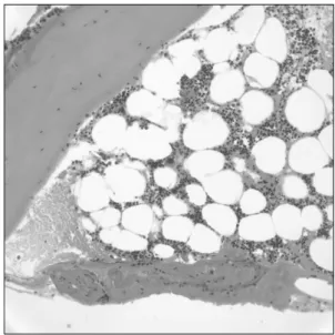
Fig. 2. Bone marrow examination reveals a slight hypo- cellular marrow with no evidence of neoplastic infiltration.
Marrow megakaryocytes are normal in number and morphology. (Hematoxylin-Eosin, 400×).
(WBC) of 7.48×10⁹/L; platelets count of 361×
109/L. Because her disease was complicated by hydronephrosis she received palliatively 45Gy of external beam radiation to the pelvis followed by ureteral stent (Double J catheter) insertion in the left ureter. Thereafter she was recommended sys- temic chemotherapy but refused. She was fol- lowed-up with ureteral stent changes every 2 to 3 months. In March 2008, her CBC showed WBC of 4.3×10⁹/L, Hb of 10.3g/dL, platelets count of 477×109/L.
In August 2008, she presented with hema- tochezia developed abruptly. She was not taking any medication and herbs. Physical examination revealed petechiae on her legs and no evidence of splenomegaly. On hematologic examination, Hb was 5.6g/dL, WBC count 4.96×10⁹/L, and plate- let count 8×10⁹/L. Serologic tests for Hepatitis B virus, Human immunodeficiency virus, and an- ti-nuclear antibody were negative. Chest radiol- ogy newly revealed multiple hematogeneous metastases in the lung. Abdomen CT showed multiple low attenuated lesions in spleen and metastasis to spleen was suggested. Biochemistry showed a normal kidney and liver function, and coagulopathy parameters were within normal ranges. The direct Coombs’ test and anti-nuclear antibody test were negative, and serum anti-plate- let antibodies were positive on immunofluorescent assay. Peripheral blood smear showed severe throm- bocytopenia with the presence of megaplatelets and lack of schistocytes (Fig. 1). Transfusions of 16 units of platelets, with together 3 units of red blood cells, were immediately administered after initial laboratory evaluation. A transient rise in platelet count after transfusion was observed, but the number decreased again to reach to 4×10⁹/L.
Bone marrow examination revealed a slight hypo- cellular marrow (about 30%) with no evidence of neoplastic infiltration. Marrow megakaryocytes were normal in number and morphology (Fig. 2).
Clinical diagnosis of ITP was made, and ther- apy with intravenous immunoglobulin (400mg/kg per day for 5 days) and steroid (intravenous
methylprednisolone 125mg per day followed by prednisone 40mg per day) was started and plate- let counts increased gradually. Four weeks after administering steroid her platelet count was 234×109/L. Thereafter prednisone was tapered to 10mg per day and subsequent platelet count re- mained stable at levels over 200×109/L.
DISCUSSION
ITP is an autoimmune bleeding disorder char-
60 Korean J Hematol Vol. 44, No. 1, March, 2009
acterized by the development of autoantibodies against platelets, which resulted in phagocytosis by spleen and thrombocytopenia. The majority of autoantibodies are directed against the GPIIb/
IIIa structural complex on the surface of the platelets.1) The common causes of immunologic thrombocytopenia are viral infection, drugs, and chronic autoimmune disorders. However, Immune- mediated thrombocytopenia is occasionally en- countered in patients with lymphoproliferative disorders, especially malignant lymphoma, chron- ic lymphocytic leukemia.1)
In patients with solid tumor, the most common cause of thrombocytopenia is myelosuppression secondary to systemic chemotherapy or radio- therapy. Especially in patients with advanced cancer, thrombotic thrombocytopenic purpura (TTP), disseminated intravascular coagulopathy (DIC), and bone marrow infiltration with malig- nant cells (myelophthisis) can be considered as a possible cause of thrombocytopenia. In this case, patient did not receive systemic chemotherapy.
On laboratory examinations, she showed no evi- dence of DIC or TTP. We initially thought that thrombocytopenia would be resulted from mye- lophthsis secondary to bone marrow infiltration with malignant cells. In myelophthisis, the blood smear is characterized by leukoerythroblastosis (immature granulocyte, nucleated red cells, and teardrop-shaped red cells). However, there was no evidence of leukorerythroblastosis on the periph- eral blood smear of our patient. Moreover, serum anti-platelet antibodies were positive, and mas- sive platelet transfusions failed to increase circu- lating platelet counts. Therefore, we also consid- ered that other factors may have caused thrombo- cytopenia in our patient. On bone marrow exami- nation, megakaryocytes were normal in number and morphology, although marrow cellularity was slightly decreased. In ITP, the marrow is usually normal, although megakaryocytes may be in- creased in number. Because marrow hypocell- ularity in our patient was thought to be asso- ciated with the history of pelvic irradiation, clin-
ical diagnosis of ITP could be made. Therapy with intravenous immunoglobulin and steroid was successful.
A literature review showed that various solid tumors, including breast, lung, prostate, skin, and ovarian cancers, have been reported to be asso- ciated with ITP,2-9) and the diagnosis of ITP was based on the clinicopathologic findings of pos- itive platelet antibody, normal number of mega- karyocyte in BM, and treatment responses to ste- roid, immunoglobulin, and splenectomy. In rela- tion to carcinoma of the uterine cervix, there were two cases of autoimmune thrombocytopenia developing after cisplatin and radiation therapy.10) In case 1, patient developed pancytopenia ten days after administrating the last course of cisplatin. Leukopenia improved immediately with granulocyte colony stimulation factor, but throm- bocytopenia was not ameliorated in despite of massive platelet transfusion. In case 2, thrombo- cytopenia alone was developed two months after chemotherapy. In both cases, the diagnosis of au- toimmune thrombocytopenia was made on the re- sults of elevated platelet-associated IgG, increased numbers of megakyrocytes on marrow examina- tion, and the response to prednisone therapy.
Because these patients had received chemo- therapy recently before developing thrombocyto- penia, platelet-associated antibodies might be in- duced by drug (cisplatin or paclitxel). On the other side, our patient had no history of chemo- therapy.
There are two explanations for the develop- ment of ITP in cervix cancer.6-9) First, as de- scribed in other solid malignancies,6,7) paraneo- plastic ITP may be triggered by the formation of anti-platelet antibodies in our patient who had newly developed distant metastases. The fact that many cases including our case had stage IV can- cer6,8,9) suggest that a high tumor burden or meta- stasis to other organ via blood stream may pro- duce an immune-response resulting in platelet destruction.8) Second, the involvement of spleen by tumor may change the splenic environmental
Hyeong Su Kim, et al: ITP in Carcinoma of the Uterine Cervix 61
milieu producing an ITP and may be heralded by ITP.9)
In conclusion, we have treated a patient with carcinoma of the uterine cervix complicated by ITP, which was not related to the chemothera- peutic agent. To our knowledge, this is the first ITP case developed in a patient with advanced cervix carcinoma in Korea.
요 약
진행성 고형암 환자에서 혈소판 감소의 원인은 대부분 항암치료나 종양의 골수 침범에 의한 골 수억제에 기인한다. 하지만, 다양한 진행성 고형 암에서 특발혈소판감소자색반병에 의한 혈소판 감소의 보고가 있었다. 저자 등은 최근 진행성 자 궁경부암 환자에서 발생한 특발혈소판감소자색 반병을 진단하였고, 면역글로불린과 스테로이드 로 성공적인 치료를 한 증례를 경험하였기에 문 헌 고찰과 함께 보고하는 바이다.
REFERENCES
1) Bussel JB. Autoimmune thrombocytopenic purpura.
Hematol Oncol Clin North Am 1990;4:179-91.
2) Chehal A, Taher A, Seoud M, Shamseddine A.
Idiopathic thrombocytopenic purpura and ovarian cancer. Eur J Gynaecol Oncol 2003;24:539-40.
3) Kim HD, Boggs DR. A syndrome resembling idio- pathic thrombocytopenic purpura in 10 patients with diverse forms of cancer. Am J Med 1979;67:371-7.
4) Schwartz KA, Slichter SJ, Harker LA. Immune-medi- ated platelet destruction and thrombocytopenia in patients with solid tumours. Br J Haematol 1982;51:17-24.
5) DiFino SM, Lachant NA, Kirshner JJ, Gottlieb AJ.
Adult idiopathic thrombocytopenic purpura. Clinical findings and response to therapy. Am J Med 1980;69:430-42.
6) Bir A, Bshara W, George M, Fakih MG. Idiopathic thrombocytopenic purpura in a newly diagnosed pancreatic adenocarcinoma. JOP 2006;7:647-50.
7) Demirer T, Celebi H, Arat M, et al. Autoimmune thrombocytopenia in a patient with small cell lung cancer developing after chemotherapy and resolving following autologous peripheral blood stem cell transplantation. Bone Marrow Transplant 1999;24:
335-7.
8) Porrata LF, Alberts S, Hook C, Hanson CA. Idio- pathic thrombocytopenic purpura associated with breast cancer: a case report and review of the cur- rent literature. Am J Clin Oncol 1999;22:411-3.
9) Cummings OW, Mazur MT. Breast carcinoma dif- fusely metastatic to the spleen. A report of two cases presenting as idiopathic thrombocytopenic purpura.
Am J Clin Pathol 1992;97:484-9.
10) Mitsuhashi A, Yamazawa K, Tanaka N, Sekiya S.
Autoimmune thrombocytopenia in women with car- cinoma of the uterine cervix developing after cispla- tin and radiation therapy. BJOG 2002;109:103-4.