Although the overall survival of women that received breast conserving therapy (BCT) and radiation therapy is equivalent to that of mastectomy (1-5), failure to maintain local control is clearly associ- ated with a reduction in long-term cancer-specific survival (6). Negative surgical margin of resection at the time of BCT for breast cancer is associated with substantially lower recurrence rates (7). In previous studies, various factors, including age, tumor size, and
INTRODUCTION
�Received; April 3, 2014�Revised; May 7, 2014
�Accepted; May 13, 2014
This work was supported by Korea Science and Engineering Foundation (KOSEF) grant funded by the Korean government (MEST) (grant code:
2012R1A1A3008621).
Corresponding author : Sun Mi Kim, M.D.
Department of Radiology, Seoul National University Bundang Hospital, 166 Gumi-ro, Bundang-gu, Seongnam-si, Gyeonggi-do 463-707, Korea.
Tel. 82-31-787-7617, Fax. 82-31-787-4011 E-mail : [email protected]
This is an Open Access article distributed under the terms of the Creative Commons Attribution Non-Commercial License (http://creativecommons.org/licenses/by- nc/3.0/) which permits unrestricted non-commercial use, distribution, and reproduction in any medium, provided the original work is properly cited.
Magnetic Resonance Imaging Factors Predicting Re-excision in Breast Cancer Patients Having
Undergone Conserving Therapy
Mijung Jang1, Sun Mi Kim1, Bo La Yun1, Sung-Won Kim2, Eun Young Kang2, So Yeon Park3, Jee Hyun Kim4, Yeongmi Kim5, Hye Shin Ahn6
1Department of Radiology, Seoul National University Bundang Hospital, Gyeonggi-do, Korea
2Department of Surgery, Seoul National University Bundang Hospital, Gyeonggi-do, Korea
3Department of Pathology, Seoul National University Bundang Hospital, Gyeonggi-do, Korea
4Department of Oncology, Seoul National University Bundang Hospital, Gyeonggi-do, Korea
5Department of Radiology, Samsung Changwon Hospital, Sungkyunkwan University, Gyeongsangnam-do, Korea
6Department of Radiology, Chung-Ang University Hospital, Seoul, Korea
Purpose : The aim of this study was to determine the magnetic resonance imaging (MRI) features associated with re-exci- sion due to the presence of a positive margin after breast conserving therapy (BCT) in breast cancer patients.
Materials and Methods: We reviewed the records of 286 consecutive breast cancer patients who received BCT between January 2006 and December 2007. Among 246 patients who had undergone BCT, 38 (15.4%) underwent immediate further surgery due to positive margin status. We analyzed the MRI findings using χ2test, Fisher’s exact test and t tests.
Multivariate logistic regression was conducted for prediction of re-excision.
Results: Tumor size (p < 0.001), lesion multiplicity (p = 0.003), and non-mass-like enhancement (NMLE) type on MRI (p <
0.001) were associated with margin involvement in BCT. On preoperative MRI, larger size (≥ 5 cm) (odds ratio = 2.96), NMLE (odds ratio = 3.81), and multifocal lesions (odds ratio = 2.54) were positively associated with re-excision. In cases involving NMLE, segmental distribution was associated with a greater likelihood of immediate re-excision.
Conclusion: Larger size, multiplicity, and NMLE on MRI are significantly associated with re-excision after BCT in breast cancer patients. For NMLE lesions, the segmental distribution pattern was predictive of re-excision.
Index words : Breast∙Breast neoplasm∙Conservation therapy∙Diagnosis, Magnetic resonance imaging (MRI) Original Article
pathologic contents, have been evaluated with regard to the re-excision rate (8-15).
Breast magnetic resonance imaging (MRI) has become a frequently employed imaging modality in the evaluation of the local extent of disease in patients with a confirmed diagnosis of breast cancer. The sensitivity of breast MRI is close to 100% in the context of preoperative evaluation. This high sensitiv- ity is presumed to improve the selection of patients for BCT and reduce the rates of re-excision. MRI detects additional foci of disease in the ipsilateral breast of patients with known breast cancer in 10-30% of cases (16). Clinically and mammographically occult cancer in the contralateral breast is detected in 3-5% of patients who undergo preoperative breast MRI (16).
Furthermore, several studies have shown alterations in surgical management planning in approximately 20- 30% of all breast cancer patients undergoing preoper- ative MRI, with an associated increase in mastectomy rates (8, 11-13, 15). Concerns have been raised that such false-positive findings may increase the use of mastectomy without any clear survival benefit. In a previous study, no significant difference was observed in the recurrence rates between the patients who underwent preoperative MRI versus those who did not. Additionally, low rates of recurrence after both BCT and mastectomy were reported (14, 17).
Therefore, the role of preoperative MRI in breast cancer patients being considered for BCT remains a matter of some controversy.
However, preoperative breast MRI has been routinely performed in certain institutions; breast MRI remains a relevant surgical planning tool. Therefore, it is necessary to determine which preoperative MRI findings are associated with re-excision. The objective of this study was to determine the MRI features associ- ated with immediate further surgery due to the presence of a positive margin after BCT in breast cancer patients.
Patients and Lesions
This study was approved by the institutional review board, which waived the requirement for informed consent. At our institute, all patients with biopsy
results indicating breast cancer underwent preopera- tive MRI. The patients’ medical records were reviewed in order to obtain patients’ information and MRI findings. We retrospectively evaluated 443 consecutive newly diagnosed breast cancer patients from January 2006 through December 2007. Among these, 286 (65%) cases underwent conservation surgery. Among these 286 cases, 40 were excluded: 20 cases in which no preoperative MRI study data were available, and 20 cases involving preoperative systemic therapy. Thus, a total of 246 BCT cases of 241 patients (mean age, 49 years; range, 25-84) were ultimately included in our study, and of these, 236 patients received unilateral conservation surgery and 5 patients received bilateral conservation surgery.
Surgical Therapy
All conservation surgeries were conducted by a single surgeon (S.W.K) with 9 years of experience in breast cancer surgery. Conserving surgery was suggested for the majority of women with stage I or II early breast cancer or with a maximum cancer diameter of less than 4 cm after review of all imaging data, including MRI data (2, 18). The expected percentage of residual breast volume after wide excision and the patient’s preferences were also considered. Subareolar tumors (23 cases) were approached by central excision with or without resection of the nipple areolar complex depending on the tumor involvement. Palpable tumors (152 cases) were excised with the acceptable margin width of more than 1 cm by intraoperative palpation without any image guidance. Non-palpable malignant lesions (94 cases) were routinely guided by breast radiologists using ultrasound- or mammography-guided hookwire needle localization. Ultrasound-guided needle localization was performed in 77 cases and mammog- raphy-guided needle localization was performed in 17 cases. In some cases with non-palpable tumors, specimen mammography was performed to confirm adequate resection. Intraoperative frozen sections for evaluation of the resection margins were not routinely collected. Pathology reports included size, histologic grade, and surgical margin status. Margins were classi- fied as involved (positive) or clear. Involved margins were defined as those with tumor cells less than 2 mm from the inked margin in invasive breast cancer, and MATERIALS AND METHODS
less than 1 mm from the inked margin in ductal carcinoma in situ (DCIS). Patients with involved margins were expected to undergo further surgery within 2 weeks. If the residual volume was sufficient and the patients desired a re-trial, they underwent another wide local excision rather than a mastectomy.
MRI sequence
All MRI examinations were conducted using 1.5 T or 3.0 T magnets (1.5 T Intera and 3.0 T Achieva, Philips Healthcare, Best, Hamburg, Germany), randomly.
Patients were placed in the prone position with both breasts imaged using a one-channel dedicated coil for 1.5 T MRI, and a four-channel dedicated coil for 3.0 T.
For 1.5 T MRI, our standard protocol consisted of a transverse T1-weighted localizer sequence through both breasts followed by a fat-saturated sagittal T2- weighted fast spin-echo acquisition (SPAIR) (TR/TE, 2300/65), and a dynamic series consisting of a T1- weighted two dimensional gradient echo pulse sequence (TR/TE, 290/4.6)], and a flip angle of 15�
through the affected breast. For the 3.0 T MRI protocol, a transverse T1-weighted localizer sequence through both breasts was followed by a fat-saturated sagittal T2-weighted fast spin-echo acquisition (SPAIR) (TR/TE, 5,000/120), and a dynamic series consisting of a T1-weighted two dimensional gradient echo pulse sequence (TR/TE, 269/2.3), and a flip angle of 12�
through the affected breast. Scanning parameters included the typical section thickness, 3-5 mm; the field of view, 26-35 cm depending on patient size; and a matrix of 344 × 345. Axial and coronal reforma- tions, in addition to maximum intensity projection (MIP), were constructed from the first contrast- enhanced image. Imaging was initiated via a bolus injection of gadodiamide at a dose of 0.1 mmol/kg of body weight (Omniscan, GE healthcare), administered through a needle into the antecubital vein, followed by a 20-mL flushing bolus of isotonic saline solution.
The total injection time was 10 seconds. Contrast- enhanced sagittal T1-weighted series were 60 seconds in duration and centered at 60, 120, 180, 240, and 300 seconds for the ipsilateral breast. After contrast- enhanced high-spatial-resolution images and their subtraction images were used for lesion detection, time-intensity plots of diagnosed breast cancer lesion dynamic images were generated using dedicated MRI
software.
MRI analysis
MRI review was retrospectively performed by two radiologists (S. M. K., M. J.) with 8 and 5 years’ breast MRI experience, by consensus. Bilateral breast MRI was interpreted with the benefit of a brief clinical history, knowledge of the initial histopathologic findings, and knowledge of the mammography and sonography results. However, both radiologists were blinded to the patient’s final pathological result and the reports of the initial MRI interpretation. We classi- fied MRI features of breast cancer based on ACR BI- RADS (Breast Imaging and Data Reporting System) MRI lexicon. (19) According to this lexicon, we categorized the lesion type on MRI as mass or non- mass-like enhancement (NMLE). In cases of mass, we additionally categorized kinetic curve delay enhance- ment characteristics as persistent, plateau, and wash out patterns based on the time-intensity plots of dynamic images. In cases of NMLE, we additionally categorized distribution as linear, ductal, segmental, regional, multiple regional, or diffuse patterns. We included mass associated with adjacent or coexistent NMLE of breast lesion. The shape, margin, internal enhancement of mass, and internal enhancement of NMLE were not included in our additional evalua- tions. We analyzed the size and number of lesions evident on MRI. Multifocal cancer was defined as the presence of two or more foci of cancer in the same quadrant that were separated by less than 2 cm of normal parenchyma. Multicentric cancer was classified as the presence of two or more foci of breast cancer in different quadrants. In cases of multifocal or multicen- tric lesions, the lesion size on MRI was determined at the largest portion. We also reviewed the lesion location in terms of distance from the nipple and distance from the chest wall on MIP images.
Data analysis
We analyzed clinical factors such as age, palpability, and breast cancer history with regard to re-excision.
We also analyzed the pathologic reports of final surgical specimens according to histologic type and the existence of a DCIS component, and conducted immunohistochemical analyses including hormone receptor and human epidermal growth factor receptor
type 2 staining scores. TNM staging was also evaluated based on the sixth American Joint Committee on Cancer (AJCC) guidelines (20).
Statistical analyses were conducted using the χ2test, Fisher’s exact test, and Student’s t-test using SPSS version 19.0 for Windows (SPSS Inc., Chicago, IL). We considered p values less than 0.05 to be indicative of a statistically significant difference. Multivariate analysis was performed using logistic regression of the variables that were found to be statistically significant through univariate analysis, and was used to estimate odds ratios (ORs).
In accordance with our surgical therapy guidelines, 38/246 (15.4%) cases subsequently underwent re- excision. Among these 38 cases, 11 (28.9%) received subsequent total mastectomy and 27 (71.1%) underwent one additional wide local excision. Among these 27 cases, 3 received one further local excision and ultimately underwent total mastectomy. Clinical
RESULTS
Table 1. Analysis of Associations Between Re-excision and Clinical and Pathologic Findings
Single BCT cases Re-excised cases p value
n = 208 n = 38
Clinical features
Age* 49.6 ± 11.3 45.7 ± 11.2 0.268
Palpability 0.591
Yes 130 (85.5) 22 (14.5)
No 78 (83.0) 16 (17.0)
Family history of
breast cancer 0.543
Yes 20 (90.9) 2 (9.1)
No 188 (83.9) 36 (16.1)
Pathologic features
Histology < 0.001
Invasive ductal 116 (87.9) 16 (12.1) carcinoma with DCIS
Invasive ductal 33 (97.1) 1 (2.9) carcinoma only
DCIS only 31 (63.3) 18 (36.7)
Invasive lobular 8 (80.0) 2 (20.0) carcinoma
Other� 20 (95.2) 1 (4.8)
Tumor stage� 0.045
T1 121 (85.8) 20 (14.2)
T2 61 (89.7) 7 (10.3)
T3 1 (100.0) 0 (0.0)
Tis 25 (69.4) 11 (30.6)
Estrogen receptor 0.598
Positive 151 (85.3) 26 (14.7)
Negative 57 (82.6) 12 (17.4)
Progesterone receptor 0.296
Positive 141 (82.9) 29 (17.1)
Negative 67 (88.2) 9 (11.8)
HER2§ 0.517
0 32 (82.1) 7 (17.9)
1+ 72 (87.8) 10 (12.2)
2+ 72 (80.9) 17 (19.1)
3+ 32 (88.9) 4 (11.1)
Note.─ Data are number of patients, with percentages in parentheses.
*Mean ± SD
�“Other” histologic type included 5 mucinous carcinoma, 3 metaplastic carcinoma, 3 apocrine carcinoma, 3 papillary carcinoma, 2 solid neuroendocrine carcinoma, 1 adenoid cystic carcinoma, 1 tubular carcinoma, and 1 malignant adenomyoepithelioma.
�According to the sixth American Joint Committee on Cancer (AJCC) guideline.
§HER2 staining scores of 0 or 1+ represent no or barely perceptible membrane staining.
HER2 2+ represents indeterminate membrane staining and HER2 gene amplification less than 25% of cells
HER2 3+ represents strong complete membrane staining in more than 10% in fluorescence in situ hybridization.
BCT = breast conserving therapy, DCIS = ductal carcinoma in situ, HER2 = human epidermal growth factor receptor type 2, NMLE = non-mass-like enhancement.
Table 2. Analysis of Associations Between Re-excision and MRI Findings
Single BCT cases Re-excised cases p value
n = 208 n = 38
MRI features
Lesion type < 0.001
Mass 168 (90.8) 17 (9.2)
NMLE 40 (65.6) 21 (34.4)
Size (mm) < 0.001
<20 99 (90.0) 11 (10.0)
≥20 to <50 106 (83.5) 21 (16.5)
≥50 3 (33.3) 6 (66.7)
Distance to 35.6 ± 18.9 26.3 ± 16.7 0.450 nipple (mm)*
Distance to chest 18.0 ± 16.3 13.2 ± 12.3 0.077 wall (mm)*
Number of lesions <0.005
Single 172 (88.2) 23 (11.8)
Multifocal 31 (73.8) 11 (26.2) Multicentric 5 (55.6) 4 (44.4)
Note.─ Data are number of patients, with percentages in parentheses.
*Mean ± SD
BCT = breast conserving therapy, DCIS = ductal carcinoma in situ, HER2 = human epidermal growth factor receptor type 2, NMLE = non-mass-like enhancement.
factors that were not significantly associated with re- excision included age (p = 0.268), palpability (p = 0.591), and breast cancer family history (p = 0.543) (Table 1). We analyzed the final pathologic report of surgical specimens with regard to re-excision. Tumor histology was significantly associated with re-excision (p < 0.001). Additionally, the coexistence of a DCIS component in invasive ductal carcinoma was signifi- cantly correlated with re-excision. In cases of pure
invasive ductal carcinoma without a DCIS component, only 2.9% (1/34) required re-excision. On the other hand, in cases of invasive ductal carcinoma involving DCIS components, 12.1% (16/132) required re- excision. In cases of pure DCIS, 36.7% (18/49) required re-excision. Twenty percent (2/10) of cases of invasive lobular carcinoma (ILC) required re-excision due to positive surgical margin. No significant correla- tions were noted between receptor status and re-
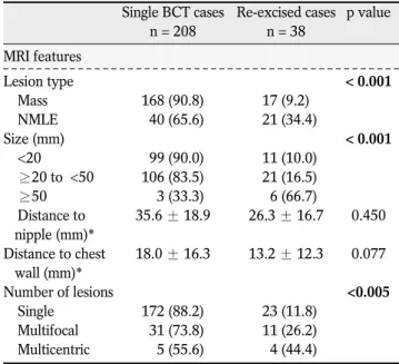
a b c
Fig. 1. Preoperative MR imaging showing a mass that was treated with single BCT. Images are of the left breast of a 34-year-old woman with confirmed invasive ductal carcinoma. Sagittal, contrast enhanced, fat-suppressed, 2D gradient (a) T1-weighted, (b) subtraction, and (c) MIP images of the left breast show a 2.1-cm-sized oval, not circumscribed, homogenous enhancing mass (arrows).
a b c
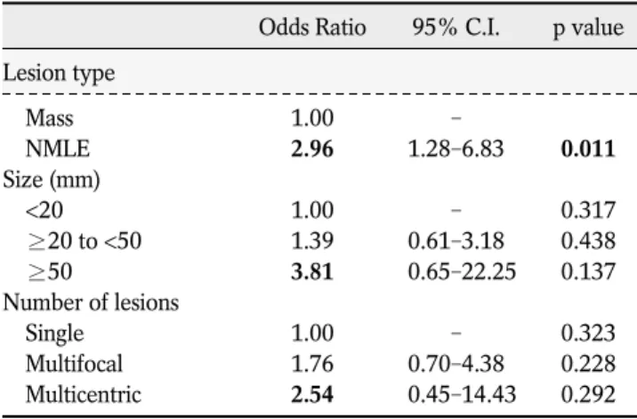
Fig. 2. Preoperative MRI showing NMLE; the patient underwent immediate re-excision after single BCT. Images are of the right breast of a 43-year-old woman with confirmed DCIS. Sagittal, contrast enhanced, fat-suppressed, 2D gradient (a) T1-weighted, (b) subtraction, and (c) MIP images of the right breast show a segmental clumped NMLE (arrows) extending 6 cm. This patient had received a lumpectomy and then underwent immediate re-excision due to positive superficial and deep margin.
excision (Table 1).
Table 2 compares the group that required immediate re-excision due to the presence of a positive margin (n
= 38) and the group in which a single BCT was sufficient (n = 208) according to MRI variables, includ- ing the type, size, and location of lesions, and the number of lesions. In cases of mass, re-excisions were conducted in 9.2% (17/185) of the patient group (Fig.
1). In the patients exhibiting NMLE on MRI the rate of re-excision was 34.4% (21/61) (Fig. 2), which was significantly higher than that of patients with masses (p
< 0.001). The mean size of malignant lesions was 20.7
± 9.2 mm (range, 9-65 mm) in single BCT cases and 29.6 ± 15.0 mm (range, 5-59 mm) in cases involving
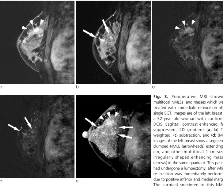
re-excision. Lesion size was positively associated with re-excision (p < 0.001). The distance from the nipple and chest wall, i.e., the lesion location, was not signifi- cantly associated with re-excision. Multiplicity of cancer identified via MRI was significantly associated with re-excision (p = 0.003) (Fig. 3). Among 61 cases exhibiting an MRI pattern indicative of NMLE, the distribution was significantly associated with re- excision (p = 0.047). In 30 NMLE cases in which segmental distribution was observed, 50% (15/30) underwent re-excision (Fig. 2) (Table 3).
Table 4. Multivariate Analysis of Associations Between Re-excision and MRI Findings
Odds Ratio 95% C.I. p value Lesion type
Mass 1.00 -
NMLE 2.96 1.28-6.83 0.011
Size (mm)
<20 1.00 - 0.317
≥20 to <50 1.39 0.61-3.18 0.438
≥50 3.81 00.65-22.25 0.137
Number of lesions
Single 1.00 - 0.323
Multifocal 1.76 0.70-4.38 0.228
Multicentric 2.54 00.45-14.43 0.292
Note.─ NMLE = non mass like enhancement, C.I. = confidence interval.
Table 5. Analysis of MRI Findings Correlated with Increased Re-excision Rate in NMLE
Odds Ratio 95% C.I. p value Size (mm)
<20 1.00 - 0.801
≥20 to <50 1.01 0.19-5.38 0.987
≥50 2.12 00.17-26.46 0.559
Distance to nipple (mm) 0.95 0.90-1.00 0.042 Distance to chest wall (mm) 01.010 0.96-1.06 0.686 Number of lesions
Single 1.00 - 0.604
Multifocal 2.47 00.42-14.50 0.315
Multicentric 1.32 00.06-27.56 0.858
Distribution
Ductal 1.00 - 0.373
Segmental 010.53 000.76-146.70 0.080
Regional 6.52 00.50-84.88 0.152
Multiple regional 7.17 000.07-765.37 0.409 Note.─ NMLE = non-mass like enhancement, C.I. = confidence interval.
Table 3. Analysis of Associations Between Re-excision and MRI Findings According to Lesion Type
Single BCT cases Re-excised cases p value NMLE (n = 61)* 40 (65.6) 21 (34.4)
Size (mm) 0.075
<20 13 (76.5) 4 (23.5)
≥20 to <50 24 (68.6) 11 (31.4)
≥50 3 (33.3) 6 (66.7)
Number of lesions 0.104
Single 28 (75.7) 9 (24.3)
Multifocal 10 (52.6) 9 (47.4)
Multicentric 2 (40) 3 (60)
Distribution 0.047
Linear 0 (0) 0 (0)
Ductal 11 (91.7) 1 (8.3)
Segmental 15 (50) 15 (50)
Regional 13 (76.5) 4(23.5)
Multiple regional 1 (50) 1 (50)
Diffuse 0 (0) 0 (0)
Mass (n = 185)* 168 (90.8) 17 (9.2)
Size (mm) 0.431
20 > 86 (92.5) 7 (7.5)
20 ≤ <50 82 (89.1) 10 (10.9)
50 ≤ 0 (0) 0 (0)
Number of lesions 0.542
Single 144 (91.1) 14 (8.9)
Multifocal 21 (91.3) 2 (8.7)
Multicentric 3 (75) 1 (25)
Enhancement pattern 0.051
Persistent 35 (81.4) 8 (18.6)
Plateau 71 (93.4) 5 (6.6)
Washout 62 (93.9) 4 (6.1)
Note.─ Data are number of patients, with percentages in parentheses. BCT = breast conserving therapy, NMLE = non- mass-like enhancement.
In multivariate logistic regression, larger size, NMLE pattern, and multiplicity on MRI were significantly associated with re-excision (Table 4). With regard to those significant MRI factors, size larger than 5 cm (OR = 3.81), NMLE type (OR = 2.96), and multifocal lesion (OR = 2.96) were independently predictive of re-excision. Among the NMLE lesions, additional multivariate logistic regression analysis was performed incorporating all variables associated with re-excision.
NMLE with segmental distribution was strongly associated with re-excision (OR =10.53) (Table 5).
With regard to breast cancer, it is well known that involved surgical margins are associated with higher local recurrence rates, (21-23) and in a recent meta- analysis, increased local recurrence was associated with poorer overall survival (6). Negative surgical margin in women undergoing BCT is universally accepted as a standard method of reducing the risk of local recurrence (24, 25). In some previous studies, the rates of margin-positive resection remain within a range of 20-70% (7, 10, 24). Additionally, re-excision after BCT is associated with increased expense,
DISCUSSION
a b c
d e
Fig. 3. Preoperative MRI showing multifocal NMLEs and masses which were treated with immediate re-excision after single BCT. Images are of the left breast of a 52-year-old woman with confirmed DCIS. Sagittal, contrast enhanced, fat- suppressed, 2D gradient (a, b) T1- weighted, (c) subtraction, and (d) (MIP) images of the left breast show a segmental clumped NMLE (arrowheads) extending 3 cm, and other multifocal 1-cm-sized irregularly shaped enhancing masses (arrows) in the same quadrant. This patient had undergone a lumpectomy, after which re-excision was immediately performed due to positive inferior and medial margin.
The surgical specimen of this NMLE confirmed that it was a 0.3-cm-sized invasive ductal carcinoma with a 4.5-cm area of DCIS (pT1a).
compromised cosmesis, patient anxiety, and delays in adjuvant therapy, and it can also be associated with compromised oncologic outcomes (26-28).
In this study, we conducted breast cancer treatment according to the NCCN practice guidelines (24).
Usually, conservation therapy of the breast is associ- ated with tumor size, extent, expected residual volume, and patient preference. Owing to the develop- ment of surgical techniques using skin flaps and nipple reconstruction, we can now attempt BCT more often in locally advanced cases. As compared to rates reported in previous studies, (26, 29-33) the overall re-excision rate of 15.4% (38/246) in this study was relatively low.
In previous studies, larger size, ILC, and the presence of DCIS (either as the principal pathology or in association with invasive cancer) were the factors that were the most consistently associated with margin positivity (26, 29-34). Younger age, high nuclear grade, and the presence of lymphovascular invasion have also been associated with a higher rate of margin positivity (26, 29, 30, 32-36). In our study, clinical factors including age, family history, and personal history of breast cancer were not significantly associ- ated with re-excision. ILC is slow-growing, and as it grows, it fails to invoke a desmoplastic reaction.
Unique histology of ILC can be the cause of detection difficulty, both clinically and radiographically. ILC is reportedly associated with higher rates of positive margins at excision than invasive ductal carcinomas, (37, 38) especially without preoperative MRI (39). In our study, 2/10 (20%) re-excised patients with ILC were treated with BCS. These results were within the range of the previously reported rates of 15.8% (29) and 51% (34). In our study, the presence of a DCIS component was significantly associated with re- excision. This result substantiated the findings of previous reports (26, 29-34) that the DCIS component is significantly associated with the presence of a positive margin. In 49 cases of DCIS only, 18 (36.7%) underwent re-excision.
In this study, the MRI factors that influenced the re- excision rate were lesion size, number of lesions, and NMLE type, particularly segmental distribution.
Associations between re-excision and multiplicity of lesions and larger tumor size have been studied previously (26, 31, 40, 41). A review of the correla-
tion between MRI-determined tumor size and pathologically determined tumor size is imperative.
MRI tumor size correlates with pathologically determined size; however, significant overestimation occurs in cases where both invasive and non-invasive tumors are present (42). In our study, tumor staging on final pathology was associated with re-excision (p = 0.045). However, this result does not imply a positive correlation. While the re-excision rate was 30.6%
(11/25) in Tis patients, the rates were 10.3% and 0%
in T2 and T3 respectively. Therefore, tumor size as measured by MRI could predict re-excision because MRI effectively detects DCIS (43). This result also explains the observation that in cases of NMLE with segmental distribution, DCIS is most often apparent, (43) which is associated with a high re-excision rate.
NMLE pattern was significantly associated with re- excision in our study. Therefore, NMLE pattern on breast MRI implies a greater likelihood of the presence of a DCIS component or an extensive intraductal component, aside from the pathologically confirmed tumor itself. Moreover, preoperative lesion localiza- tion using ultrasound or mammography guidance, not MRI, would be problematic in cases of NMLE lesions.
NMLE patterns with multiple regional distributions were also positively associated with re-excision.
However, there were only 2 cases of NMLE patterns with multiple regional distribution, limiting the interpretation of it as a risk factor. Lesion multiplicity was identified as a risk factor for re-excision. Of the 42 cases of multifocal lesions, 11 (26.6%) involved re- excision, and 4/9 (44.4%) of the cases of multicentric lesions involved re-excision. If a multiple or multicen- tric tumor is suspected, careful planning and perfor- mance of BCT are always required. Multiple bracket- ing wires using image-guided localization could make it possible to approach multiple lesions from the perspective of conservation.
The lesion location with regard to the distance to the nipple and chest wall was not significantly associated with re-excision. We did not consider the relationship between the tumor and the nipple or the tumor and the chest wall to be significant factors with regard to decisions relating to conservation or surgical techniques such as central lumpectomy with or without nipple areolar complex, or modified pectoralis muscle approaches. Even in cases with nipple involve-
ment, localized lesions could be excised, as has been previously reported (44, 45).
Our study had some limitations. The total number of re-excision cases was relatively small. Another limitation is selection bias, which is inherent in any retrospective study. We focused on the outcomes of patients who underwent BCT, and this could have influenced the results of the study. We have utilized 1 channel or 4 channel coil. Low channel number of receiver coil may affect the signal to noise ratio and reduce the quality of signal. Additionally, we used two different magnetic resonance systems randomly.
In this study, larger size, lesion multiplicity, and the presence of an NMLE pattern on MRI were associated with involved margin. These factors should be consid- ered to ensure proper surgical management and to lower the re-excision rate. BCT can be planned initially even in cases that involve multiple lesions, larger size, and NMLE pattern on MRI. However, when we plan BCT, these findings may prove useful for preoperative patient counseling and may help guide the surgeon in their decision as to whether to perform a wider excision in these patients. Further investigation is required with regard to the MRI factors that are associated with higher rates of re-excision.
In conclusion, larger size, multiplicity, and NMLE on MRI are positively associated with re-excision after BCT in breast cancer patients. In the NMLE lesion type, the segmental distribution pattern is predictive of re-excision.
References
1. Fisher B, Anderson S, Bryant J, et al. Twenty-year follow-up of a randomized trial comparing total mastectomy, lumpectomy, and lumpectomy plus irradiation for the treatment of invasive breast cancer. N Engl J Med 2002;347:1233-1241
2. Jacobson JA, Danforth DN, Cowan KH, et al. Ten-year results of a comparison of conservation with mastectomy in the treatment of stage I and II breast cancer. N Engl J Med 1995;332:907-911
3. Morris AD, Morris RD, Wilson JF, et al. Breast-conserving therapy vs mastectomy in early-stage breast cancer: a meta- analysis of 10-year survival. Cancer J Sci Am 1997;3:6-12 4. van Dongen JA, Voogd AC, Fentiman IS, et al. Long-term
results of a randomized trial comparing breast-conserving therapy with mastectomy: European Organization for Research and Treatment of Cancer 10801 trial. J Natl Cancer Inst 2000;92:1143-1150
5. Veronesi U, Cascinelli N, Mariani L, et al. Twenty-year follow- up of a randomized study comparing breast-conserving surgery
with radical mastectomy for early breast cancer. N Engl J Med 2002;347:1227-1232
6. Clarke M, Collins R, Darby S, et al. Effects of radiotherapy and of differences in the extent of surgery for early breast cancer on local recurrence and 15-year survival: an overview of the randomised trials. Lancet 2005;366:2087-2106
7. Singletary SE. Surgical margins in patients with early-stage breast cancer treated with breast conservation therapy. Am J Surg 2002;184:383-393
8. Bedrosian I, Mick R, Orel SG, et al. Changes in the surgical management of patients with breast carcinoma based on preoperative magnetic resonance imaging. Cancer 2003;98:468- 473
9. Berg WA, Gutierrez L, NessAiver MS, et al. Diagnostic accuracy of mammography, clinical examination, US, and MR imaging in preoperative assessment of breast cancer. Radiology 2004;233:830-849
10. Camp ER, McAuliffe PF, Gilroy JS, et al. Minimizing local recurrence after breast conserving therapy using intraoperative shaved margins to determine pathologic tumor clearance. J Am Coll Surg 2005;201:855-861
11. Chung A, Saouaf R, Scharre K, Phillips E. The impact of MRI on the treatment of DCIS. Am Surg 2005;71:705-710
12. Del Frate C, Borghese L, Cedolini C, et al. Role of pre-surgical breast MRI in the management of invasive breast carcinoma.
Breast 2007;16:469-481
13. Fischer U, Kopka L, Grabbe E. Breast carcinoma: effect of preoperative contrast-enhanced MR imaging on the therapeutic approach. Radiology 1999;213:881-888
14. Solin LJ, Orel SG, Hwang WT, Harris EE, Schnall MD.
Relationship of breast magnetic resonance imaging to outcome after breast-conservation treatment with radiation for women with early-stage invasive breast carcinoma or ductal carcinoma in situ. J Clin Oncol 2008;26:386-391
15. Tillman GF, Orel SG, Schnall MD, Schultz DJ, Tan JE, Solin LJ.
Effect of breast magnetic resonance imaging on the clinical management of women with early-stage breast carcinoma. J Clin Oncol 2002;20:3413-3423
16. Mann RM, Kuhl CK, Kinkel K, Boetes C. Breast MRI: guidelines from the European Society of Breast Imaging. Eur Radiol 2008;18:1307-1318
17. Pettit K, Swatske ME, Gao F, et al. The impact of breast MRI on surgical decision-making: are patients at risk for mastectomy? J Surg Oncol 2009;100:553-558
18. Fisher B, Anderson S, Redmond CK, Wolmark N, Wickerham DL, Cronin WM. Reanalysis and results after 12 years of follow- up in a randomized clinical trial comparing total mastectomy with lumpectomy with or without irradiation in the treatment of breast cancer. N Engl J Med 1995;333:1456-1461
19. Radiology ACo. Breast Imaging Reoprting and Data System(BI- RADS). Reston, VA. American College of Radiology, 4th ed.
2003
20. AJCC cancer staging manual 6th ed. New York (NY) : Springer 2002
21. Anscher MS, Jones P, Prosnitz LR, et al. Local failure and margin status in early-stage breast carcinoma treated with conservation surgery and radiation therapy. Ann Surg 1993;218:22-28
22. Leong C, Boyages J, Jayasinghe UW, et al. Effect of margins on ipsilateral breast tumor recurrence after breast conservation
therapy for lymph node-negative breast carcinoma. Cancer 2004;100:1823-1832
23. Silverstein MJ, Lagios MD, Groshen S, et al. The influence of margin width on local control of ductal carcinoma in situ of the breast. N Engl J Med 1999;340:1455-1461
24. Carlson RW, McCormick B. Update: NCCN breast cancer Clinical Practice Guidelines. J Natl Compr Canc Netw 2005;3 Suppl 1:S7-11
25. Morrow M, Strom EA, Bassett LW, et al. Standard for breast conservation therapy in the management of invasive breast carcinoma. CA Cancer J Clin 2002;52:277-300
26. Aziz D, Rawlinson E, Narod SA, et al. The role of reexcision for positive margins in optimizing local disease control after breast- conserving surgery for cancer. Breast J 2006;12:331-337 27. Kuhl C, Kuhn W, Braun M, Schild H. Pre-operative staging of
breast cancer with breast MRI: one step forward, two steps back? Breast 2007;16 Suppl 2:S34-44
28. O’Sullivan MJ, Li T, Freedman G, Morrow M. The effect of multiple reexcisions on the risk of local recurrence after breast conserving surgery. Ann Surg Oncol 2007;14:3133-3140 29. Chagpar AB, Martin RC, 2nd, Hagendoorn LJ, Chao C,
McMasters KM. Lumpectomy margins are affected by tumor size and histologic subtype but not by biopsy technique. Am J Surg 2004;188:399-402
30. Dillon MF, Hill AD, Quinn CM, McDermott EW, O’Higgins N.
A pathologic assessment of adequate margin status in breast- conserving therapy. Ann Surg Oncol 2006;13:333-339
31. Kurniawan ED, Wong MH, Windle I, et al. Predictors of surgical margin status in breast-conserving surgery within a breast screening program. Ann Surg Oncol 2008;15:2542-2549 32. Miller AR, Brandao G, Prihoda TJ, Hill C, Cruz AB, Jr., Yeh IT.
Positive margins following surgical resection of breast carcinoma: analysis of pathologic correlates. J Surg Oncol 2004;86:134-140
33. Smitt MC, Horst K. Association of clinical and pathologic variables with lumpectomy surgical margin status after preoper- ative diagnosis or excisional biopsy of invasive breast cancer.
Ann Surg Oncol 2007;14:1040-1044
34. Moore MM, Borossa G, Imbrie JZ, et al. Association of infiltrat- ing lobular carcinoma with positive surgical margins after
breast-conservation therapy. Ann Surg 2000;231:877-882 35. Dzierzanowski M, Melville KA, Barnes PJ, MacIntosh RF,
Caines JS, Porter GA. Ductal carcinoma in situ in core biopsies containing invasive breast cancer: correlation with extensive intraductal component and lumpectomy margins. J Surg Oncol 2005;90:71-76
36. Mai KT, Chaudhuri M, Perkins DG, Mirsky D. Resection margin status in lumpectomy specimens for duct carcinoma of the breast: correlation with core biopsy and mammographic findings. J Surg Oncol 2001;78:189-193
37. Hussien M, Lioe TF, Finnegan J, Spence RA. Surgical treatment for invasive lobular carcinoma of the breast. Breast 2003;12:23- 35
38. Raje D, Bollard R, Wilson A. Invasive lobular cancer of the breast--is breast conservation surgery a good option? Breast J 2006;12:574-575
39. Mann RM, Loo CE, Wobbes T, et al. The impact of preoperative breast MRI on the re-excision rate in invasive lobular carcinoma of the breast. Breast Cancer Res Treat 2010;119:415-422 40. Cabioglu N, Hunt KK, Sahin AA, et al. Role for intraoperative
margin assessment in patients undergoing breast-conserving surgery. Ann Surg Oncol 2007;14:1458-1471
41. Ramanah R, Pivot X, Sautiere JL, Maillet R, Riethmuller D.
Predictors of re-excision for positive or close margins in breast- conservation therapy for pT1 tumors. Am J Surg 2008;195:770- 774
42. Onesti JK, Mangus BE, Helmer SD, Osland JS. Breast cancer tumor size: correlation between magnetic resonance imaging and pathology measurements. Am J Surg 2008;196:844-848;
discussion 849-850
43. Raza S, Vallejo M, Chikarmane SA, Birdwell RL. Pure ductal carcinoma in situ: a range of MRI features. AJR Am J Roentgenol 2008;191:689-699
44. Clough KB, Lewis JS, Couturaud B, Fitoussi A, Nos C, Falcou MC. Oncoplastic techniques allow extensive resections for breast-conserving therapy of breast carcinomas. Ann Surg 2003;237:26-34
45. Rainsbury RM. Surgery insight: oncoplastic breast-conserving reconstruction--indications, benefits, choices and outcomes. Nat Clin Pract Oncol 2007;4:657-664
통신저자 : 김선미, (463-707) 경기도 성남시 분당구 구미로 166, 분당서울대학교병원 영상의학과 Tel. (031) 787-7617 Fax. (031) 787-4011 E-mail: [email protected]
유방보존술을 시행받는 유방암환자에서 재절제 예측의 자기공명영상인자
1분당서울대학교병원 영상의학과
2분당서울대학교병원 외과
3분당서울대학교병원 병리과
4분당서울대학교병원 종양학과
5성균관대학교 삼성창원병원 영상의학과
6중앙대학교병원 영상의학과
장미정1∙김선미1∙윤보라1∙김성원2∙강은영2∙박소연3∙김지현4∙김영미5∙안혜신6
목적: 유방암 환자의 수술 전 자기공명영상 소견에서 유방 부분절제술 실패와 관련된 영상 소견을 알아보고자 하였 다.
대상과 방법: 2006년 1월부터 2007년 12월까지 유방 보존술을 시행받은 286명의 유방암 환자를 대상으로 하였 다. 이들 중 38(15.4%)명은 수술 직후 보존술 부위 경계에 유방암 양성 소견이 있어 추가 수술을 시행 받았다. 수술 전 시행한 자기공명영상 소견을 보존술 실패에 따른 재수술 여부와 비교하여 평가하였다. 재수술을 예측할 수 있는 인 자를 보기 위해 다중 회귀 분석을 시행하였다.
결과: 수술 전 자기공명영상에서 유방암의 크기가 5 cm이상일 때 (p < 0.001) (odds ratio = 2.96), 비종괴성 조 영증강소견으로 나타날때(p < 0.001) (odds ratio = 3.81), 그리고 다발성 병변일때(p = 0.003) (odds ratio
= 2.54) 재수술의 빈도가 높았다. 비종괴성 조영증강소견의 경우에는 분절성 분포를 보일때 다른 분포와 비교하여 유방 보존술이 실패할 가능성이 높았다.
결론: 수술전 자기공명영상에서 유방암 크기가 클때, 다발성 병변일 때 그리고 비종괴성 조영증강소견으로 보일때 유 방 보존술 실패 확률이 높은 것으로 나타났다. 분절성 분포를 가지는 비종괴성 조영증강소견의 경우 유방 보존술이 실 패할 가능성이 높을 것으로 예측할 수 있었다.
대한자기공명의과학회지 18:133-143(2014)