pISSN 2093-596X · eISSN 2093-5978
Article
Expression of Glucagon-Like Peptide 1 Receptor during Osteogenic Differentiation of Adipose-Derived Stem Cells
Yun Kyung Jeon1,2,*, Min Jung Bae3,*, Ju In Kim2, Joo Hyoung Kim3,4, Soo Jong Choi3,4, Su Kyoung Kwon5, Joon Hyop An6, Sang Soo Kim1,2, Bo Hyun Kim1,2, Yong Ki Kim3, In Joo Kim1,2
1Department of Internal Medicine, Pusan National University School of Medicine; 2Biomedical Research Institute, Pusan National University; 3Kim Yong Ki Internal Medicine Clinic; 4Department of Plastic and Reconstructive Surgery, Pusan National University Hospital; 5Division of Endocrinology and Metabolism, Department of Internal Medicine, Kosin University College of Medicine; 6Department of Internal Medicine, Good Moonhwa Hospital, Busan, Korea
Background: Glucagon-like peptide 1 (GLP-1), an incretin hormone well known for its glucose-lowering effect, was recently re- ported to exert an anabolic effect on bone. Although the exact mechanism is not known, it likely involves the GLP-1 receptor (GLP-1R), which is expressed in some osteoblastic cell lines. Adipose-derived stem cells (ADSCs) have mesenchymal stem cell- specific characteristics, including osteoblastic differentiation potential. We evaluated the expression of GLP-1R during osteogen- ic differentiation of ADSCs.
Methods: ADSCs were isolated from subcutaneous adipose tissue obtained from three male donors during plastic surgery and were subjected to osteogenic induction. Mineralization was assessed by Alizarin Red staining on day 21. Expression of alkaline phosphatase (ALP), osteocalcin (OC), and GLP-1R was measured by real-time polymerase chain reaction in triplicate for each patient on days 0, 7, 14, and 21. Target mRNA expression levels were normalized to that of β-actin.
Results: ADSCs were fibroblast-like in morphology, adhered to plastic, and had multipotent differentiation potential, as assessed using specific antigen markers. The osteogenic markers ALP and OC were notably upregulated at 21 days. Osteogenic differenti- ation resulted in a time-dependent increase in the expression of GLP-1R (P=0.013).
Conclusion: We demonstrated upregulation of GLP-1R gene expression during osteogenic differentiation of ADSCs. This find- ing suggests that GLP-1 may induce osteogenic differentiation in bone tissue.
Keywords: Glucagon-like peptide 1; Glucagon-like peptide 1 receptor; Adipose-derived stem cell; Osteogenesis
INTRODUCTION
Glucagon-like peptide 1 (GLP-1) is a 30-amino-acid incretin hormone produced by intestinal L-cells. Although it is well
known to exert beneficial effects by lowering postprandial glu- cose levels, GLP-1 has been reported to have multiple other functions, including modulation of cell proliferation, differenti- ation, and apoptosis in various tissues [1]. Many recent studies
Received: 10 March 2014, Revised: 24 April 2014, 13 May 2014, Accepted: 22 May 2014
Corresponding author: In Joo Kim
Division of Endocrinology and Metabolism, Department of Internal Medicine, Pusan National University School of Medicine, 179 Gudeok-ro, Seo-gu, Busan 602-739, Korea
Tel: +82-51-240-7224, Fax: +82-51-254-3237, E-mail: [email protected]
*These authors contributed equally to this work.
Copyright © 2014 Korean Endocrine Society
This is an Open Access article distributed under the terms of the Creative Com- mons Attribution Non-Commercial License (http://creativecommons.org/
licenses/by-nc/3.0/) which permits unrestricted non-commercial use, distribu- tion, and reproduction in any medium, provided the original work is properly cited.
reported an anabolic effect of GLP-1 on bone, with one study showing that GLP-1 reversed hyperlipidemia-related osteope- nia in a rat model [2]. Another study reported an insulin-inde- pendent anabolic effect of GLP-1 in an insulin-resistant rat model [3]. However, the exact mechanism of this effect has not been established. Results concerning expression of the GLP-1 receptor (GLP-1R) in osteoblastic cells are inconsistent. GLP- 1R expression has been reported in various osteoblastic cell lines, albeit at differing levels [4,5]. Adipose-derived stem cells (ADSCs) are reported to be multipotent and capable of osteo- genic differentiation [6-8], and studies suggest that they could be an abundant, accessible, and replenishable cell source for bone cell therapy applications [9,10]. In an animal study, criti- cal-size mouse calvarial defects were healed using scaffolds seeded with ADSCs [11].
To our knowledge, no studies have described the expression of GLP-1R in ADSCs. The aim of this study was to evaluate the expression of GLP-1R during the osteogenic differentiation of ADSCs.
METHODS
Design and participants
ADSCs were isolated from subcutaneous abdominal adipose tissue obtained from three male donors (mean age, 40 years) during plastic surgery. Body mass indices of the donors were 22.6, 26, and 23.6 kg/m2. They had received no medications, including antilipidemic or antidiabetic agents. The donors pro- vided written informed consent, and the study protocol was ap- proved by the Institutional Review Board of Pusan National University Hospital (IRB number 2013-8).
Isolation and culture of cells
Knife biopsies of adipose tissue were immediately placed in minimum essential medium-alpha (MEM α, Life Technolo- gies, Carlsbad, CA, USA) supplemented with 100 U/mL peni- cillin and 100 µg/mL streptomycin (Life Technologies). Sam- ples were transported to the laboratory and processed within 30 minutes of excision. Using a sterile technique, the tissue was finely minced and digested with 0.075% type I collage- nase (Sigma-Aldrich, St. Louis, MO, USA) for 30 minutes at 37°C, with vigorous shaking. Then, 25 mL of MEM α contain- ing 10% fetal bovine serum (FBS) were added to neutralize the collagenase, and the suspension was centrifuged at 3,000 rpm for 10 minutes. Next, samples were filtered through a 70- µm nylon cell strainer (BD Biosciences, San Diego, CA,
USA), washed with phosphate-buffered saline (PBS), and cen- trifuged at 1,600 rpm for 10 minutes. The isolated ADSCs were maintained and expanded in ADSC culture medium consisting of MEM α containing 10% FBS, 100 U/mL penicillin, and 100 µg/mL streptomycin at 37°C in 5% CO2. This initial cul- ture was referred to as passage zero. The medium was replaced twice per week. When the monolayer of adherent cells reached 80% to 90% confluence, cells were trypsinized using 0.25%
trypsin-EDTA (Life Technologies) and subcultured to passage three.
Characterization of cells
Flow cytometry was used to characterize the surface marker expression of passage three ADSCs. Briefly, the medium was aspirated and the cell layer was washed with PBS before incu- bation with 1-mL 0.25% trypsin-EDTA for 3 minutes. Mono- clonal antibodies against CD90, CD44, CD73, CD105, CD31, CD19, CD11b, and HLA-DR were used (all from BD Biosci- ences). Cells were analyzed using a FACSAria flow cytometer (BD Biosciences). Analysis was performed on 10,000 cells per sample and unstained cell samples were used to compensate for background autofluorescence levels.
Cell differentiation
For osteogenic induction, ADSCs were seeded on six-well plates at 3×10⁵ cells/well. Next, the medium was replaced with osteogenic medium consisting of MEM α supplemented with 10% FBS, 100 U/mL penicillin, 100 µg/mL streptomycin, L-glutamine, 0.1 µM dexamethasone, 50 µM ascorbate- 2-phosphate, and 10 mM β-glycerophosphate (Sigma-Aldrich).
The medium was changed twice weekly.
Alizarin red S staining
On day 21 of osteogenic induction, the medium was removed and cells were washed with PBS. Cells were fixed with 70%
iced ethanol for 15 minutes at 4°C and washed with distilled wa- ter. Alizarin red S staining solution was prepared by dissolving 1 g of Alizarin red S (Sigma-Aldrich) in 100 mL of distilled wa- ter, mixing, and adjusting the pH to 4.12 with 0.1% NH4OH.
Images of Alizarin red S-stained cells were captured with a DS- U2 digital sight camera (Nikon, Tokyo, Japan).
Real-time polymerase chain reaction
Total RNA was extracted using TRIzol reagent (Life Technolo- gies) according to the manufacturer’s instructions. Each sam- ple, containing 2 µg of RNA, was heated at 65°C for 15 min-
utes, before addition of reverse transcriptase. cDNA was pre- pared through incubation at 50°C for 60 minutes using the Dia- Star RT Kit (SolGent, Seoul, Korea), and real-time polymerase chain reaction (PCR) was performed using a LightCycler in- strument (Roche Applied Science, Indianapolis, IN, USA).
LightCycler DNA Master SYBR-Green I (Roche Applied Sci- ence), cDNA template, primer pairs, and 25 mM MgCl2 were added to microcapillary tubes to give a final volume of 20 µL.
PCR was conducted for 40 to 50 cycles, each consisting of prede- naturation at 95°C for 10 seconds, 5 seconds at a specific anneal- ing temperature, and primer extension at 72°C for 20 seconds.
The expression level of the target gene was normalized to the β-actin expression level. Melting curves were visually inspect- ed to confirm the specificity of product detection. The primer sequences were as follows: GLP-1r, TCAAGGTCAACGGCT- TATTAG (forward) and TAACGTGTCCCTAGATGAACC (reverse); osteocalcin (OC), AGCAAAGGTGCAGCCTTTGT (forward) and GCGCCTGGGTCTCTTCACT (reverse); and al- kaline phosphatase (ALP), CCCCCGTGGCAACTCTATCT (forward) and GATGGCAGTGAAGGGCTTCTT (reverse).
Western blot analysis
Cells were isolated using PRO-PREPTM Protein Extraction Solution (iNtRON Biotechnology, Seoul, Korea). Protein con- centrations were determined using the BCA protein assay kit (iNtRON). GLP-1R protein was separated by 10% SDS-PAGE and electroblotted onto a Hybond-ECL nitrocellulose mem- brane (Amersham Biosciences, Little Chalfont, UK). The mem- brane was blocked with 5% skim milk and incubated with GLP-1R antibody (1:1,000 dilution; Abcam, Cambridge, UK), and subsequently with horseradish peroxidase-conjugated rab- bit anti-mouse IgG (Cell Signaling Technology, Danvers, MA, USA). Immunoreactive bands were detected using the West- One Western Blot Detection System (iNtRON).
Statistical analyses
All statistical analyses were performed using SPSS version 17.0 (SPSS Inc., Chicago, IL, USA). For all variables, descrip- tive statistics, including the mean and standard deviation, were determined for each day. Relative GLP-1R gene expression levels were compared by repeated measures one-way analysis of variance to test for time-dependency. P<0.05 was consid- ered to indicate statistical significance.
RESULTS
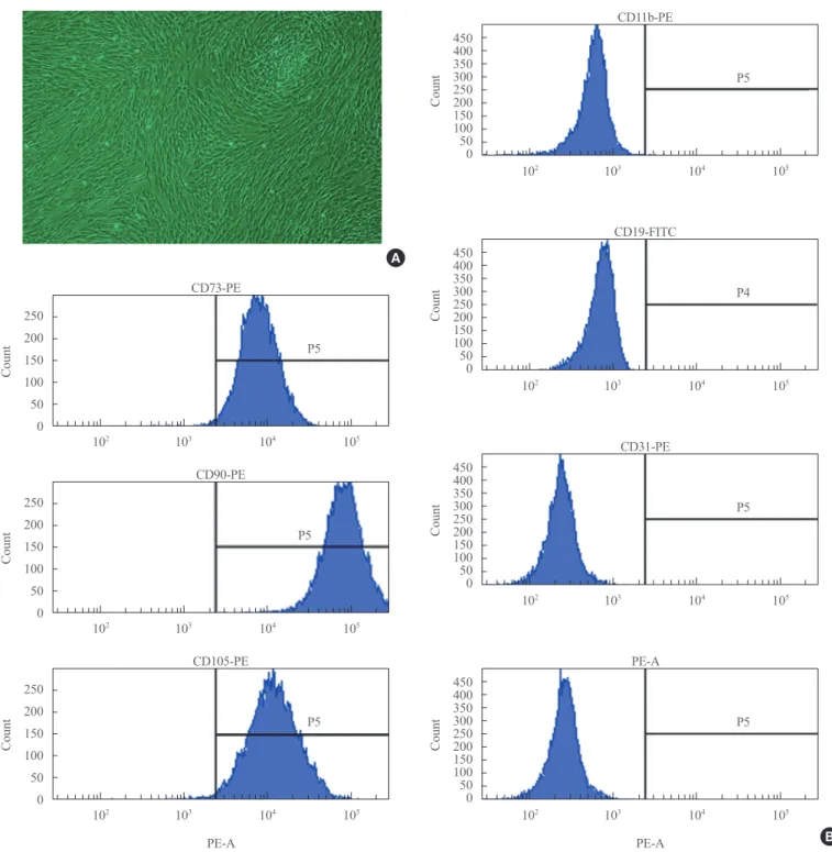
Flow cytometric surface marker expression analysis ADSCs exhibited fibroblast-like morphology and adhered to plastic (Fig. 1A). To confirm their multipotent differentiation potential, specific surface antigen expression was assessed ac- cording to the minimal criteria established by the International Society for Cellular Therapy [12]. The cells showed positive expression (≥95%) of mesenchymal stem cell (MSC)-specific antigen markers, including CD90, CD73, and CD105. Con- versely, expression of CD11b (a hematopoietic cell marker prominently expressed on monocytes and macrophages), CD19 (a B cell marker), CD31 (a hematopoietic cell marker ex- pressed on the surfaces of platelets, monocytes, and neutro- phils), CD34 (a marker for primitive hematopoietic progeni- tors and endothelial cells), and HLA-DR was not detected (Fig. 1B).
Osteoblastic differentiation of ADSCs
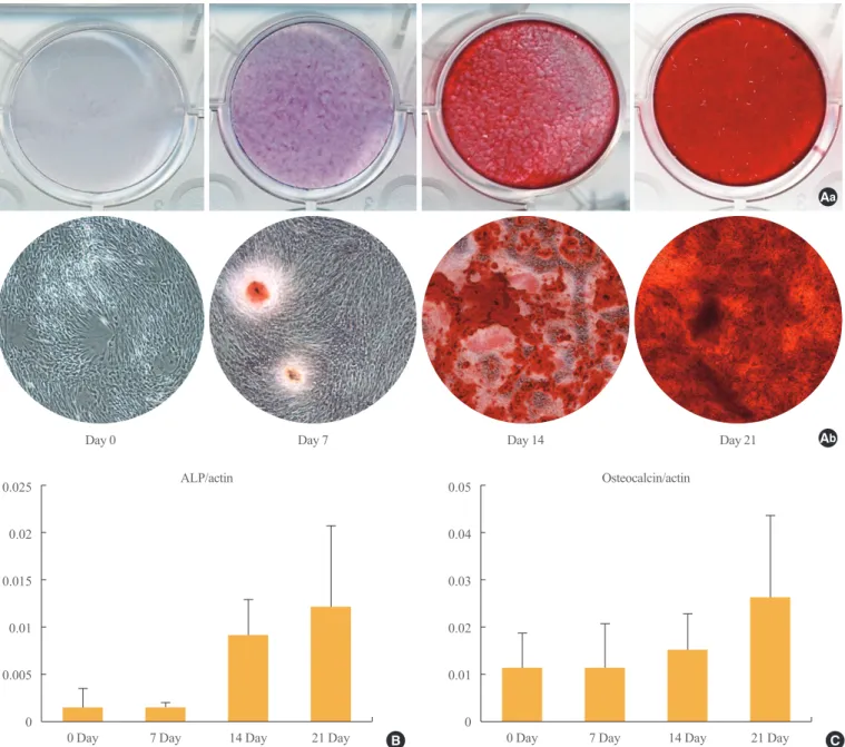
Osteogenic differentiation was confirmed by cytochemical staining and gene expression analysis. After 21 days of osteo- genic induction, positive Alizarin red S staining consistent with matrix calcification was observed (Fig. 2A). ALP is a com- monly used marker for early osteogenic differentiation. In our study, ALP was notably upregulated at the 21 days (Fig. 2B).
Expression of the late osteogenic marker OC is known to be upregulated immediately prior to mineralization, and we ob- served significant increases in OC mRNA levels by day 21 of differentiation (Fig. 2C).
Assessment of GLP-1R expression by Western blotting and PCR
Up-regulation of GLP1-R protein expression during the osteo- blastic developmental sequence was confirmed by Western blot data for day 21 (Fig. 3A). Real-time PCR was used to as- sess the time-course of GLP-1R mRNA expression during os- teogenic differentiation. For each time-point (0, 7, 14, and 21 days), the GLP-1R mRNA level of each sample was normal- ized to that of β-actin. The mean GLP-1R activity of ADSCs increased significantly in a time-dependent manner that was related to the degree of differentiation (P=0.013) (Fig. 3B).
DISCUSSION
In this study, we demonstrated that GLP-1R is expressed on ADSC-derived osteogenic cells, and that the level of expres-
sion increases in a time-dependent manner during differentia- tion. GLP-1 has been consistently reported to have a beneficial effect on bone in rodents [2,3]. Also, it is reported to have no adverse effects on bone, despite inducing weight loss in hu- mans [13]. A GLP-1R agonist has been reported to prevent os-
teopenia by promoting bone formation and by suppressing bone resorption in aged, ovarectomised rats [14]. Furthermore, a GLP-1 agonist increased bone mineral density in type 2 dia- betic rats [15]. The mechanism underlying this phenomenon is not clear. One possible explanation is that GLP-1Rs expressed
A
Fig. 1. Adipose-derived stem cells (ADSCs). (A) ADSCs exhibited fibroblast-like morphology and adhered to plastic, as assessed by light microscopy (×40) at passage three. (B) Flow cytometry using mesenchyme-specific antigen markers showed that ADSCs express CD73, CD90, and CD105, but not CD11b, CD19, CD31, and HLA-DR.
B 250
200 150 100 50 0
Count
102 103 104 105
CD73-PE
P5
450400 350300 250200 150100 500
450400 350300 250200 150100 500
450400 350300 250200 150100 500
450400 350300 250200 150100 500
CountCountCountCount
102
102
102
102
103
103
103
103
104
104
104
104
105
105
105
105 CD11b-PE
CD19-FITC
CD31-PE
PE-A
P5
P4
P5
P5 250
200 150 100 50 0
Count
102 103 104 105
CD90-PE
P5
250 200 150 100 50 0
Count
102 103 104 105
CD105-PE
PE-A
P5
PE-A
on thyroid C cells promote calcitonin secretion, which inhibits bone resorption; however, GLP-1R was not detected in osteo- blasts [16]. A second possibility is the presence of a functional receptor independent of the cAMP-linked GLP-1R. A final possibility is the existence of a GLP-1R.
The downstream effect of GLP-1 is mediated by a G-pro- tein-coupled receptor that is expressed in various tissues, such as the pancreas, stomach, and vascular system [17]. Expres- sion of GLP-1R by osteoblasts, however, has not been con-
firmed. GLP-1R has been reported to be expressed on thyroid C cells, which affect bone resorption by promoting calcitonin secretion, but the study did not detect GLP-1R expression in osteoblasts [16]. Conversely, expression of GLP-1R was re- ported in MC3T3-E1 cells, a well-known mouse osteoblastic cell line [4]. The authors suggested that this result indicates that, like other classic receptors, GLP-1R ha nd temperature- dependent. Another study reported that GLP-1R is expressed by osteoblastic cell lines, but that the expression level is cell
Day 21
ALP/actin Osteocalcin/actin
Ab Aa
Day 0 Day 7 Day 14
Fig. 2. Osteogenic differentiation of adipose-derived stem cells. (A) Calcification, detected by Alizarin red staining and indicative of mineralization, was assessed by (Aa) gross appearance and (Ab) light microscopy (×40) at days 0, 7, 14, and 21. Real-time polymerase chain reaction showed increased expression of (B) alkaline phosphatase (ALP) and (C) osteocalcin mRNA at day 21. Data are presented as means±standard deviation.
0.025 0.02 0.015 0.01 0.005 0
0.05 0.04 0.03 0.02 0.01
0 Day 7 Day 14 Day 21 Day B 0 0 Day 7 Day 14 Day 21 Day C
line-dependent, perhaps reflecting different stages of osteo- blast differentiation [5]. In that study, GLP-1R was expressed by young osteoblasts but not by mature osteoblasts derived from the Saos-2 osteosarcoma cell line. It was suggested that GLP-1R is expressed by osteoblasts, but that the expression decreases with maturation. However, GLP-1R expression was recently reported in osteocytes, which are derived from osteo- blasts [15]. In addition, GLP-1R expression may differ among species [18,19].
We also evaluated the levels of OC and ALP in our ADSC samples. One of the most common methods used to examine mineralization is Alizarin red S staining, which indicates ex- tracellular calcification. Alizarin red staining alone, however, may not be sufficient to confirm osteogenic differentiation [20,21]. ALP is a well-known indicator of the early stage of os- teogenic differentiation of MSCs, and expression of ALP in- creases with osteoblast maturation [22]. In this study, ALP levels increased in a time-dependent manner during osteogen- ic differentiation of ADSCs. OC is a noncollagenous protein found in bone and dentin and is generally considered to be a relatively late-stage marker for the period of osteoblastic dif- ferentiation immediately prior to mineralization [23,24]. In our study, expression of both ALP and OC was elevated on day 21 of differentiation, confirming that osteogenic differen- tiation of ADSCs had occurred.
In summary, we demonstrated GLP-1R expression during osteogenic differentiation of ADSCs. Even though it is not yet clear whether GLP-1 has a role in bone metabolism, our results
suggest that it may induce osteogenic differentiation in bone tissue. Further studies are needed to explore this possibility.
CONFLICTS OF INTEREST
No potential conflict of interest relevant to this article was re- ported.
ACKNOWLEDGMENTS
This study was supported by a Biomedical Research Institute Grant (2013-10) from Pusan National University Hospital.
REFERENCES
1. Drucker DJ. Glucagon-like peptides: regulators of cell pro- liferation, differentiation, and apoptosis. Mol Endocrinol 2003;17:161-71.
2. Nuche-Berenguer B, Lozano D, Gutierrez-Rojas I, Moreno P, Marinoso ML, Esbrit P, Villanueva-Penacarrillo ML.
GLP-1 and exendin-4 can reverse hyperlipidic-related os- teopenia. J Endocrinol 2011;209:203-10.
3. Nuche-Berenguer B, Moreno P, Esbrit P, Dapia S, Caeiro JR, Cancelas J, Haro-Mora JJ, Villanueva-Penacarrillo ML. Effect of GLP-1 treatment on bone turnover in nor- mal, type 2 diabetic, and insulin-resistant states. Calcif Tis- sue Int 2009;84:453-61.
4. Nuche-Berenguer B, Portal-Nunez S, Moreno P, Gonzalez
GLP-1R expression (×104)
6 5 4 3 2 1
0 0 Day 7 Day 14 Day 21 Day
a
B Fig. 3. Expression of glucagon-like peptide 1 receptor (GLP-1R) during osteogenic differentiation of adipose-derived stem cells (ADSCs).
(A) Western blotting for GLP-1R in differentiating ADSCs at days 0, 7, 14, and 21 after differentiation. Data are presented as means±
standard deviation. (B) The expression level of GLP-1R mRNA increased significantly in a time-dependent manner during osteogenic dif- ferentiation (P=0.013). GAPDH, glyceraldehyde 3-phosphate dehydrogenase. aP<0.05.
0 Day 7 Day 14 Day 21 Day A
GLP-1R 53 KDa
GAPDH 37 KDa
N, Acitores A, Lopez-Herradon A, Esbrit P, Valverde I, Villanueva-Penacarrillo ML. Presence of a functional re- ceptor for GLP-1 in osteoblastic cells, independent of the cAMP-linked GLP-1 receptor. J Cell Physiol 2010;225:
585-92.
5. Pacheco-Pantoja EL, Ranganath LR, Gallagher JA, Wilson PJ, Fraser WD. Receptors and effects of gut hormones in three osteoblastic cell lines. BMC Physiol 2011;11:12.
6. Zuk PA, Zhu M, Ashjian P, De Ugarte DA, Huang JI, Mizuno H, Alfonso ZC, Fraser JK, Benhaim P, Hedrick MH. Human adipose tissue is a source of multipotent stem cells. Mol Biol Cell 2002;13:4279-95.
7. Rodriguez AM, Elabd C, Amri EZ, Ailhaud G, Dani C.
The human adipose tissue is a source of multipotent stem cells. Biochimie 2005;87:125-8.
8. Gimble JM, Guilak F. Differentiation potential of adipose derived adult stem (ADAS) cells. Curr Top Dev Biol 2003;
58:137-60.
9. Witkowska-Zimny M, Walenko K. Stem cells from adi- pose tissue. Cell Mol Biol Lett 2011;16:236-57.
10. Izadpanah R, Trygg C, Patel B, Kriedt C, Dufour J, Gimble JM, Bunnell BA. Biologic properties of mesenchymal stem cells derived from bone marrow and adipose tissue. J Cell Biochem 2006;99:1285-97.
11. Cowan CM, Shi YY, Aalami OO, Chou YF, Mari C, Thom- as R, Quarto N, Contag CH, Wu B, Longaker MT. Adi- pose-derived adult stromal cells heal critical-size mouse calvarial defects. Nat Biotechnol 2004;22:560-7.
12. Dominici M, Le Blanc K, Mueller I, Slaper-Cortenbach I, Marini F, Krause D, Deans R, Keating A, Prockop Dj, Hor- witz E. Minimal criteria for defining multipotent mesen- chymal stromal cells. The International Society for Cellular Therapy position statement. Cytotherapy 2006;8:315-7.
13. Blonde L, Klein EJ, Han J, Zhang B, Mac SM, Poon TH, Taylor KL, Trautmann ME, Kim DD, Kendall DM. Interim analysis of the effects of exenatide treatment on A1C, weight and cardiovascular risk factors over 82 weeks in 314 overweight patients with type 2 diabetes. Diabetes Obes Metab 2006;8:436-47.
14. Ma X, Meng J, Jia M, Bi L, Zhou Y, Wang Y, Hu J, He G, Luo X. Exendin-4, a glucagon-like peptide-1 receptor ago-
nist, prevents osteopenia by promoting bone formation and suppressing bone resorption in aged ovariectomized rats. J Bone Miner Res 2013;28:1641-52.
15. Kim JY, Lee SK, Jo KJ, Song DY, Lim DM, Park KY, Bone- wald LF, Kim BJ. Exendin-4 increases bone mineral den- sity in type 2 diabetic OLETF rats potentially through the down-regulation of SOST/sclerostin in osteocytes. Life Sci 2013;92:533-40.
16. Yamada C, Yamada Y, Tsukiyama K, Yamada K, Udagawa N, Takahashi N, Tanaka K, Drucker DJ, Seino Y, Inagaki N. The murine glucagon-like peptide-1 receptor is essen- tial for control of bone resorption. Endocrinology 2008;
149:574-9.
17. Holst JJ. The physiology of glucagon-like peptide 1. Physi- ol Rev 2007;87:1409-39.
18. Bjerre Knudsen L, Madsen LW, Andersen S, Almholt K, de Boer AS, Drucker DJ, Gotfredsen C, Egerod FL, Hegelund AC, Jacobsen H, Jacobsen SD, Moses AC, Molck AM, Nielsen HS, Nowak J, Solberg H, Thi TD, Zdravkovic M, Moerch U. Glucagon-like Peptide-1 receptor agonists acti- vate rodent thyroid C-cells causing calcitonin release and C-cell proliferation. Endocrinology 2010;151:1473-86.
19. Hirsch PF, Baruch H. Is calcitonin an important physiolog- ical substance? Endocrine 2003;21:201-8.
20. Boyan BD, Schwartz Z, Boskey AL. The importance of mineral in bone and mineral research. Bone 2000;27:341-2.
21. Declercq HA, Verbeeck RM, De Ridder LI, Schacht EH, Cornelissen MJ. Calcification as an indicator of osteoin- ductive capacity of biomaterials in osteoblastic cell cul- tures. Biomaterials 2005;26:4964-74.
22. Aubin JE, Liu F, Malaval L, Gupta AK. Osteoblast and chon- droblast differentiation. Bone 1995;17(2 Suppl):77S-83S.
23. Aronow MA, Gerstenfeld LC, Owen TA, Tassinari MS, Stein GS, Lian JB. Factors that promote progressive devel- opment of the osteoblast phenotype in cultured fetal rat calvaria cells. J Cell Physiol 1990;143:213-21.
24. Koshihara Y, Kawamura M, Endo S, Tsutsumi C, Kodama H, Oda H, Higaki S. Establishment of human osteoblastic cells derived from periosteum in culture. In Vitro Cell Dev Biol 1989;25:37-43.