24
Assessment of Respiratory Tumor Movement using 4D Computed Tomography for Stereotactic Radiosurgery in Lung Tumor
Purpose: To assess the respiratory tumor movement using 4D-CT (4-dimensio- nal computed tomography) for minimizing setup and target volume uncertainty of body-frame based stereotactic radiosurgery (SRS) in lung tumor. Materials and Methods: Fifty-seven stereotactic radiation therapies with respiratory gating system in 44 patients (two targets in seven patients and three in three patients) were executed in Asan Medical Center from May 2005 to June 2006.
We used respiratory gating system consisted of RPM (Real-time Positioning Management system, Varian, USA) and 4D-CT (GE healthcare, USA), if tumor movement was exceeding 5 mm by respiration on fluoroscopy. Accurate tumor movement on reconstructed 4D-CT image was determined for respiratory gated therapy. Respiratory gated therapy was done if tumor movement was exceeding 5 mm, and non-gated therapy was done if it was below 5 mm.
Results: Forty-five tumors were treated with supine position, and the other twelve were with prone position. Median tumor movement (3-dimensional) by respiration was 8.78±5.30 mm, and it was mostly affected by superior-inferior movement (8.53±5.23 mm). Tumor movements were different by tumor location, whether upper (5.38±2.85 mm) or lower (10.12±5.08 mm) lobe (p=0.015). Tumor movement was exceeding 5 mm in 27 (47.3%) tumors, and below 5 mm in 30 tumors in 4D-CT evaluation. Tumor movements on adopted respiratory gated phase were wholly below 5 mm, and its median value was 3.70±1.13 mm. Conclusion: Assessment of respiratory tumor movement using 4D-CT and gating system was helpful for minimizing target volume uncertainty.
As a result, image-guided radiation therapy could improve the treatment accuracy of high precision stereotactic radiosurgery. (J Lung Cancer 2007;
6(1):24 28)
Key Words: Respiratory tumor movement, 4D-CT, Respiratory gated radiation therapy
Si Yeol Song, M.D.1 Sung Ho Park, Ph.D.1 Sang Min Yoon, M.D.1 Young Seok Kim, M.D.1 Jong Hoon Kim, M.D.1 Seung Do Ahn, M.D.1 Seong Soo Shin, M.D.1 Sang-wook Lee, M.D.1 Charn Il Park, M.D.2 and Eun Kyung Choi, M.D.1
Department of Radiation Oncology,
1Asan Medical Center, College of Me- dicine, University of Ulsan, Seoul, Korea
2Seoul National University Hospital, Seoul, Korea
Received: June 7, 2007 Accepted: June 18, 2007
Address for correspondence Eun Kyung Choi, M.D.
Department of Radiation Oncology, Asan Medical Center, College of Me- dicine, University of Ulsan, 388-1, Pungnap 2-dong, Songpa-gu, Seoul 138-736, Korea
Tel: 82-2-3010-4432 Fax: 82-2-486-7258 E-mail: [email protected]
서 론
정위적 방사선수술요법(Stereotactic Radiosurgery; SRS)은 주위 정상 조직의 손상을 최소화하면서 종양에만 국한하여 고 선량의 방사선을 조사하여 무혈적으로 종양을 제거하는 방법이며, 최근 뇌 종양, 폐 종양, 간 종양의 치료에서 점점 영역을 넓혀가고 있는 치료 방법이다(1). 병기 1기의 초기 폐암 또는 단일(solitary) 전이성 폐 종양의 치료는 수술이 가장 좋은 치료 방법이나 고령 또는 내과적 질환 등의 이유 로 수술이 불가능한 경우에는 정위적 방사선술이 시행되고
있으며 좋은 결과가 보고 되고 있다(2∼6).
정위적 방사선수술은 적은 용적의 종양에 많은 양의 방 사선을 집중적으로 조사하여 치료하는 방법이므로 높은 정 확성이 요구되며, 정상 조직의 파괴를 최소화하기 위하여 종양의 위치와 움직임을 정확하게 파악하는 것이 중요하지 만 예전부터 사용된 투시영상(fluoroscopy)은 많은 한계가 있었던 것이 사실이다. 특히 폐 종양은 호흡에 의한 종양의 움직임으로 인하여 정위적 방사선수술의 정확성에 한계점 이 있었다. 이전부터 호흡으로 인한 폐 종양의 움직임을 최 소화하기 위하여 다양한 방법이 연구되었으며 ABC (Active
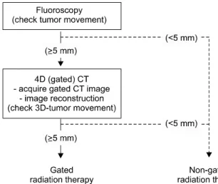
Fig. 1. Treatment flow: gated radiation therapy.
Breathing Control), AIB (Air-injected Blanket), Diaphragm Controller 등의 여러 방법(7∼9)들이 모색되었으나 모두 환 자의 상태와 협조여부에 따라서 효과에 큰 차이가 있어서 보편적으로 이용하기에는 어려운 점이 있었다. 이러한 단 점을 극복하기 위하여 최근에는 영상획득 단계에서 호흡에 의한 종양의 움직임을 관찰하고 호흡주기를 선택할 수 있 는 4차원 전산화단층 촬영(4D-CT) 장비와 호흡감시장치가 도입되어 방사선치료에 이용되고 있으며, 이전 장비와의 가장 큰 차이점은 환자가 정상적인 호흡을 하면서 선택적 인 호흡주기의 영상주기만을 획득하여 치료하므로 환자에 대한 인위적인 조작이 필요 없어 재현성과 보편성이 월등 하다는 점이다(10∼18). 4D-CT를 이용한 호흡동기 방사선 치료(Respiratory Gated Radiation Therapy)를 시행하여 얻을 수 있는 장점으로는 첫째, 호흡에 따른 종양의 움직임을 정 확히 파악하여 치료의 정확성을 확인할 수 있고, 둘째, 호흡 주기를 선택적으로 획득하여 치료범위를 최소화함으로써 정상 폐 조직의 손상을 최소화 할 수 있다는 것이다. 저자들 은 본 연구에서 4D-CT를 실제 임상적으로 폐 종양에 대한 정위적 방사선치료에 도입하여 종양의 위치에 따른 정확한 폐 종양의 움직임을 확인하고, 정위적 방사선치료를 위한 방사선치료의 범위를 최소화할 수 있는지에 대하여 알아보 고자 하였다.
대상 및 방법
1) 4 dimensional computed tomography (4D-CT)
호흡주기에 따른 영상을 획득하기 위하여 GE 사의 LightSpeed 4D-CT/RT (4 dimensional computed tomography/
radiation therapy)가 이용되었으며, 환자의 호흡을 감시하여 4D-CT에 호흡주기를 전달하기 위한 호흡추적장치로 Real- time Positioning Management system (RPM, Varian, USA)가 4D-CT에 부착되었다. 호흡주기 영상을 재구성하기 위하여 Advantage 4D software (GE, WC, USA)가 사용되었다.
2) 호흡동기영상(Respiratory Gated Image) 획득
4D-CT 촬영 전 RPM을 통하여 환자의 호흡주기를 관찰 하고 결정된 호흡주기 내에서 10 sets의 CT 영상을 얻었다.
Advantage 4D software를 이용하여 CT 영상을 최대 흡기 (full inspiration)에 해당하는 영상을 0, 90% phase에 각각 할 당하고 최대 호기(full expiration)에 해당하는 영상을 50%
phase에 할당하는 방식으로 전체 호흡주기(full respiration) 영상을 총 10 phase로 구분하였다. 호흡주기에 따른 각 phase의 영상을 3차원적으로 재구성하여 실제 호흡에 따른
종양의 움직임을 동영상으로 만들었으며, 이 영상에서 호 흡에 따른 종양의 움직임을 측정하였다.
3) 종양의 움직임 측정
호흡에 따른 종양의 움직임은 x, y, z 축 방향과 3차원 재 구성영상에서 3차원(3-dimensional) 방향의 움직임을 측정 하였다. Advantage 4D software 상에서 최대호기 영상인 영 상과 최대 흡기 영상 간의 종양의 위치를 지정하여 호흡에 따른 움직임을 측정하였으며, 1명의 물리학자와 1명의 방 사선종양학과 의사가 각각 측정한 후 결과를 취합하여 측 정결과를 분석하였다.
4) 호흡동기 정위적 방사선수술(Respiratory Gated Ste- reotactic Radiosurgery)
4D-CT를 시행하기 전에 투시영상 (fluoroscopy)을 통하여 종양의 x, y, z 축 방향의 움직임을 미리 측정한 후 가장 움 직임이 큰 방향의 움직임이 5 mm 미만인 경우에는 4D-CT 를 촬영하지 않고 spiral CT를 시행하여 영상을 얻었으며, 5 mm 이상의 움직임이 관찰되는 경우에는 RPM과 4D-CT 를 시행하여 4차원 영상을 획득하여 위에서 언급된 방법으 로 재구성을 한 후 다시 종양의 움직임을 측정하였다.
4D-CT 영상에서 재확인하여 5 mm 이상의 움직임을 보이는 경우에만 호흡동기 방사선치료를 시행하였다(Fig. 1).
정위적 방사선수술은 4일간 연속적으로 시행하였으며, 매 번 12 Gy의 방사선을 조사하여 총 48 Gy의 방사선을 종양 에 조사하였다. 치료의 정확성을 위하여 vacuum cushion이 부착된 전신 정위적 고정 틀(Stereotactic body frame, Elekta, USA)에 환자를 눕히고 환자의 자세를 고정하였다. Planning
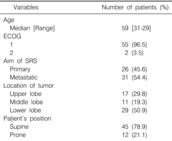
Table 1. Patient Characteristics
Variables Number of patients (%)
Age
Median [Range] 59 [31-29]
ECOG
1 55 (96.5)
2 2 (3.5)
Aim of SRS
Primary 26 (45.6)
Metastatic 31 (54.4)
Location of tumor
Upper lobe 17 (29.8)
Middle lobe 11 (19.3)
Lower lobe 29 (50.9)
Patient’s position
Supine 45 (78.9)
Prone 12 (21.1)
Table 2. Tumor Movement and Treatment Method Tumor Number of
movement (mm) p value tumors
(mean±SD) Fluoroscopy (z-axis)
Group 1* 5 3.40±2.70
Group 2† 25 9.28±2.81
Group 3‡ 27 13.06±5.79
<0.001§ 4D-CT (3-dimension)
Group 2 25 5.67±2.18
Group 3 27 11.75±5.62
*Tumor movement<5 mm on fluoroscopy, 4D-CT (), no gated therapy, †Tumor movement ≥ 5 mm on fluoroscopy, but<5 mm on 4D-CT, no gated therapy, ‡Tumor movement≥
5 mm on both fluoroscopy and 4D-CT, gated therapy,
§one-way ANOVA test, SD: standard deviation target volume (PTV)는 5 mm 미만의 작은 움직임을 보인 경
우에는 x, y 축으로는 5 mm 그리고 z 축으로는 7.5 mm의 margin을 두고 치료를 시행하였다. 4D-CT영상에서 5 mm 이상의 비교적 큰 움직임이 관찰된 경우에는 10 phase의 호 흡영상 중 호기(expiration)에 해당하는 30∼70% 영상만을 3차원적으로 재구성하여 최대호기 상태인 50% phase의 영 상에서 30% 또는 70%의 영상까지의 종양의 움직임을 측정 하여 PTV margin을 고려하거나, 또는 30∼70% 영상을 최대 강도투영영상(Maximum Intensity Projection Image: MIP ima- ge)을 생성하여 setup 오차만을 고려한 PTV margin을 두는 것으로 하였다.
5) 대상환자
병기 1기의 원발성 폐암 또는 전이성 폐 종양으로 SRS를 시행 받은 환자를 대상으로 분석하였다. 일반적인 방사선 치료와 비교하여 비교적 장시간의 고정자세가 필요한 것을 고려하여 환자의 활동성(performance)은 ECOG performance scale 2 이하인 경우로 제한하였으며, 의료진의 지시에 따라 규칙적인 호흡을 할 수 있는 환자를 대상으로 하였다. 정위 적 방사선수술을 위하여 종양의 크기는 5 cm 미만으로 제 한하였다.
결 과 1) 환자 특성
2005년 5월부터 2006년 6월까지 서울아산병원에서 SRS를 시행 받은 1기 병기의 폐암 또는 전이성 폐 종양 환자 44명 에서 57개의 종양을 대상으로 분석을 시행하였다. 7명의 환
자에서 2개, 3명의 환자에서는 3개의 폐 종양에 대하여 SRS 를 시행하였다. 환자의 중앙연령은 59세였으며, 범위는 31∼
79세였다. 대부분의 환자의 활동성은 ECOG 1이었으며, 2명 의 환자만이 ECOG 2 상태였다. SRS의 목적은 26개의 종양 에서 1기 병기의 폐암에 대한 완치 목적의 치료였고, 31개 의 종양에서는 원격전이에 따른 고식적 목적의 치료로 분 석되었다. 종양의 크기는 중앙값이 17 mm이었으며, 범위는 9∼42 mm이었다. 종양의 위치 별로 분석해 보면 폐 상엽 (upper lobe)이 17개(29.8%), 폐 중엽(middle lobe)이 11개 (19.3%), 그리고 폐 하엽(lower lobe)이 29개(50.9%)로 조사 되었으며, 치료를 위한 환자의 자세는 대부분이 앙와위 (supine) 자세였으며, 12개(21.1%)에서는 호흡에 따른 종양 의 움직임을 최소화하거나 또는 방사선조사의 편의를 위하 여 배위(prone) 자세를 취하였다(Table 1).
2) 종양의 움직임 측정
4D-CT 시행 전 투시영상(fluoroscopy)를 이용하여 측정한 결과 5개 (8.8%)의 종양에서는 5 mm 미만의 움직임을 보여 spiral CT를 시행하였으며(Group 1), 25개(43.9%)의 종양에 서는 5 mm 이상의 움직임이 관찰되어 4D-CT를 시행하였으 나 재구성된 CT 영상에서는 5 mm 미만으로 움직임이 측정 되어 호흡동기 방사선치료는 시행하지 않았다(Group 2). 나 머지 27개(47.3%)의 종양에서는 5 mm 이상의 움직임이 관 찰되어 호흡동기방사선치료를 시행하였다(Group 3). 각 군 의 종양의 움직임은 Table 2에 보여지는 바와 같으며 Group 2에서는 투시영상과 4D-CT 영상에서 측정된 종양의 움직 임에 차이가 있는 것을 알 수 있었다. 4D-CT를 시행하였던 52개의 종양에서 전체 호흡에서의 종양의 움직임을 보면
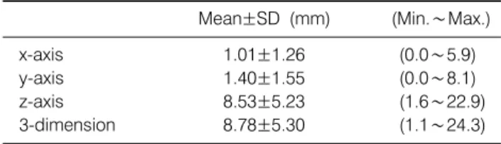
Table 3. Tumor Movement with Full Respiration
Mean±SD (mm) (Min.∼Max.)
x-axis 1.01±1.26 (0.0∼5.9)
y-axis 1.40±1.55 (0.0∼8.1)
z-axis 8.53±5.23 (1.6∼22.9)
3-dimension 8.78±5.30 (1.1∼24.3)
SD: standard deviation
Table 4. Tumor Movement by Location of Tumor with Full Respiration (3-dimension)
Mean±SD (mm) p value
Upper lobe 5.38±2.85
Middle lobe 10.01±6.81 0.015*
Lower lobe 10.12±5.08
*one-way ANOVA test, SD: standard deviation
x 축으로는 1.01±1.26 mm, y 축으로는 1.40±1.55 mm, z 축으 로는 8.53±5.23 mm의 움직임이 관찰 되었고, 3차원 재구성 영상에서의 3차원적인 움직임은 8.78±5.30 mm이었다(Table 3). 결과적으로 종양의 움직임 중 대부분의 요소는 z 축 (longitudinal)으로의 움직임이라는 것을 CT 영상을 통하여 확인할 수 있었으며, 치료방법의 기준도 z 축 방향의 움직 임으로 정하였다. 종양의 폐엽 위치에 따라서도 움직임의 차이를 평가해 보았으며 폐 상엽의 경우 5.38±2.85 mm (3-di- mensional)의 움직임을 보여 폐 중엽 또는 폐 하엽의 종양과 는 움직임에 차이가 있는 것을 알 수 있었다(Table 4).
3) 호흡동기 방사선치료
Group 3에 해당하는 27개의 종양에 대해서는 모두 호흡 동기방사선치료를 시행하였으며 앞서 기술한 대로 종양의 움직임을 최소화하기 위하여 4D-CT 재구성 영상에서 호기 에 해당하는 30∼70% 영상만을 선택적으로 재구성하여 종 양의 움직임을 측정하였으며, 측정결과에 따라 PTV margin 을 조정하였다. 호흡동기를 하지 않은 전체호흡(full respi- ration)에서는 11.75±5.62 mm의 움직임을 보였으나 30∼
70%로 제한된 호흡에서는 3.70±1.13 mm의 움직임만 있는 것으로 측정되었으며, 통계적으로 의미 있는 차이를 보였 다(p<0.001, one-way ANOVA).
고안 및 결론
폐 종양의 방사선치료에서 가장 큰 문제점은 호흡에 의
하여 발생하는 종양의 움직임으로 인한 치료의 부정확성과 실제 방사선치료의 용적이 되는 PTV가 커진다는 것이다.
1기 병기의 폐암환자에서 수술을 받지 않고 정위적 방사선 수술을 받는 환자는 폐기능 또는 내과적인 질환의 문제가 있는 경우가 대부분이며, 폐 전이로 SRS를 받는 경우에도 전신기능의 저하를 동반하고 있는 경우가 많다. 따라서 일 반적인 방사선치료에 비하여 SRS의 경우에는 정상 폐 조직 의 손상을 최소화하는 것이 더욱 필요하며, 동시에 5 cm 미 만의 작은 종양에 대하여 고 선량의 방사선을 조사하므로 치료의 정확성이 중요한 문제이다. 4D-CT의 가장 큰 장점 은 호흡에 따른 종양의 움직임을 CT 영상을 이용하여 직접 확인할 수 있어 각각의 종양에 대한 ITV (internal target volume)를 정확하게 측정할 수 있으며, 결과적으로 PTV에 는 setup 오차만을 고려하면 된다는 것이다. 이전에는 투시 영상에서 확인한 대략적인 움직임만으로 ITV를 고려한 후 PTV를 설정함으로써 불확실성을 감안하여 PTV 용적이 종 양의 크기에 비하여 과도하게 커지는 경우가 많았으나, ITV를 정확하게 설정함으로써 치료의 불확실성을 없앨 수 있게 되었다. Underberg 등(19)의 보고에 따르면 종양에 일 반적으로 10 mm margin을 두는 것과 비교하면 4D-CT를 이 용하여 호흡동기방사선치료를 시행하였을 때 PTV 용적을 33.3%까지 감소시킬 수 있는 것으로 보고 하였고, 동시에 정상 폐 조직에 조사되는 방사선량도 39.1%까지 줄여 줄 수 있다고 보고 하였다. 결과적으로 폐 종양의 치료에서 모 든 환자에 동일하게 10 mm margin을 두고 치료하는 것은 불필요하게 방사선을 조사 받는 폐 조직이 늘어나게 하여 폐 독성을 증가시킨다고 주장하였다. Liu 등(20)은 III 또는 IV 병기의 비소세포성 폐암환자에서 166개의 종양을 분석 하여 호흡에 따른 종양의 움직임의 대부분의 요소는 횡격 막의 운동에 의한 상하(superior-inferior) 움직임이며, 대부분 의 경우 정상호흡에서 10 mm 이상의 움직임을 보이지는 않는 것으로 보고하였고, 95%의 환자에서 종양의 상하 움 직임이 13.4 mm 미만이라고 주장하였다.
본 연구의 결과에서도 보면 x축(좌-우), y축(전-후) 움직 임은 평균값이 1 mm 내외로 움직임이 거의 없는 것을 관찰 할 수 있었고, z축(상-하) 움직임은 평균값이 8.53 mm로 호 흡에 의한 움직임의 거의 대부분을 차지하는 것을 알 수 있었다. 횡경막과 가까운 폐 하엽에 위치한 종양일수록 움 직임이 커져서 10.12 mm의 평균적인 움직임이 있는 것을 확인할 수 있었으며, 투시영상을 통하여 종양의 움직임을 판단하는 것은 실제 결과와는 다를 수도 있음을 확인할 수 있었다.
4D-CT를 이용하여 종양의 움직임을 파악하는 것은 정위
적 방사선수술의 정확성을 높여 줄 수 있으며, 호흡동기 방 사선치료는 선택된 호흡주기에서 종양의 움직임을 최소화 할 수 있어 고 선량의 방사선에 따른 폐 손상을 최소화할 수 있을 것으로 기대된다.
REFERENCES
1. Wulf J, Hadinger U, Oppitz U, et al. Stereotactic radiotherapy of targets in the lung and liver. Strahlenther Onkol 2001;
177:645-655.
2. Nagata Y, Negoro Y, Aoki T, et al. Clinical outcomes of 3D conformal hypofractionated single high-dose radiotherapy for one or two lung tumors using a stereotactic body frame. Int J Radiat Oncol Biol Phys 2002;52:1041-1046.
3. Nakagawa K, Aoki Y, Tago M, et al. Megavoltage CT-assisted stereotactic radiosurgery for thoracic tumors: original research in the treatment of thoracic neoplasms. Int J Radiat Oncol Biol Phys 2000;48:449-457.
4. Hof H, Herfarth KK, Munter M, et al. Stereotactic single-dose radiotherapy of stage I non-small-cell lung cancer (NSCLC).
Int J Radiat Oncol Biol Phys 2003;56:335-341.
5. Timmerman R, Papiez L, McGarry R, et al. Extracranial ste- reotactic radioablation: results of a phase I study in medically inoperable stage I non-small cell lung cancer. Chest 2003;
124:1946-1955.
6. SM Yoon, EK Choi, SW Lee, et al. Clinical results of ste- reotactic body frame based fractionated radiation therapy for primary or metastatic thoracic tumors. Acta Oncol 2006;45:
1108-14.
7. Dawson LA, Brock KK, Kazanjian S, et al. The reproducibility of organ position using active breathing control (ABC) during liver radiotherapy. Int J Radiat Oncol Biol Phys 2001;51:
1410-1421.
8. Wilson EM, Williams FJ, Lyn BE, et al. Validation of active breathing control in patients with non-small-cell lung cancer to be treated with CHARTWEL. Int J Radiat Oncol Biol Phys 2003;57:864-874.
9. YL Suh, BY Yi, SD Ahn, et al. A feasibility study on the abdomen immobilization with air injected balloon blanket. Kor J Med Phys 2002;13:176-180.
10. Shirato H, Shimizu S, Kitamura K, et al. Four-dimensional treatment planning and fluoroscopic real-time tumor tracking radiotherapy for moving tumor. Int J Radiat Oncol Biol Phys 2000;48:435-442.
11. Shirato H, Shimizu S, Kunieda T, et al. Physical aspects of a real-time tumor-tracking system for gated radiotherapy. Int J Radiat Oncol Biol Phys 2000;48:1187-1195.
12. Underberg RW, Lagerwaard FJ, Cuijpers JP, et al. Four-di- mensional CT scans for treatment planning in stereotactic radiotherapy for stage I lung cancer. Int J Radiat Oncol Biol Phys 2004;60:1283-1290.
13. Lu W, Ruchala KJ, Chen ML, et al. Real-time respiration mo- nitoring using the radiotherapy treatment beam and four-di- mensional computed tomography (4DCT)--a conceptual study.
Phys Med Biol 2006;51:4469-4495.
14. Rietzel E, Liu AK, Doppke KP, et al. Design of 4D treatment planning target volumes. Int J Radiat Oncol Biol Phys 2006;
66:287-295.
15. Slotman BJ, Lagerwaard FJ, Senan S. 4D imaging for target definition in stereotactic radiotherapy for lung cancer. Acta Oncol 2006;45:966-972.
16. Britton KR, Starkschall G, Tucker SL, et al. Assessment of gross tumor volume regression and motion changes during radiotherapy for non-small-cell lung cancer as measured by four-dimensional computed tomography. Int J Radiat Oncol Biol Phys 2007;68:1036-1046.
17. Haasbeek CJ, Lagerwaard FJ, Cuijpers JP, et al. Is adaptive treatment planning required for stereotactic radiotherapy of stage I non-small-cell lung cancer? Int J Radiat Oncol Biol Phys 2007;67:1370-1374.
18. Nelson C, Starkschall G, Balter P, et al. Assessment of lung tumor motion and setup uncertainties using implanted fidu- cials. Int J Radiat Oncol Biol Phys 2007;67:915-923.
19. Underberg RW, Lagerwaard FJ, Slotman BJ, et al. Benefit of respiration-gated stereotactic radiotherapy for stage I lung cancer: an analysis of 4DCT datasets. Int J Radiat Oncol Biol Phys 2005;62:554-560.
20. Liu HH, Balter P, Tutt T, et al. Assessing respiration-induced tumor motion and internal target volume using four-dimen- sional computed tomography for radiotherapy of lung cancer.
Int J Radiat Oncol Biol Phys 2007;68:531-540.