Abstract (J Korean Assoc Oral Maxillofac Surg 2010;36:375-9)
Ⅰ. 서 론
교정치료를 동반한 수술적 방법으로 상악골과 하악골의 골격적인 부조화를 가진 환자의 심미적, 기능적인 면을 회
복시키기 위한 여러 노력들이 오랫동안 시도되었으며, 근 본적인 골격적 부조화를 개선시키기 위해 1900년대에 처 음으로 하악골절제술을 통한 하악골전돌증의 치료가 시도 되었다1. 악안면기형의 외과적인 처치에 있어서 하악골만 을 후방 또는 전방위치 시키는 것이 일반적인 방법으로 시 행할 수 있지만 상하악의 골격적인 차이가 심하거나 상악 전치부의 치축의 변화가 필요한 경우, 장안모를 동반하여 전체적인 안모의 길이의 감소가 필요한 경우 또는 교합평 면의 변형이 심한 경우 등에는 상하악골의 동시수술을 시 행하는 경우가 많다. 특히 최근 악골의 부조화가 있는 환자 에 있어서 기능적인 개선뿐만 아니라 심미적인 개선을 함 김 철 훈
602-812부산광역시 서구 동대신동3가1번지 동아대학교 의료원 치과학교실 구강악안면외과 Chul-Hoon Kim
Department of Oral & Maxillofacial surgery, Department of Dentistry, Dong-A University Medical Center
Dong dae shin-dong 3-1, Seo-gu, Busan, 602-812, Korea Tel: +82-51-240-5470 Fax: +82-51-241-5475 E-mail: [email protected]
Le Fort I 골절단술에서 posterior impaction의 양과
occlusal plane angle, incisor inclination의 변화 관계에 관한 연구
김복주∙김민구∙김정한∙김철훈 동아대학교 의료원 치과학교실 구강악안면외과
Study about the relationship between the amount of posterior impaction and the change of occlusal plane angle and incisor inclination in Le Fort I osteotomy
Bok-Joo Kim, Min-Gu Kim, Jung-Han Kim, Chul-Hoon Kim
Department of Oral and Maxillofacial surgery, Department of Dentistry, Dong-A University Medical Center, Busan, Korea
Introduction:In the management of dentofacial deformities, variable movement of the maxilla can be made possible by a Le Fort I osteotomy.
Posterior impaction of the maxilla necessary for rotation of the maxillomandibular complex enhances the functions and esthetic results. In cases of posterior impaction of the maxilla, an increase in the figure of the occlusal plane angle and incisor inclination can occur. This study reports the rela- tionship between the amount of posterior impaction and the change in the occlusal plane angle and incisor inclination in a Le Fort I osteotomy by pre- operative and postoperative lateral cephalograms.
Materials and Methods:Twenty patients who had undergone orthognathic surgery in Dong-A University Medical Center participated in this study.
Lateral cephalometrics, within 3 weeks prior to surgery and 3 days after surgery, were used for analysis. Pre and postoperative measurements of the occlusal plane angle and incisal inclination based on the Frankfort horizontal (FH) plane were performed. X and Y were defined as the amount of ver- tical change in the upper incisor tip and the amount of vertical change in the upper first molar mesial cup tip through the operation. The amount of final posterior maxillary impaction was determined by subtracting Y from X, which is the difference in vertical height. According to the amount of posterior maxillary impaction, the change in the occlusal plane angle and incisal inclination was measured.
Results:The average posterior maxillary impaction was 2.91 mm and the average change in the occlusal plane angle and incisal inclination was 6.54�after surgery. As a result, each mm of posterior maxillary impaction changed the occlusal plane angle and incisal inclination by 2.25�.
Statistically, there was high significance. Two cases were observed: one with the same amount of posterior maxillary impaction performed on both the right and left showing 2.20�, and the other with a different amount of posterior maxillary impaction performed showing 2.35�. In this case, there was no significance difference between the two cases.
Conclusion:Each mm of posterior maxillary impaction changes the occlusal plane angle and incisal inclination by an average of 2.25�. In posterior maxillary impaction, there was no significant difference in the amount of change in the occlusal plane angle and incisal inclination regardless of whether there was an equal amount of posterior maxillary impaction on both sides. This study is expected to help in the presurgical orthodontic prepa- ration and presurgical treatment planning.
Key words:Maxilla, Le Fort Osteotomy, Malocclusion, Impacted tooth, Incisor
[paper submitted 2010. 7. 2 / revised 2010. 10. 15 / accepted 2010. 10. 20 ]
*본 연구는 동아대학교 연구비 지원에 의해 이루어졌음.
께 요구하는 환자들이 늘어감에 따라, 양악수술의 적응증 이 되는 환자도 꾸준히 늘어나고 있는 실정이다. Epker와 Fish2는 하악전돌증의 경우에 외과적으로 교정해야 하는 양이 전후방으로 12 mm 이상인 경우에는 양악수술을 고려 해야 한다고 하였고 실제로 하악전돌증 환자 26명에 대하 여 상하악골의 동시이동술(simultaneous mobilization)을 시 행하여 적절한 기능적, 심미적인 결과를 얻을 수 있었음을 보고하였다. 따라서, Le Fort I 골절단술을 이용한 상하악골 의 동시이동술은, 골격성 제 III급 부정교합을 비롯한 대부 분의 악안면기형의 수정에 사용하는 수술로3,4 광범위한 수술에도 불구하고 술후 합병증이 적은 편이며5악안면기 형 환자를 한번에 3차원적으로 교정가능하고 안면골을 보 다 적절한 위치로 이동시켜 술후에 더욱 심미적일 뿐만 아 니라 술후교정도 또한 용이하게 한다6-8.
상하악골의 동시이동술에서 상악 posterior impaction의 정의는 상악의 전방부보다 후방부의 상방 이동이며, 다른 말로 상하악 복합체의 clockwise rotation으로도 표현한다.
상악 posterior impaction의 효과는 Frankfort horizontal (FH) plane에 대한 occlusal plane angle의 증가와 incisor inclina- tion의 감소를 가져올 수 있고 chin 즉, pogonion point의 후 방 이동을 이끌어낼 수 있으며, 또한 상하악 복합체의 cra- nial base에 대한 관계를 향상시킬 수 있고, 골격성 III급 부 정교합 환자의 악교정 수술에서 하악의 후방 이동량을 증 가시킴으로써 심미적인 개선에 대한 기대치를 좀 더 높일 수 있다. Sarver 등9은 1993년 hypodivergent skeletal pattern 을 가진 환자에서 clockwise rotation을 통해 우수한 안정성 과 심미적 결과를 얻었음을 보고하였으며, Naini 등10은 2003년 differential maxillary impaction에서 maxillary incisor inclination을 구하는 공식을 제시하였으나, 단점으로 공식 이 복잡하여 임상적 적용에 한계가 있다.
따라서, 본 연구는 상악의 후상방 이동양에 대한 occlusal plane angle, incisor inclination의 임상적, 통계학적으로 변화 한 수치를 확인하여 상악의 posterior impaction에 의한 교합 평면 및 상악전치부 치축의 변화량을 정량적으로 확인해 보고자 한다.
Ⅱ. 연구대상 및 방법 1. 연구대상
골격성 II, 또는 III급 부정교합으로 진단받고 술전교정을 시행한 뒤 동아대학교 의료원 구강악안면외과학교실에서 1명의 숙련된 의사에 의해 2008년 10월부터 2009년 6월까 지 악교정 수술을 시행받은 환자 중에서 6개월 이상 추적 가능한 환자로, 상악 후상방 이동을 시행한 환자 20명을 대 상으로 연구를 시행하였다. 여성은 11명 남성은 9명이었으 며 평균연령은 22 (18-31)세였다. 20례 모두 금속판을 이용 하여 고정하였으며, 1주간의 악간고정이 시행되었다.
1명의 환자는 앵글씨 분류에 의한 골격성 II급 부정교합 으로 단지 상악 Le Fort I 골절단술만 시행한 후 하악의 autorotation만을 시행하였고, 19명은 앵글씨 분류에 의한 골격성 III급 부정교합으로 양악의 동시이동술을 시행하였 다. Le Fort I 골절단술을 시행할 경우 posterior impaction과 함께 상악의 다양한 이동이 가능한데, 단지 posterior impaction만을 시행한 환자는 3명이였으며 나머지 17명의 환자는 상악의 advancement, setback, leveling 등의 이동을 동반하여 수술을 시행하였다. 또한, 상악을 후상방으로 rotation 시킬 때 rotation의 center는 A-point를 기준으로 하 였다. 20명의 환자 중 좌우 같은 양의 posterior impaction을 시행한 환자는 10명이었고 좌우 다른 양의 posterior impaction (differential posterior impaction)을 시행한 환자 또 한 10명이었다.
2. 연구방법
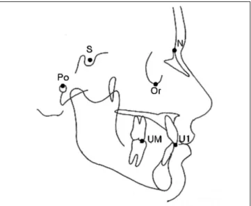
술전 3주 이내, 술후 3일 이내 채득한 측모방사선사진을 분석에 이용하였다. 얻어진 방사선 사진을 교정용 영상진 단 프로그램(V-cephTM 4.0, Cyber-Med Inc., Seoul, Korea) 을 이용하여 Fig. 1과 같이 6개의 계측점을 설정하였다. 이 때 측정에 대한 신뢰도를 평가하기 위해 처음 계측 후 10일 후 동일한 계측자가 다시 반복 계측을 하였다. 두 번의 계 측을 Pearson 상관분석을 이용해 비교한 결과 반복 측정 시 모든 계측항목에서 높은 상관관계를 나타내어, 검사와 재 검사 측정치 간의 일치도가 높음을 알 수 있었다. 또한 trac- ing 후 확대한 상에서 오차를 수정하였으며 이러한 과정을 통해 술전과 술후의 landmark로 sella nasion (SN) plane과 FH plane이가 동일하게 표시되도록 하였다.
Fig. 1.Anatomic landmark used in this study.
(S: sella, N: nasion, Po: porion, Or: orbitale, UM: upper fist molar mesial cusp tip, U1: upper incisor cusp tip)
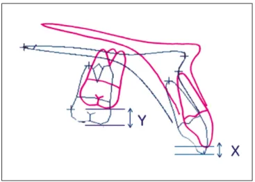
술전 및 술후 FH plane에 대해 occlusal plane angle, incisal inclination을 측정하였으며, 술전, 술후 상악전치 tip의 수직 적 높이 변화량을 X라 하고, 상악 제1대구치의 mesial cusp tip의 수직적 변화량 Y라고 정하였으며 Y-X, 즉 수직적 높 이 차이를 통해 최종 posterior impaction 양을 결정하였 다.(Fig. 2)
3. 통계분석
이번 연구의 주요 목적은 상악의 posterior impaction의 양 에 따른 occlusal plane angle, incisor inclination의 변화를 알 아보는 것이다. 각각의 환자에서 posterior impaction의 양을 측정하고, 이에 따른 occlusal plane angle, incisor inclination 의 변화량을 측정하여 posterior impaction 1 mm당 occlusal plane angle, incisor inclination의 변화값의 평균값을 구하였 으며 이에 관한 유의성은 Pearson’s correlation coefficients 을 통해 검정하였다. 좌우 같은 양 및 다른 양의 posterior impaction을 시행하였을 때 occlusal plane angle, incisal incli- nation의 변화값 사이의 유의성은 Mann-Whitney U-test을 통해 검정하였다. 통계분석은 SPSS version 12.0 (SPSS Inc., Chicago, IL, USA)을 이용하였고 유의수준은 P<0.05에서 검정하였다.
Ⅲ. 결 과
환자 20명의 술전 평균 occlusal plane angle은 7.2�이었으 며 가장 낮은 환자가 -1.75�로 측정되었다. 술전 평균 상악 중절치 incisal inclination은 122.46�였다.(Table 1)
평균 posterior impaction의 양은 2.91 mm이였으며 최대치 는 상악전방부의 inferior positioning을 동반하여 posterior impaction을 시행한 환자로 7.75 mm였다. 술후 평균
occlusal plane angle, incisal inclination 변화량은 6.54°이며, occlusal plane angle, incisal inclination은 상악의 회전과 함 께 변화하는 수치이므로 변화량은 동일하였다.(Table 2) 결 과적으로 posterior impaction 1 mm당 occlusal plane angle, incisal inclination 변화량은 2.25°이였으며 높은 통계학적 유의성을 보이는 것으로 나타났다.(Pearson’s correlation coefficients: 0.969, P<0.01)
한편, 좌우 같은 양 및 다른 양의 posterior impaction을 시 행하였을 때 occlusal plane angle, incisal inclination의 변화 를 살펴본 결과, 좌우 같은 양의 posterior impaction에서는 2.20°이었으며, 좌우 다른 양의 posterior impaction에서는 2.35°로 두 수치 간에 통계학적으로 차이는 없었다.(Table 3)
Ⅳ. 고 찰
Le FortⅠ골절단술은 1864년 Cheever가 비인두강의 종괴 절제를 위한 상악골의 down fracture를 최초로 소개한 이후, 1921년 Wasmund에 의해 안면기형의 수정을 위한상악골절 단술로 이용되었다. 그러나 기술적인 어려움 등으로 주로 하악골수술만으로 악안면기형을 해결하다가, 1965년경에 이르러서야 Wilmar, Obwegeser, Bell 등에 의해 악안면기형 의 해결을 위한 상악골절단술의 술후 안정성이 보고된 이 후 보편화되었다.
양악수술의 적응증으로 첫째 악골전후방의 skeletal
Fig. 2.Measuring parameters.
(X: vertical change of upper incisor, Y: vertical change of upper first molar, Y-X: amount of posterior Mx. impaction)
Table 1.Average occlusal plane angle and incisal inclina- tion in presurgical state
Mean Range
occlusal plane angle 7.2 -1.75-12.24
incisal inclination 122.46 114.31-132.7
Table 2. Average amount of posterior impaction and average amount of change of occlusal plane angle, incisal inclination after surgery
Amount Mean Range SD
PI 2.91 0.26-7.75 1.78
OPA, II change 6.540.5-16 3.77
OPA, II change due to PI 2.25* 1.5-3.1 0.39 (PI: posterior impaction, OPA: occlusal plane angle, II: incisal incli- nation, *: Pearson's correlation coefficients, 0.969, P<0.01)
Table 3. Average amount of change of occlusal plane angle, incisal inclination in same posterior impaction and differential posterior impaction
Mean SD N P value
Same posterior impaction 2.2 0.27 10
0.393*
Differential posterior impaction 2.35 0.38 10 (*: Mann-Whitney U-test)
deformity가 전후방적으로 12 mm 이상인 심한 하악전돌증, 둘째 수직상악골과다증(vertical maxillary excess)를 동반한 하악전돌증, 셋째 수직상악골과소증(vertical maxillary defi-
ciency)를 동반한 하악전돌증을 들었다11,12. 이러한 적응증
은 최근에 이르러서도 적용되는 기준이며 근래에 와서는 수술기구와 수술방법의 발전 그리고 심미성을 중시하는 환자와 교정의사들의 요구가 늘어나 조금 더 복잡한 양상으 로 상악의 3차원적인 이동이 필요한 경우가 늘어나고 있다.
양악 동시수술에서 Le Fort I 골절단술 시 상악 posterior impaction의 적응증은 상악 교합 평면이 후방으로 canting 되어 있는 경우나 anterior open bite의 경우에 좀 더 나은 심 미적인 결과를 얻기 위해서 시행하기도 하며 하악 이동양 의 증가가 필요할 때 적용 가능하고 또한, 골격성 III급 부 정교합에서 술전교정이 불충분하여 치성보상이 부족한 경 우에도 상악전치각도를 증가시키기 위해 필요한 경우가 있다. 실제로 약간의 전치부 개교를 동반한 골격성 III급 부 정교합의 양악수술에서 교합평면 자체를 시계방향으로 회 전시켜주는 상악의 posterior impaction을 동반하지 않는다 면 후안면고경의 증가로 인한 pterygomasseteric sling과 suprahyoid muscle group의 긴장을 야기하여 술후 불안정을 야기한다고 보고된 바가 있다13. 그와 동반하여 안면고경 자체의 감소가 제한적으로 이루어질 수 있으며 이것은 하 악골의 chin 부위에서의 reduction genioplasty를 부가적으로 시행해야 할 수도 있는 여지를 남기게 되는 경우가 있게 된다.
또한, 상악골의 후방부를 올려서 교합평면의 경사도가 증가하는 시계방향의 회전이 그 반대인 경우인 반시계 방 향의 회전에 비해 유리하고 이 때 하악골의 pogonion 부위 의 후상방 이동으로 인해서 이부가 돌출된 III급 부정교합 의 수술 시 부가적으로 심미적인 증진을 가져올 수 있다고
하였고14,15, Swinnen 등16은 전치부 개교합을 가진 환자에서
Le Fort I 골절단술 시 posterior impaction을 시행한 결과 술 후 안정성이 높게 나타났다고 보고하였다.
Le Fort I 상악골절단술 시 posterior impaction의 비적응증 으로는 상악의 후상방 이동으로 인해 occlusal plane angle과 incisor inclination이 증가하며 이미 교합평면의 경사가 큰 경우에 만약 occlusal plane angle이 temporal bone의 articular eminence의 inclination 보다 커지게 되면 canine guidance와 incisal guidance의 상실 및 작업측과 비작업측의 구치부 교 합 간섭을 유발할 수 있고 또한, 상악전치의 설측 경사로 인한 심미성 상실, lip support의 부족으로 인해 nosolabial angle의 증가를 가져올 수 있다.
이상의 내용을 종합해 볼 때 상악의 posterior impaction은 환자의 심미개선과 높은 안정성을 가진 술식이며 필연적 으로 occlusal plane angle, incisal inclination의 변화를 가져 온다. 따라서 occlusal plane angle과 incisor inclination의 증 가에 따른 문제를 예방하기 위해 술전 occlusal plane angle, incisal inclination을 고려하여 posterior impaction의 양을 결 정해야하며, 현재 시행하는 악교정수술의 술전 surgical
treatment objective (STO)에서도 상악골의 이동양을 결정하 는 기준으로 상악절치의 위치나 각도를 기준으로 하는 경 우가 많다. 하지만 posterior impaction에 따른 occlusal plane angle, incisal inclination의 변화량에 대한 임상적 연구가 부 족한 것은 사실이며, 이는 실제로 술전 STO를 통한 paper surgery를 통해서 어느 정도 상악절치의 위치나 각도의 변 화를 예상가능하기 때문일 것이라 생각한다. 그럼에도 불 구하고 이번 연구에서 확인한 상악의 posterior impaction에 따른 occlusal plane angle, incisal inclination의 변화량은 술 전 교정치료의 양을 결정하는데 도움이 되리라 생각한다.
특히 술전 교정단계에서 골격성 III급 부정교합 환자의 증 가되어 있는 상악 incisal inclination을 상악의 posterior impaction을 통해 감소, 즉 치성보상(decompensation)을 시 킬 수 있으므로 술전 교정 시 incisal inclination의 양을 결정 하는데 도움이 된다. 또한 상악의 posterior impaction에 따 른 occlusal plane angle, incisal inclination의 변화량에 임상 적 평균값을 통해 model surgery 및 수술 중에 일어날 수 있 는 posterior impaction 양의 오차에 대해서 상악절치의 각도 변화를 수술 중에도 수치적으로 예상할 수 있는 자료가 됨 으로써 능동적인 대처가 가능할 것으로 생각한다.
한편 각각의 환자에서 posterior impaction의 양에 따른 occlusal plane angle, incisal inclination의 변화량의 차이가 생기는 원인으로 상악의 전후방 길이 즉, 본 연구에서 측정 한 central incisor에서 제1대구치 사이의 거리의 차이가 변 수로 작용한 것으로 생각한다. 각 환자에서 central incisor 에서 제1대구치 사이의 거리가 다른 원인으로 술전 교정 시 상악의 발치 교정의 여부, 각 환자의 상악악궁 크기, 악 궁 형태가 있다. 따라서 발치 교정을 시행한 경우 central incisor에서 제1대구치 사이의 거리가 짧아져 상악의 poste- rior impaction의 양에 따른 occlusal plane angle, incisal incli- nation의 변화량은 증가할 것이다.
좌우 같은 양 및 다른 양의 posterior impaction을 시행하였 을 때 occlusal plane angle, incisal inclination의 변화 결과에 서 두 결과 간에 통계학적 차이는 없었다. 이는 계측에 있 어서의 오차 때문이라 생각하며 조사한 환자 수가 많았다 면 유의성이 있었으리라 생각한다.
Ⅴ. 결 론
본 연구에서 동아대학교 의료원 구강악안면외과학 교실 에서 시행한 20명의 악교정수술 환자에서 상악골의 poste- rior impaction의 양에 따른 occlusal plane angle, incisor incli- nation 변화량을 계측한 연구결과는 다음과 같다.
1. 상기 연구를 통해 상악골의 posterior impaction 1 mm당 occlusal plane angle과 incisal inclination은 평균 2.25°
의 변화를 보였다.
2. 상악의 posterior impaction 시 좌우 같은 양과 좌우 다른 양의 수술을 시행한 경우에도 occlusal plane angle과
incisal inclination의 변화량에는 큰 차이가 없었다.
3. Occlusal plane angle, incisal inclination 변화량에 미치는 변수로 고려되는 것은 각 환자의 central incisor에서 제 1대구치 사이의 거리 즉, 상악의 전후방 길이의 차이가 변수로 작용할 것으로 판단된다.
이 연구의 결과를 통해 pre-surgical orthodontic preparation 과 pre-surgical treatment planning에 도움이 되리라 생각하 며 또한, posterior impaction의 양에 따른 chin의 후방 이동 양에 대한 수치적인 상관관계를 파악한다면 treatment plan- ning에 더욱 도움이 되리라 여겨진다.
References
1. Blair VP. Operations on the jaw bone and face: a study of etiolo- gy and pathological anatomy of developmental malrelations of the maxilla and mandible to each other and to facial outline and of their operative treatment when beyond the scope of the ortho- dontist. Surg Gynecol Obstet 1907;4:67-78.
2. Epker BN, Fish LC. The surgical orthodontic correction of class III skeletal open-bite. Am J orthod 1978;73:601-18.
3. Choi SW, Park HS, Cha IH. A study on accuracy of the maxillary repositioning in orthognathic surgery by the external measuring technique. J Korean Assoc Oral Maxillofac Surg 1996;22:537- 43.
4. Rotter BE, Zeitler DL. Stability of the Le Fort Ⅰ maxillary os- teotomy after rigid internal fixation. J Oral Maxillofac Surg 1999;57:1080-8; discussion 1089.
5. Moser K, Freihofer HP. Long-term experience with simultaneous movement of the upper and lower jaw. J Maxillofac Surg 1980;8:
271-7.
6. Choi BH, Yoon JH. Soft tissue changes associated with Le Fort I
maxillary advancement. J Korean Assoc Oral Maxillofac Surg 1984;10:175-82.
7. Epker BN, Turvey T, Fish LC. Indication for simultaneous mobi- lization of the maxilla and mandible for the correction of dentofa- cial deformities. Oral Surg Oral Med Oral Pathol 1982;54:369- 81.
8. Turvey TA. Simultaneous mobilization of the maxilla and mandible: surgical technique and results. J Oral Maxillofac Surg 1982;40:96-9.
9. Sarver DM, Weissman SM, Johnston MW. Diagnosis and treat- ment planning of hypodivergent skeletal pattern with clockwise occlusal plane rotation. Int J Adult Orthodon Orthognath Surg 1993;8:113-21.
10. Naini FB, Hunt NP, Moles DR. The relationship between maxil- lary length, differential maxillary impaction, and the change in maxillary incisor inclination. Am J Orthod Dentofacial Orthop 2003;124:526-9.
11. Turvey T, Hall DJ, Fish LC, Epker BN. Surgical-orthodontic treatment planning for simultaneous mobilization of the maxilla and mandible in the correction of dentofacial deformities. Oral Surg Oral Med Oral Pathol 1982;54:491-8.
12. Wolford LM, Chemello PD, Hilliard FW. Occlusal plane alter- ation in orthognathic surgery. J Oral Maxillofac Surg. 1993;51:
730-40.
13. Lee JH, Lee HJ. Stability of occlusal plane after Le Fort I maxil- lary osteotomy in patients with skeletal class III maloccusion. J Korean Assoc Oral Maxillofac Surg 1996;22:429-36.
14. Reyneke JP, Evans WG. Surgical manipulation of the occlusal plane. Int J Adult Orthodon Orthognath Surg 1990;5:99-110.
15. Reitzik M. Skeletal and dental changes after surgical correction of mandibular prognathism. J Oral surg 1980;38:109-16.
16. Swinnen K, Politis C, Willems G, de Bruyne I, Fieuws S, Heidbuchel K, et al. Skeletal and dento-alveolar stability after surgical orthodontic treatment of anterior open bite: a retrospec- tive study. Eur J Orthod 2001;23:547-57.