www.jpis.org
pISSN 2093-2278 eISSN 2093-2286 Copyright © 2010 Korean Academy of PeriodontologyThis is an Open Access article distributed under the terms of the Creative Commons Attribution Non-Commercial License (http://creativecommons.org/licenses/by-nc/3.0/).
A simple approach to preserve keratinized mucosa around implants using a pre-fabricated implant-
retained stent: a report of two cases
Jung-Chul Park1, Ki-Bin Yang2, Youna Choi1, Yong-Tae Kim1, Ui-Won Jung1, Chang-Sung Kim1, Kyoo-Sung Cho1, Jung-Kiu Chai1, Chong-Kwan Kim1, Seong-Ho Choi1*
1Department of Periodontology, Research Institute for Periodontal Regeneration, Yonsei University College of Dentistry, Seoul, Korea
2Goodmorning Dental Clinic, Seoul, Korea
Purpose: There is no consensus regarding the relationship between the width of keratinized mucosa and the health of peri- implant tissues, but clinicians prefer to provide enough keratinized mucosa around dental implants for long-term implant main- tenance. An apically positioned flap during second stage implant surgery is the chosen method of widening the keratinized zone in simple procedures. However, the routine suture techniques used with this method tend to apply tension over the pro- visional abutments and decrease pre-existing keratinized mucosa. To overcome this shortcoming, a pre-fabricated implant-re- tained stent was designed to apply vertical pressure on the labial flap and stabilize it in a bucco-apical direction to create a wide keratinized mucous zone.
Methods: During second stage implant surgery, an apically displaced, partial thickness flap with a lingualized incision was re- tracted. A pre-fabricated stent was clipped over the abutments after connecting to the provisional abutment. Vertical pressure was applied to displace the labial flap. No suture was required and the stent was removed after 10 days.
Results: A clinically relevant amount of keratinized mucosa was achieved around the dental implants. Buccally displaced ke- ratinized mucosa was firmly attached to the underlying periosteum. A slight shrinkage of the keratinized zone was noted after the healing period in one patient, but no discomfort during oral hygiene was reported. Clinically healthy gingiva with enough keratinized mucosa was achieved in both patients.
Conclusions: The proposed technique is a simple and time-effective technique for preserving and providing keratinized tissue around dental implants
Keywords: Dental esthetics, Dental implants, Gingiva.
INTRODUCTION
The significance of keratinized tissue in implant mainte- nance is a controversial issue, and there is a lack of consen- sus in the literature regarding the relationship between the width of the keratinized mucosa and the health of peri-im- plant tissues. Several authors have claimed that there is no
correlation between implant success rate and the presence of keratinized tissue in the peri-implant soft tissue [1-3]. On the other hand, some studies have reported that the presence of an adequate band of keratinized tissue adjacent to the implant reduces inflammation [4,5], hyperplasia [5], and retraction of the marginal peri-implant soft tissues [1,5,6]. Despite the fact that a lack of keratinized tissues does not influence the long- Received: May. 27, 2010; Accepted: Jul. 20, 2010
*Correspondence: Seong-Ho Choi
Department of Periodontology, Yonsei University College of Dentistry, 134 Shinchon-dong, Seodaemun-gu, Seoul 120-752, Korea E-mail: [email protected], Tel: +82-2-2228-3189, Fax: +82-2-392-0398
keratinized tissue around implants may help facilitate restor- ative procedures and improve aesthetics [7,8]. In addition, pro- viding enough keratinized tissue around implants enables patients to maintain good oral hygiene without irritation or discomfort during routine oral hygiene [9]. Therefore, clini- cians usually seek methods that provide a keratinized mucous zone that is as wide as possible around implants.
Various techniques have been proposed for obtaining ade- quate amounts of keratinized tissue. Apically or laterally posi- tioned flaps are two techniques focused on preserving exist- ing keratinized tissue [10,11]. When there is not enough kera- tinized tissue, a free gingival graft (FGG) can be used [10,11]. In shallow vestibules with minimal keratinized tissue, the com- bination of an apically positioned flap (APF) and FGG can be utilized [12]. However, these techniques have certain limita- tions, such as a limited quantity of donor tissue [13], frequent infections, patient discomfort, post-surgical pain, paresthesia, bleeding from the donor area, and unpredictable collapse. Most importantly, these methods are highly technique sensitive and time consuming, and cannot be used routinely by gener- al clinicians. To overcome these shortcomings, an easier tech- nique with fewer complications was considered, and a pre-fab- ricated implant-retained stent (Louis Button®, Dentis, Seoul, Korea) clipped on provisional abutments was developed to easily displace pre-existing keratinized mucosa in the bucco- apical direction, preserving the width of the keratinized mu- cosa and providing flap stabilization without time-consum- ing and difficult suture techniques. Using this stent, vertical pressure is applied over the displaced buccal flap, reducing the dead space under the flap, helping the displaced gingiva quick- ly attach to the underlying periosteum, and preserving the width of the pre-existing keratinized mucosa.
The present case report describes a new and simple tech- nique for increasing the width of keratinized mucosa around implants using a pre-fabricated implant-retained stent clipped over provisional abutments.
CASES DECRIPTION
Case 1
Patient and site description
A 52-year-old woman was referred by the Department of Prosthodontics to the Department of Periodontology, Yonsei University Dental Hospital in April 2008 with a chief complaint of increased mobility on the right second mandibular molar.
The patient was in good general health and was a non-smok- er. The right second mandibular molar had attachment loss of over 8 mm on the lingual side and degree III mobility. The tooth was diagnosed as a hopeless tooth and was extracted.
Table 1 shows the patient information and surgical procedures.
Surgical procedures
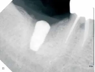
Implant surgery was performed by routine procedures in August 2008. After sequential osteotomies, a threaded sand- blasted large grit acid-etched (SLA) surface root-form implant (Dentium®, Implantium, Seoul, Korea) was placed (Fig. 1). The implant fixture was submerged and left to heal. The surgery and subsequent healing were uneventful.
Second stage surgery was performed 4 months after the first stage surgery in December 2008. The mobile mucosa was par- tially covering the area above the implants with a decreased keratinized tissue width (Fig. 2). An APF with a partial thick- ness flap was chosen for using a pre-fabricated implant-re- tained stent in order to provide a wider zone of keratinized tissue around the implant. The protocol was approved by the Yonsei Institutional Review Board and informed consent was obtained from the patient after careful explanation of the sur- gical procedure, prognosis, and possible complications. After the lingualized incision, leaving 4 mm of keratinized mucosa on the buccal flap, a partial thickness flap was reflected. An additional vertical incision was made to maximize the apical displacement of the existing keratinized mucosa (Fig. 3).
The provisional abutment (ø5.5 × 5 mm) was connected to the fixture, and the pre-fabricated implant-retained stent (Fig. 4) was pushed down over the provisional abutment. A vertical force was applied to stabilize the labial flap and remove the dead space. The lack of stent rotation was confirmed. Origi- nally, the manufacturer recommended that no suture was required. However, simple interrupted sutures (Vicryl® 5.0, Polyglactin 910, Ethicon, Johnson & Johnson, Edinburgh, UK) were made without penetrating the periosteum on the distal
Figure 1. Radiographic photo after first stage implant surgery.
imize stabilization (Fig. 5). However, the displaced flap was mainly stabilized by the stent. The patient was post-operatively administered amoxicillin plus clavulanic acid (Augmentin,
days. The sutures and stent were removed 10 days after sur- gery, and the patient was recalled for a check-up 1 and 2 months after surgery for post-operative care. At the 1-month
Figure 4. Clinical photograph and three dimensional schematic drawing of the pre-fabricated implant-retained stent.
Figure 3. After lingualized incision and retraction of the partial thickness flap, the implant fixture was uncovered.
Figure 2. Clinical photo taken 4 months post-implantation. There was a decreased keratinized mucous zone and the buccal side of
#31 was partially covered with mucosa.
7 mm
Figure 5. The stent was stabilized over the healing abutment. Addi-
tional sutures were placed on the mesial and distal side. Figure 6. Clinical photo taken 1 month post-surgery. Slight shrink- age of the keratinized mucous zone was noted.
3 mm
firm and stabilized over the underlying tissue. However, a 4-mm-wide band of keratinized mucosa on the buccal side was decreased to 3 mm (Fig. 6). Due to the vertical incision, a step was made on the mesiobuccal side of the implant that happened to show the amount of displacement. At the 2-month check up, the patient was referred to the Department of Prosth- odontics for restoration over the implant and increased kera- tinized mucosa was found to be well maintained at the 6-month check up (Fig. 7).
Case 2
Patient and site description
A 49-year-old woman was referred by the Department of Prosthodontics to the Department of Periodontology in Sep- tember 2008 with a chief complaint of missing state on #28,
#29, and #30. The patient was in good general health and was a non-smoker. An intraoral examination revealed an accept- able oral hygiene status. Initial therapy, including scaling and
Surgical procedures
At the time of the implant surgery, the keratinized mucosa was very narrow and the mucosa was partially covering the buccal side of the edentulous area (Fig. 8). FGG was recom- mended to the patient, but the patient refused as the proce- dure could be painful. Therefore, an APF with a partial thick- ness flap was planned to preserve the pre-existing keratinized mucosa and displace the 3-mm-wide band of keratinized mu- cosa to the labial side. Informed consent was obtained from the patient. The same surgery protocols were used as in Case 1. Information regarding implant fixture size (Dentium®, Im- plantium, Seoul, Korea) and the provisional abutment is pro- vided in Table 1. No suture was used in this case and the dis- placed flap was solely stabilized by the stent. The implant sur- Table 1. Patient information and procedures.
Patient 1 Patient 2
Age 52 49
Gender Female Female
PMH N-S N-S
Implant 1st stage August 19, 2008 October 16, 2008 surgery date
Tooth number #31 #28 #30
Fixture
information Ø 4.8 × 10 mm SLA surface root-form implant
Ø 3.8 × 12 mm SLA surface root-form implant
Ø 4.3 × 10 mm SLA surface root-form implant Implant 2nd stage
surgery date 4 months after
1st stage surgery 4 months after 1st stage surgery Provisional
abutment Ø 5.5 × 5 mm Ø 5.5 × 5 mm Ø 5.5 × 5 mm information
Restoration date 2 months after
2nd surgery 2 months after 2nd surgery PMH: past medical history, SLA: sandblasted large grit acid-etched.
Figure 7. The final restoration was seated 3 months post-surgery.
The apically positioned keratinized mucosa was well maintained.
Figure 8. Clinical photo of the implant area (A) before first stage implant surgery and (B) 4 months after first stage surgery. (C) Clinical pho- to of the provisional abutment connection with pre-fabricated implant-retained stent and (D) 2 months after second stage surgery.
A B C D
3 mm
5 mm
gery and subsequent healing were uneventful.
The stent was removed after 10 days and there was no sign of infection. The displaced labial flap was already secured over the underlying periosteum. The patient was referred to the Department of Prosthodontics for restoration after 2 months.
DISCUSSION
Lang and Loe [14] determined how much keratinized gingi- va is required to maintain gingival health; inflammation per- sists in areas with less than 2.0 mm of keratinized gingiva, re- gardless of the patient’s oral health. However, de Trey and Bernimoulin [15] proposed that the width cannot be the only factor deciding the adequacy of the attached gingiva. Other factors, such as the patient’s age, oral hygiene capability, aes- thetic considerations, and patient’s expectations, should be considered, too [16].
Despite reports that a lack of keratinized mucosa may not influence the long-term survival rate of implants [17,18], the presence and reconstruction of keratinized mucosa around implants seems to reduce the discomfort and irritation of pa- tients during oral hygiene. Thus, clinicians consider it essen- tial to provide enough keratinized mucosa in the long-term maintenance of implants, especially in patients who are not able to maintain adequate oral hygiene [9].
To our knowledge, no specific amount of keratinized muco- sa has been recommended as an adequate amount [19]. Cli- nicians usually choose an appropriate technique to maximize the width of keratinized mucosa from the various available methods, such as an APF, laterally positioned flap, FGG, apical- ly positioned partial thickness flap, or connective tissue graft.
However, some of these methods are highly technique sen- sitive and time-consuming, and the suturing procedures are difficult and complicated. Therefore, APF with a partial thick-
ness flap is preferred in general clinics when a minimum band of existing keratinized mucosa exists. This technique requires no graft or complicated sutures and causes less pain to the patient. The method also reduces the overall operation time and produces acceptable results. However, the periosteal su- tures used in this method to stabilize the displaced flap are technique sensitive and time-consuming, and also lack verti- cal pressure over the displaced flap, creating dead space. This context delays the healing process and sometimes induces necrosis. Therefore, the pre-fabricated implant-retained stent was designed to overcome these shortcomings (Fig. 9) and to be used easily in general clinics without any laboratory equip- ment, such as other stents used in mucogingival surgeries [20-25].
In these reported cases, the pre-existing narrow band of ke- ratinized mucosa was moved bucco-apically by the APF with a partial thickness flap and stabilized by the stent. The first clinical case had a vertical incision on the mesial side of the implant during the second stage surgery and additional su- tures were placed. We decided to make two additional sutures, mesially and distally, to provide additional anchorage. The labial flap, however, was mainly held stable by the stent.
In the second case, the keratinized mucous zone was quite narrow. The remaining band of the keratinized zone was 5 mm wide for #28 and 3 mm wide for #30. The lingualized in- cision would seem to provide 2-3 mm of keratinized mucosa on the labial flap. After retracting the partial thickness flap, the stents were clipped on the provisional abutment. The labial flap was pushed bucco-apically and the collapsed gingival con- tour was restored. The increased keratinized mucosa was firm- ly maintained at a 6-month check up.
Further controlled investigations on a larger patient popu- lation are needed to define the efficacy of this stent. Within the limitation of these cases, the pre-fabricated implant-re- Figure 9. Comparison of two techniques using an apically positioned flap (APF) with a partial thickness flap around the dental implants. (A) Periosteal and vertical line sutures. Dead space is created under the displaced flap and sutures are difficult and time-consuming. (B) The pre- fabricated implant-retained stent in the APF with partial thickness flap. Dead space is eliminated by vertical pressure from the stent, and no suture is required on vertical incisions.
Dead
space Time consuming
Technique sensitive Preservation of
keratinized gingiva No dead
space Less time
simple and easy Maxized preservation of keratinized gingiva
placed the labial flap bucco-apically with the existing keratin- ized mucous band and secured its position; 2) the flap was flat- tened over the underlying periosteum and prevented shrink- age and reduction of the keratinized mucosa; 3) the operation time and effort was critically reduced due to no need for peri- osteal or vertical line sutures; 4) the secondary healing area was partially covered by the stent and food impaction or pain was decreased; and 5) the size of the stent was pre-fabricated and standardized according to the size of the provisional abut- ments the clinician used.
Even though no complication was noted in these reported cases, the authors have agreed that there might be certain dis- advantages to this procedure that require precautions before or during usage: 1) dental implant stability must be obtained to securely attach the stent over the provisional abutment; 2) application in the aesthetic region should be avoided; 3) the stent increases the risk of infection in patients with poor oral hygiene or who smoke, so professional prophylaxis must be provided; and 4) there is a chance the stent may rotate or slip out of the abutment. When these precautions are followed properly, this technique could be potentially used in routine implant procedures to provide enough keratinized mucosa.
CONFLICT OF INTEREST
No potential conflict of interest relevant to this article was reported.
ACKNOWLEDGMENTS
This research was supported by the Basic Science Research Program through the National Research Foundation of Ko- rea (NRF) funded by the Ministry of Education, Science and Technology (No. R13-2003-013-04002-0).
REFERENCES
1. Adell R, Lekholm U, Rockler B, Branemark PI, Lindhe J, Er- iksson B, et al. Marginal tissue reactions at osseointegrat- ed titanium fixtures (I). A 3-year longitudinal prospective study. Int J Oral Maxillofac Surg 1986;15:39-52.
2. Lekholm U, Adell R, Lindhe J, Branemark PI, Eriksson B, Rockler B, et al. Marginal tissue reactions at osseointegrat- ed titanium fixtures. (II) A cross-sectional retrospective study. Int J Oral Maxillofac Surg 1986;15:53-61.
3. Schou S, Holmstrup P, Hjorting-Hansen E, Lang NP.
Plaque-induced marginal tissue reactions of osseointe- grated oral implants: a review of the literature. Clin Oral Implants Res 1992;3:149-61.
peri-implantitis in the presence or absence of keratinized mucosa. An experimental study in monkeys. Clin Oral Im- plants Res 1995;6:131-8.
5. Zarb GA, Schmitt A. The longitudinal clinical effectiveness of osseointegrated dental implants: the Toronto study. Part III: Problems and complications encountered. J Prosthet Dent 1990;64:185-94.
6. Adell R, Lekholm U, Rockler B, Branemark PI. A 15-year study of osseointegrated implants in the treatment of the eden- tulous jaw. Int J Oral Surg 1981;10:387-416.
7. Block MS, Kent JN. Factors associated with soft- and hard- tissue compromise of endosseous implants. J Oral Maxil- lofac Surg 1990;48:1153-60.
8. Buser D, Weber HP, Lang NP. Tissue integration of non- submerged implants. 1-year results of a prospective study with 100 ITI hollow-cylinder and hollow-screw implants.
Clin Oral Implants Res 1990;1:33-40.
9. Albrektsson T, Zarb G, Worthington P, Eriksson AR. The long-term efficacy of currently used dental implants: a re- view and proposed criteria of success. Int J Oral Maxillofac Implants 1986;1:11-25.
10. Langer B, Sullivan DY. Osseointegration: its impact on the interrelationship of periodontics and restorative dentistry:
Part I. Int J Periodontics Restorative Dent 1989;9:84-105.
11. Langer B, Langer L. Overlapped flap: a surgical modifica- tion for implant fixture installation. Int J Periodontics Re- storative Dent 1990;10:208-15.
12. Landi L, Sabatucci D. Plastic surgery at the time of mem- brane removal around mandibular endosseous implants:
a modified technique for implant uncovering. Int J Peri- odontics Restorative Dent 2001;21:280-7.
13. Reiser G, Brun J, Mahan P, Larkin L. The subepithelial con- nective tissue graft palatal donor site:Anatomic consider- ations for surgeons. Int J Periodontics Restorative Dent 1996;16:130-7.
14. Lang NP, Loe H. The relationship between the width of keratinized gingiva and gingival health. J Periodontol 1972;
43:623-7.
15. de Trey E, Bernimoulin JP. Influence of free gingival grafts on the health of the marginal gingiva. J Clin Periodontol 1980;7:381-93.
16. Hall WB. Present status of soft tissue grafting. J Periodon- tol 1977;48:587-97.
17. Wennstrom JL, Bengazi F, Lekholm U. The influence of the masticatory mucosa on the peri-implant soft tissue condi- tion. Clin Oral Implants Res 1994;5:1-8.
18. Bengazi F, Wennstrom JL, Lekholm U. Recession of the soft tissue margin at oral implants. A 2-year longitudinal prospective study. Clin Oral Implants Res 1996;7:303-10.
plant dentistry. Chicago: Quintessence Publishing Co.;
2003.
20. Moore JR. A modification of stent design for preprosthet- ic surgery. J Oral Surg 1970;28:263-6.
21. Sanders B, Starshak TJ. Modified technique for palatal mu- cosal grafts in mandibular labial vestibuloplasty. J Oral Surg 1975;33:950-2.
22. Firtell DN, Oatis GW, Curtis TA, Sugg WE Jr. A stent for a split-thickness skin graft vestibuloplasty. J Prosthet Dent 1976;36:204-10.
ness skin grafting and placement of endosteal implants in the edentulous mandible: a preliminary report. J Oral Max- illofac Surg 1992;50:448-51.
24. Nystrom E, Kahnberg KE, Albrektsson T. Treatment of the severely resorbed maxillae with bone graft and titanium implants: histologic review of autopsy specimens. Int J Oral Maxillofac Implants 1993;8:167-72.
25. Brygider RM, Bain CA. Custom stent fabrication for free gingival grafts around osseointegrated abutment fixtures.
J Prosthet Dent 1989;62:320-2.