In the treatment of recurrent anterior instability of the shoulder, surgeons frequently encounter bipolar bone de- fects—glenoid bone defects combined with humeral bone defects.1) The reported incidence of bipolar bone defects in patients with fewer than five dislocation events is 44%;
however, the incidence increases up to 82% with the in- crease in the number of dislocations.1) Bipolar bone defects are known as one of the most important risk factors for postoperative recurrence of anterior shoulder instability and surgical failure.2-4) Therefore, it is important to deter- mine optimal surgical procedures by preoperatively assess-
ing the size of a bipolar bone defect. Generally, a bipolar bone defect is considered substantial and increases the risk of recurrence if a glenoid bone defect is greater than 20%–
25% and an off-track Hill-Sachs lesion is present.2,3,5,6) In 2000, Burkhart and De Beer2) reported a recurrence rate of 67% after arthroscopic Bankart repair in patients with an inverted pear-shaped glenoid. They recommended bony procedures such as the Latarjet procedure to im- prove postoperative stability in patients with large glenoid bone defects. However, recent studies have demonstrated satisfactory clinical outcomes in patients with large bone defects after arthroscopic Bankart repair using additional procedures such as the remplissage procedure or capsu- lar plication.7-10) Still, the 20%–25% glenoid bone defect is often considered the critical cutoff in the literature. In this review article, the authors provide an overview of the effect of glenoid and humeral bone defects on shoulder instability and treatment strategies for recurrent anterior instability of the shoulder with bone defects.
Effects of Glenoid and Humeral Bone Defects on Recurrent Anterior Instability of the Shoulder
In Park, MD, Min-Joon Oh, MD*, Sang-Jin Shin, MD
Department of Orthopedic Surgery, Ewha Womans University Seoul Hospital, Ewha Womans University School of Medicine, Seoul,
*Department of Orthopedic Surgery, Ewha Womans University Mokdong Hospital, Ewha Womans University School of Medicine, Seoul, Korea
For proper treatment of recurrent anterior instability of the shoulder with a bone defect, the defect size should be assessed pre- operatively with three-dimensional computed tomography or magnetic resonance imaging. In general, the risk of postoperative recurrence of instability is estimated on the basis of preoperative imaging of bipolar bone defects: more than 20%–25% glenoid bone loss and off-track Hill-Sachs lesions have been considered risk factors for recurrence. In patients with a glenoid bone defect more than 20%–25%, a bone graft procedure, such as the Latarjet procedure, is preferred regardless of the glenoid track concept, because compared with arthroscopic stabilization procedure, it provides greater postoperative stability. For patients with a border- line glenoid bone defect (around 20%), surgeons should discuss surgical options with the patients, considering their demand and physical activity level. In addition, the surgeon should take care to prevent postoperative instability and long-term complications.
Arthroscopic soft-tissue reconstruction including labral repair and capsular plication combined with the additional remplissage procedure is an anatomical procedure and could be considered as one of the primary treatment methods for patients with glenoid bone defects around 20%. Therefore, treatment strategies for recurrent anterior shoulder instability combined with bone defects should be determined more flexibly on the basis of the patient’s individual condition.
Keywords: Shoulder, Joint instability, Glenoid cavity, Humeral head, Bankart lesions
Copyright © 2020 by The Korean Orthopaedic Association
This is an Open Access article distributed under the terms of the Creative Commons Attribution Non-Commercial License (http://creativecommons.org/licenses/by-nc/4.0) which permits unrestricted non-commercial use, distribution, and reproduction in any medium, provided the original work is properly cited.
Clinics in Orthopedic Surgery • pISSN 2005-291X eISSN 2005-4408 Received April 24, 2019; Accepted August 26, 2019
Correspondence to: Sang-Jin Shin, MD
Department of Orthopedic Surgery, Ewha Womans University Seoul Hospital, Ewha Womans University School of Medicine, 260 Gonghang- daero, Gangseo-gu, Seoul 07804, Korea
Tel: +82-2-2650-5143, Fax: +82-2-2642-0349 E-mail: [email protected]
EFFECT OF GLENOID BONE DEFECTS
Glenoid bone defects are known as one of the important risk factors for recurrence after surgical treatment of in- stability.2,4) The size of a glenoid bone defect should be preoperatively estimated to determine the optimal surgical procedure for recurrent shoulder instability. On the basis of the defect size, surgeons choose either an arthroscopic soft-tissue stabilization procedure or a bone graft pro- cedure.3) However, the cutoff value for determining the surgical procedure remains controversial. The general consensus is that greater than 20%–25% glenoid bone loss requires a bony procedure such as the Latarjet proce- dure.2,3,11) Bigliani et al.12) first emphasized the importance of bone defects that affect more than 25% of the glenoid width in the choice of a treatment method for recurrent anterior shoulder instability; they recommended bony reconstruction procedures as primary surgical treatment.
In a recent clinical study including 223 patients who un- derwent arthroscopic Bankart repair, the postoperative re- currence rate increased up to 21% when the glenoid defect was more than 20% of the glenoid width.13) However, some recent studies have reported that the critical cutoff value should be lower than 20%.14-16) Shaha et al.14) evaluated clinical outcomes and recurrence rates among 72 patients with anterior shoulder instability divided according to the extent of bone defects. They suggested that a glenoid bone defect above 13.5% led to a clinically significant decrease in the Western Ontario Shoulder Instability score even in patients without postoperative recurrence. Another clini- cal study also proposed that the cutoff value of glenoid bone defects for surgical failure should be 17.3% based on the assessment of 169 patients with anterior glenoid erosion.15) In the study, compared to patients with a bone defect of less than 17.3%, patients with a glenoid bone defect of more than 17.3% showed a significantly higher rate of surgical failure. These results were also supported biomechanically by cadaveric studies. In a biomechanical study, Shin et al.17) simulated 10%, 15%, 20%, and 25% gle- noid bone defects with osteotomies. After Bankart repair of each shoulder, they found that shoulders with a glenoid bone defect of 15% or more had significantly higher ante- rior glenohumeral translation than shoulders with less gle- noid loss. Therefore, even a glenoid bone defect less than 20% should be considered as a critical amount of bone defect that requires surgical procedures.
EFFECT OF HUMERAL BONE DEFECTS
Humeral bone defects are frequently accompanied by
glenoid bone defects especially in patients who undergo recurrent instability events.1) Because of their interaction during shoulder abduction and external rotational move- ment, it is important to address these two lesions simul- taneously. The glenoid track concept is an excellent tool to evaluate bipolar bone defects and predict postoperative recurrence by using three-dimensional computed to- mography (3D CT) or magnetic resonance imaging.5,18-20) Metzger et al.21) showed that the glenoid track concept is a good preoperative predictor of humeral head engagement and may help to guide surgical decision making. They classified patients on the basis of the glenoid track con- cept and then compared with clinical evidence of humeral head engagement found during arthroscopic examina- tion. Among patients with an off-track lesion, 84.5% had clinical evidence of engagement, whereas only 12.4% of patients with an on-track lesion showed clinical engage- ment. Many studies regarding the glenoid track concept demonstrated that the off-track lesion is a strong predictor of postoperative recurrence. One clinical study showed a 75% recurrence rate in eight patients with off-track lesions and only an 8% recurrence rate in 49 patients with on- track lesions.6) The positive predictive value of off-track measurement for postoperative recurrence was 75%—the value was more than 20% higher than that of glenoid bone defects (44%), although the sample size of that study was relatively small. In another study evaluating 100 patients who underwent an arthroscopic stabilization procedure, the postoperative recurrence rate was significantly higher in patients with off-track lesions (33%) than in patients with on-track lesions (6%).22) Therefore, Di Giacomo et al.3) recommended arthroscopic Bankart repair with ad- ditional remplissage procedure in patients with off-track lesions if glenoid bone loss was less than 20%. Park et al.22) evaluated clinical outcomes of 23 patients with off-track lesions after arthroscopic Bankart repair combined with additional remplissage procedure.23) They performed the additional remplissage procedure only in patients with an engaging humeral head in arthroscopic examination after Bankart repair with capsular plication. As a result, com- pared with patients with on-track lesions, patients with off-track lesions showed satisfactory clinical outcomes and recurrence rates after arthroscopic Bankart repair with the remplissage procedure. They emphasized the greater importance of the glenoid bone defect than the off-track lesion as a predictor of postoperative surgical failure.
There would be some weaknesses of the glenoid track concept, because on- or off-track lesions are deter- mined only by the bone defect size of the humeral head and glenoid although soft-tissue conditions such as cap-
sulolabrum and glenohumeral ligaments are also impor- tant factors for postoperative stability.9,10) In patients with high-quality anterior capsule, the anterior capsule usually becomes tight and the humeral head rotates internally after Bankart repair with capsular plication. The location of the Hill-Sachs lesion is then altered far from the glenoid anterior margin, leaving little chance to reach the glenoid anterior margin. On the other hand, in patients with poor- quality anterior capsule, this phenomenon would not oc- cur even after proper capsular plication. The shape of Hill- Sachs lesions would also affect the glenoid track concept.
In patients with a wide and shallow Hill-Sachs lesion, there would be little possibility of humeral head engage- ment, even if it is an off-track lesion by the glenoid track concept.24)
TREATMENT STRATEGY FOR RECURRENT ANTERIOR INSTABILITY WITH BONE DEFECTS
The size of a glenoid bone defect could be calculated on the en face view of preoperative 3D CT by using the Suga- ya method.25) Surgeons should determine whether a bony procedure is needed on the basis of the size of a glenoid bone defect. The critical value of a glenoid defect size has been widely considered as 20%–25%; however, that value should be lowered to 13%–17% according to recent clinical and biomechanical studies.14-17) In the treatment of patients with borderline glenoid bone defects, surgeons should discuss surgical options with patients because compared with arthroscopic Bankart repair, bony procedures have inherent advantages and disadvantages. In a recent study comparing clinical outcomes after the Latarjet procedure and arthroscopic Bankart repair in patients with a bor- derline glenoid bone loss of 15%–20%, both procedures
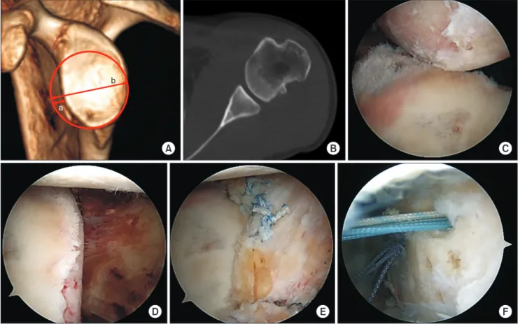
Fig. 1. A 38-year-old male with recurrent anterior instability of the left shoulder. All arthroscopic images were taken from an anterosuperior portal. (A) The glenoid defect size was calculated as 16.3% (a) of the widest glenoid width (b) on the en face view of three-dimensional computed tomography (CT). (B) The wide Hill-Sachs lesion was observed on the axial view of CT. (C) The Hill-Sachs lesion was engaging the anterior margin of the glenoid in the anterior apprehension position (the arm in 90° abduction and 90° external rotation). (D) The anterior capsuloligamentous complex was contracted and medially retracted without appropriate tension. (E) The capsuloligamentous complex regained appropriate tension after mobilization and repair using suture anchors. (F) Additional remplissage procedure using two suture anchors was performed because of the engaging Hill-Sachs lesion even after Bankart repair.
b
a
A B C
D E F
provided satisfactory clinical outcomes and pain relief;
however, the recurrence rate was significantly lower after the Latarjet procedure than arthroscopic Bankart repair.26) For patients with more than 20%–25% glenoid bone loss, many clinical studies recommend bony procedures such as the Latarjet procedure because of the low recurrence rate:
5%–17% after the Latarjet procedure vs 11%–16% after the arthroscopic stabilization procedure.7,8,27,28) According to a meta-analysis study comparing clinical outcomes and recurrence rates after the Latarjet procedure and Bankart repair, the Latarjet procedure offered greater postoperative stability although differences in the glenoid bone defect size were not considered in the study. However, compared with arthroscopic stabilization surgery, the bony proce- dure has been associated with a higher incidence of surgi- cal complications. One meta-analysis reported a 13.4%
overall complication rate after glenoid bone grafting, such as iatrogenic nerve palsy, graft nonunion, fracture, hema- toma formation, and screw loosening.29) Compared to the Latarjet procedure, arthroscopic Bankart repair showed a lower incidence of postoperative complications (0%–3.1%) even when combined with a complex bone incorporation procedure.30,31) Therefore, patient’s demand and physical activity level should be considered first when the surgeon determines surgical options in patients with a glenoid defect around 20%. A recent clinical analysis provided sat- isfactory clinical outcomes after an arthroscopic stabiliza- tion procedure combined with the remplissage procedure, which is considered as a primary treatment option even in patients with a glenoid bone defect more than 20%.11) The authors of the study proposed that compared to nonana- tomic reconstruction such as bony procedures, anatomi- cal reconstruction using soft-tissue procedures including labral repair with capsular plication would result in less complications.
For the Hill-Sachs lesion, the engagement into the anterior edge of the glenoid was the important factor for determining the necessity of additional procedures such as the remplissage procedure in the past. Nowadays, treat- ment of the Hill-Sachs lesion is based on the combined glenoid defect. It would be important to determine wheth- er the patient has an on- or off-track lesion. According to the glenoid track concept, the width of the glenoid track and the width of the Hill-Sachs lesion are used to deter- mine whether the patient has an on- or off-track lesion.
The width of the glenoid track is calculated as 83% of the normalized glenoid width minus the glenoid bone defect width.19) If the width of the Hill-Sachs lesion is greater than the width of the glenoid track, the patient is considered to have an off-track lesion, whereas an on-track lesion is con- sidered present in the opposite case. Generally, in patients with recurrent instability who have an on-track lesion and less than 20% glenoid bone loss, isolated Bankart repair is recommended; the additional remplissage procedure is recommended in patients with an off-track lesion and less than 20% glenoid bone loss.3) Bone graft procedures are necessary in patients who have a glenoid bone defect of more than 20% regardless of the glenoid track lesion. This treatment algorithm was supported by a recent study com- paring clinical outcomes after arthroscopic Bankart repair with selective remplissage procedure between patients with and without off-track lesions.23) In the study, patients with an off-track lesion showed clinical outcomes and re- currence rates comparable to those in patients with an on- track lesion after arthroscopic Bankart repair with selective remplissage procedure. Selective remplissage procedure, performed only when engagement of the humeral head is observed in arthroscopic examination after arthroscopic Bankart repair with capsular plication, should be also con- sidered as a treatment option for off-track lesions (Fig. 1).
CONCLUSION
In the treatment of recurrent anterior instability of the shoulder, the proper treatment strategy is determined on the basis of the evaluation of glenoid and humeral bone defects. However, there are various measurement methods, and the critical value of glenoid and humeral bone defects to determine surgical procedures has yet to be established.
Therefore, the treatment strategy for recurrent anterior instability of the shoulder should be determined more flexibly by the patient’s condition. For more individualized treatment, surgeons should discuss surgical options with patients, considering their demand and physical activity level.
CONFLICT OF INTEREST
No potential conflict of interest relevant to this article was reported.
REFERENCES
1. Nakagawa S, Ozaki R, Take Y, Iuchi R, Mae T. Relationship Between glenoid defects and Hill-Sachs lesions in shoulders
with traumatic anterior instability. Am J Sports Med. 2015;
43(11):2763-73.
2. Burkhart SS, De Beer JF. Traumatic glenohumeral bone defects and their relationship to failure of arthroscopic Ban- kart repairs: significance of the inverted-pear glenoid and the humeral engaging Hill-Sachs lesion. Arthroscopy. 2000;
16(7):677-94.
3. Di Giacomo G, Itoi E, Burkhart SS. Evolving concept of bi- polar bone loss and the Hill-Sachs lesion: from "engaging/
non-engaging" lesion to "on-track/off-track" lesion. Ar- throscopy. 2014;30(1):90-8.
4. Nakagawa S, Mae T, Sato S, Okimura S, Kuroda M. Risk fac- tors for the postoperative recurrence of instability after ar- throscopic Bankart repair in athletes. Orthop J Sports Med.
2017;5(9):2325967117726494.
5. Itoi E. 'On-track' and 'off-track' shoulder lesions. EFORT Open Rev. 2017;2(8):343-51.
6. Shaha JS, Cook JB, Rowles DJ, Bottoni CR, Shaha SH, To- kish JM. Clinical validation of the glenoid track concept in anterior glenohumeral instability. J Bone Joint Surg Am.
2016;98(22):1918-23.
7. Kim SJ, Kim SH, Park BK, Chun YM. Arthroscopic stabi- lization for recurrent shoulder instability with moderate glenoid bone defect in patients with moderate to low func- tional demand. Arthroscopy. 2014;30(8):921-7.
8. Mologne TS, Provencher MT, Menzel KA, Vachon TA, Dewing CB. Arthroscopic stabilization in patients with an inverted pear glenoid: results in patients with bone loss of the anterior glenoid. Am J Sports Med. 2007;35(8):1276-83.
9. Ozbaydar M, Elhassan B, Diller D, Massimini D, Higgins LD, Warner JJ. Results of arthroscopic capsulolabral repair:
Bankart lesion versus anterior labroligamentous periosteal sleeve avulsion lesion. Arthroscopy. 2008;24(11):1277-83.
10. Shin SJ, Ko YW, Lee J. Intra-articular lesions and their rela- tion to arthroscopic stabilization failure in young patients with first-time and recurrent shoulder dislocations. J Shoul- der Elbow Surg. 2016;25(11):1756-63.
11. Park I, Park CJ, Lee JH, Hyun HS, Park JY, Shin SJ. Clinical outcomes and recurrence rates after arthroscopic stabiliza- tion procedures in young patients with a glenoid bone ero- sion: a comparative study between glenoid erosion more and less than 20. Arthroscopy. 2018;34(8):2287-93.
12. Bigliani LU, Newton PM, Steinmann SP, Connor PM, Mcllveen SJ. Glenoid rim lesions associated with recurrent anterior dislocation of the shoulder. Am J Sports Med. 1998;
26(1):41-5.
13. Park I, Lee JH, Hyun HS, Oh MJ, Shin SJ. Effects of bone in- corporation after arthroscopic stabilization surgery for bony
Bankart lesion based on preoperative glenoid defect size.
Am J Sports Med. 2018;46(9):2177-84.
14. Shaha JS, Cook JB, Song DJ, et al. Redefining "critical" bone loss in shoulder instability: functional outcomes worsen with "subcritical" bone loss. Am J Sports Med. 2015;43(7):
1719-25.
15. Shin SJ, Kim RG, Jeon YS, Kwon TH. Critical value of ante- rior glenoid bone loss that leads to recurrent glenohumeral instability after arthroscopic bankart repair. Am J Sports Med. 2017;45(9):1975-81.
16. Yamamoto A, Massimini DF, DiStefano J, Higgins LD. Gle- nohumeral contact pressure with simulated anterior labral and osseous defects in cadaveric shoulders before and after soft tissue repair. Am J Sports Med. 2014;42(8):1947-54.
17. Shin SJ, Koh YW, Bui C, et al. What is the critical value of glenoid bone loss at which soft tissue bankart repair does not restore glenohumeral translation, restricts range of mo- tion, and leads to abnormal humeral head position? Am J Sports Med. 2016;44(11):2784-91.
18. Momaya AM, Tokish JM. Applying the glenoid track con- cept in the management of patients with anterior shoulder instability. Curr Rev Musculoskelet Med. 2017;10(4):463-8.
19. Omori Y, Yamamoto N, Koishi H, et al. Measurement of the glenoid track in vivo as investigated by 3-dimensional mo- tion analysis using open MRI. Am J Sports Med. 2014;42(6):
1290-5.
20. Trivedi S, Pomerantz ML, Gross D, Golijanan P, Provencher MT. Shoulder instability in the setting of bipolar (glenoid and humeral head) bone loss: the glenoid track concept.
Clin Orthop Relat Res. 2014;472(8):2352-62.
21. Metzger PD, Barlow B, Leonardelli D, Peace W, Solomon DJ, Provencher MT. Clinical application of the "glenoid track"
concept for defining humeral head engagement in anterior shoulder instability: a preliminary report. Orthop J Sports Med. 2013;1(2):2325967113496213.
22. Park I, Kang JS, Jo YG, Kim SW, Shin SJ. Off-track Hill- Sachs lesions do not increase postoperative recurrent insta- bility after arthroscopic Bankart repair with selective Rem- plissage procedure. Knee Surg Sports Traumatol Arthrosc.
2019;27(12):3864-70.
23. Kurokawa D, Yamamoto N, Nagamoto H, et al. The preva- lence of a large Hill-Sachs lesion that needs to be treated. J Shoulder Elbow Surg. 2013;22(9):1285-9.
24. Sugaya H, Moriishi J, Dohi M, Kon Y, Tsuchiya A. Glenoid rim morphology in recurrent anterior glenohumeral insta- bility. J Bone Joint Surg Am. 2003;85(5):878-84.
25. Jeon YS, Jeong HY, Lee DK, Rhee YG. Borderline glenoid bone defect in anterior shoulder instability: latarjet proce-
dure versus bankart repair. Am J Sports Med. 2018;46(9):
2170-6.
26. Mizuno N, Denard PJ, Raiss P, Melis B, Walch G. Long-term results of the Latarjet procedure for anterior instability of the shoulder. J Shoulder Elbow Surg. 2014;23(11):1691-9.
27. Yang JS, Mazzocca AD, Cote MP, Edgar CM, Arciero RA.
Recurrent anterior shoulder instability with combined bone loss: treatment and results with the modified latarjet proce- dure. Am J Sports Med. 2016;44(4):922-32.
28. Beran MC, Donaldson CT, Bishop JY. Treatment of chronic glenoid defects in the setting of recurrent anterior shoulder instability: a systematic review. J Shoulder Elbow Surg. 2010;
19(5):769-80.
29. An VV, Sivakumar BS, Phan K, Trantalis J. A systematic review and meta-analysis of clinical and patient-reported outcomes following two procedures for recurrent traumatic anterior instability of the shoulder: Latarjet procedure vs.
Bankart repair. J Shoulder Elbow Surg. 2016;25(5):853-63.
30. Sugaya H, Moriishi J, Kanisawa I, Tsuchiya A. Arthroscopic osseous Bankart repair for chronic recurrent traumatic an- terior glenohumeral instability. J Bone Joint Surg Am. 2005;
87(8):1752-60.
31. Sugaya H, Moriishi J, Kanisawa I, Tsuchiya A. Arthroscopic osseous Bankart repair for chronic recurrent traumatic an- terior glenohumeral instability. J Bone Joint Surg Am. 2005;
87(8):1752-60.